BrainSCUBA: Fine-Grained Natural Language Captions of Visual Cortex Selectivity
Abstract
Understanding the functional organization of higher visual cortex is a central focus in neuroscience. Past studies have primarily mapped the visual and semantic selectivity of neural populations using hand-selected stimuli, which may potentially bias results towards pre-existing hypotheses of visual cortex functionality. Moving beyond conventional approaches, we introduce a data-driven method that generates natural language descriptions for images predicted to maximally activate individual voxels of interest. Our method – Semantic Captioning Using Brain Alignments (“BrainSCUBA”) – builds upon the rich embedding space learned by a contrastive vision-language model and utilizes a pre-trained large language model to generate interpretable captions. We validate our method through fine-grained voxel-level captioning across higher-order visual regions. We further perform text-conditioned image synthesis with the captions, and show that our images are semantically coherent and yield high predicted activations. Finally, to demonstrate how our method enables scientific discovery, we perform exploratory investigations on the distribution of “person” representations in the brain, and discover fine-grained semantic selectivity in body-selective areas. Unlike earlier studies that decode text, our method derives voxel-wise captions of semantic selectivity. Our results show that BrainSCUBA is a promising means for understanding functional preferences in the brain, and provides motivation for further hypothesis-driven investigation of visual cortex.
Andrew F. Luo
Carnegie Mellon University
afluo@cmu.edu
Margaret M. Henderson
Carnegie Mellon University
mmhender@cmu.edu
Michael J. Tarr
Carnegie Mellon University
michaeltarr@cmu.edu
Leila Wehbe
Carnegie Mellon University
lwehbe@cmu.edu
1 Introduction
The recognition of complex objects and semantic visual concepts is supported by a network of regions within higher visual cortex. Past research has identified the specialization of certain regions in processing semantic categories such as faces, places, bodies, words, and food (Puce et al., 1996; Kanwisher et al., 1997; McCarthy et al., 1997; Maguire, 2001; Epstein & Kanwisher, 1998; Grill-Spector, 2003; Downing et al., 2001; Khosla et al., 2022; Pennock et al., 2023; Jain et al., 2023). Notably, the discovery of these regions has largely relied on a hypothesis-driven approach, whereby the researcher hand-selects stimuli to study a specific hypothesis. This approach risk biasing the results as it may fail to capture the complexity and variability inherent in real-world images, which can lead to disagreements regarding a region’s functional selectivity (Gauthier et al., 1999).
To better address these issues, we introduce BrainSCUBA (Semantic Captioning Using Brain Alignments), an approach for synthesizing per-voxel natural language captions that describe voxel-wise preferred stimuli. Our method builds upon the availability of large-scale fMRI datasets (Allen et al., 2022) with a natural image viewing task, and allows us to leverage contrastive vision-language models and large-language models in identifying fine-grained voxel-wise functional specialization in a data-driven manner. BrainSCUBA is conditioned on weights from an image-computable fMRI encoder that maps from image to voxel-wise brain activations. The design of our encoder allows us to extract the optimal encoder embedding for each voxel, and we use a training-free method to close the modality gap between the encoder-weight space and natural images. The output of BrainSCUBA describes (in words) the visual stimulus that maximally activates a given voxel. Interpretation and visualization of these captions facilitates data-driven investigation into the underlying feature preferences across various visual sub-regions in the brain.
In contrast to earlier studies that decode text from the brain activity related to an image, we demonstrate voxel-wise captions of semantic selectivity. Concretely, we show that our method captures the categorical selectivity of multiple regions in visual cortex. Critically, the content of the captions replicates the field’s pre-existing knowledge of each region’s preferred category. We further show that BrainSCUBA combined with a text-to-image model can generate images semantically aligned with targeted brain regions and yield high predicted activations when evaluated with a different encoder backbone. Finally, we use BrainSCUBA to perform data-driven exploration for the coding of the category “person”, finding evidence for person-selective regions outside of the commonly recognized face/body domains and discovering new finer-grained selectivity within known body-selective areas.
2 Related Work
Several recent studies have yielded intriguing results by using large-scale vision-language models to reconstruct images and text-descriptions from brain patterns when viewing images (Takagi & Nishimoto, 2022; Chen et al., 2022; Doerig et al., 2022; Ferrante et al., 2023; Ozcelik & VanRullen, 2023; Liu et al., 2023), or to generate novel images that are predicted to activate a given region (Ratan Murty et al., 2021; Gu et al., 2022; Luo et al., 2023). Broadly speaking, these approaches require conditioning on broad regions of the visual cortex, and have not demonstrated the ability to scale down and enable voxel-level understanding of neural selectivity. Additionally, these methods produce images rather than interpretable captions. Below we review some of this work in more detail.
Semantic Selectivity in Higher Visual Cortex.
Higher visual cortex in the human brain contains regions which respond selectively to specific categories of visual stimuli, such as faces, places, bodies, words, and food (Desimone et al., 1984; Puce et al., 1996; Kanwisher et al., 1997; McCarthy et al., 1997; Maguire, 2001; Epstein & Kanwisher, 1998; Grill-Spector, 2003; Downing et al., 2001; Cohen et al., 2000; Khosla et al., 2022; Pennock et al., 2023; Jain et al., 2023). These discoveries have predominantly relied on the use of hand-selected stimuli designed to trigger responses of distinct regions. However the handcrafted nature of these stimuli may misrepresent the complexity and diversity of visual information encountered in natural settings (Gallant et al., 1998; Felsen & Dan, 2005). In contrast, the recent progress in fMRI encoders that map from stimulus to brain response have enabled data-driven computational tests of brain selectivity in vision (Naselaris et al., 2011; Huth et al., 2012; Yamins et al., 2014; Eickenberg et al., 2017; Wen et al., 2018; Kubilius et al., 2019; Conwell et al., 2023; Wang et al., 2022), language (Huth et al., 2016; Deniz et al., 2019), and at the interface of vision and language (Popham et al., 2021). Here, based on Conwell et al. (2023)’s evaluation of the brain alignment of various pre-trained image models, we employ CLIP as our encoder backbone.
Image-Captioning with CLIP and Language Models.
Vision-language models trained with a contrastive loss demonstrate remarkable capability across many discriminative tasks (Radford et al., 2021; Cherti et al., 2023; Sun et al., 2023). However, due to the lack of a text-decoder, these models are typically paired with an adapted language model in order to produce captions. When captioning, some models utilize the full spatial CLIP embedding (Shen et al., 2021; Li et al., 2023a), whilst others use only the vector embedding (Mokady et al., 2021; Tewel et al., 2022; Li et al., 2023b). By leveraging the multi-modal latent space learned by CLIP, we are able to generate voxel-wise captions without human-annotated voxel-caption data.
Brain-Conditioned Image and Caption Generation.
There are two broad directions when it comes to brain conditioned generative models for vision. The first seeks to decode (reconstruct) visual inputs from the corresponding brain acitvations, including works that leverage retrieval, variational autoencoders (VAEs), generative adversarial networks (GANs), and score/energy/diffusion models (Kamitani & Tong, 2005; Han et al., 2019; Seeliger et al., 2018; Shen et al., 2019; Ren et al., 2021; Takagi & Nishimoto, 2022; Chen et al., 2023; Lu et al., 2023; Ozcelik & VanRullen, 2023). Some approaches further utilize or generate captions that describe the observed visual stimuli (Doerig et al., 2022; Ferrante et al., 2023; Liu et al., 2023; Mai & Zhang, 2023).
The second approach seeks to generate stimuli that activates a given region rather than exactly reconstructing the input (Walker et al., 2019; Bashivan et al., 2019). Some of these approaches utilize GANs or Diffusion models to constrain the synthesized output (Ponce et al., 2019; Ratan Murty et al., 2021; Gu et al., 2022; Luo et al., 2023). BrainSCUBA falls under the broad umbrella of this second approach. But unlike prior methods which were restricted to modeling broad swathes of the brain, our method can be applied at voxel-level, and can output concrete interpretable captions.
3 Methods
We aim to generate fine-grained (voxel-level) natural language captions that describe a visual scene which maximally activate a given voxel. We first describe the parameterization and training of our voxel-wise fMRI encoder which goes from images to brain activations. We then describe how we can analytically derive the optimal CLIP embedding given the encoder weights. Finally, we describe how we close the gap between optimal CLIP embeddings and the natural image embedding space to enable voxel-conditioned caption generation. We illustrate our framework in Figure 1.

3.1 Image-to-Brain Encoder Construction
An image-computable brain encoder is a learned function that transforms an image to voxel-wise brain activation beta values represented as a vector of brain voxels , where . Recent work identified models trained with a contrastive vision-language objective as the highest performing feature extractor for visual cortex, with later CLIP layers being more accurate for higher visual areas (Wang et al., 2022; Conwell et al., 2023). As we seek to solely model higher-order visual areas, we utilize a two part design for our encoder. First is a frozen CLIP (Radford et al., 2021) backbone which outputs a dimensional embedding vector for each image. The second is a linear probe with bias , which transform a unit-norm image embedding to brain activations.
| (1) |
After training with MSE loss, we evaluate the encoder on the test set in Figure 2(a) and find that our encoder can achieve high .
3.2 Deriving the Optimal Embedding and Closing the Gap
The fMRI encoder we construct utilizes a linear probe applied to a unit-norm CLIP embedding. It follows from the design that the maximizing embedding for a voxel can be derived efficiently from the weight, and the predicted activation is upper bounded by when
| (2) |
In practice, a natural image that achieves does not typically exist. There is a modality gap between the CLIP embeddings of natural images and the optimal embedding derived from the linear weight matrix. We visualize this gap in Figure 2(b) in a joint UMAP (McInnes et al., 2018) fitted on CLIP ViT-B/32 embeddings and fMRI encoder weights, both normalized to unit-norm. To close this modality gap, we utilize a softmax weighted sum to project the voxel weights onto the space of natural images. Let the original voxel weight be , which we will assume to be unit-norm for convenience. We have a set with natural images . For each image, we compute the CLIP embedding . Given , we use cosine similarity followed by softmax with temperature to compute a score that sums to across all images. For each weight and example image :
| (3) |
We parameterize using a weighted sum derived from the scores, applied to the norms and directions of the image embeddings:
| (4) |
In Figure 2(c) we show the cosine similarity between and as we increase the size of . This projection operator can be treated as a special case of dot-product attention (Vaswani et al., 2017), with , and value equal to norm or direction of . A similar approach is leveraged by Li et al. (2023b), which shows a similar operator outperforms nearest neighbor search for text-only caption inference. As lies in the space of CLIP embeddings for natural images, this allows us to leverage any existing captioning system that is solely conditioned on the final CLIP embedding of an image. We utilize a frozen CLIPCap network, consisting of a projection layer and finetuned GPT-2 (Mokady et al., 2021).

4 Results

In this section, we utilize BrainSCUBA to generate voxel-wise captions and demonstrate that it can capture the selectivity in different semantic regions in the brain. We first show that the generated nouns are interpretable across the entire brain and exhibit a high degree of specificity within pre-identified category-selective regions. Subsequently, we use the captions as input to text-to-image diffusion models to generate novel images, and confirm the images are semantically consistent within their respective regions. Finally, we utilize BrainSCUBA to analyze the distribution of person representations across the brain. These results illustrate BrainSCUBA’s ability to characterize human visual cortical populations, rendering it a promising framework for exploratory neuroscience.
4.1 Setup
We utilize the Natural Scenes Dataset (NSD; Allen et al. (2022)), the largest whole-brain 7T human visual stimuli dataset. Of the subjects, subjects viewed the full image set repeated . We use these subjects, S1, S2, S5, S7, for experiments in the main paper, and present additional results in the appendix. The fMRI activations (betas) are computed using GLMSingle (Prince et al., 2022), and further normalized so each voxel’s response is on a session basis. The response across repeated viewings of the same image is averaged. The brain encoder is trained on the unique images for each subject, while the remaining images viewed by all are used to validate .
The unpaired image projection set is a 2 million combination of LAION-A v2 ( subset) and Open Images (Schuhmann et al., 2022; Kuznetsova et al., 2020). We utilize OpenAI’s ViT-B/32 for the encoder backbone and embedding computation as this is the standard for CLIP conditioned caption generation. For image generation, we use the same model as used by Luo et al. (2023) in BrainDiVE, stable-diffusion-2-1-base with steps of second order DPM-Solver++. In order to ensure direct comparability with BrainDiVE results, OpenCLIP’s CoCa ViT-L/14 is used for image retrieval and zero-shot classification. We define face/place/body/word regions using independent category localizer data provided with the NSD by Allen et al. (2022) (threshold of ), and use the masks provided by Jain et al. (2023) to define the food regions. For details on the human study, please see the appendix.
4.2 Voxel-Wise Text Generations
In this section, we first investigate how BrainSCUBA outputs conceptually tile the higher visual cortex. We perform part-of-speech (POS) tagging and lemmatization of the BrainSCUBA output for four subjects, and extract the top-50 nouns. To extract noun specific CLIP embeddings, we reconstitute them into sentences of the form ”A photo of a/an [NOUN]” as suggested by CLIP. Both the noun embeddings and the brain encoder voxel-wise weights are projected to the space of CLIP image embeddings and normalized to the unit-sphere for UMAP. We utilize UMAP fit on the encoder weights for S1. Results are shown in Figure 3. We observe that the nouns generated by BrainSCUBA are conceptually aligned to pre-identified functional regions. Namely, voxels in extrastriate body area (EBA) are selective to nouns that indicate bodies and activities (green), fusiform face area (FFA-1/FFA-2) exhibits person/body noun selectivity (blue-green), place regions – retrosplenial cortex (RSC), occipital place area (OPA), and parahippocampal place area (PPA) – show selectivity for scene elements (magenta), and the food regions (yellow; Jain et al. (2023)) surrounding FFA exhibit selectivity for food-related nouns. These results show that our framework can characterize the broad semantic selectivity of visual cortex in a zero-shot fashion.

We further quantify the top-10 nouns within each broad category selective region (Figure 4). We observe that BrainSCUBA generates nouns that are conceptually matched to the expected preferred category of each region. Note the multimodal selectivity for words/people/food within the word region has also been observed by Mei et al. (2010); Khosla & Wehbe (2022).
4.3 Text-Guided Brain Image Synthesis
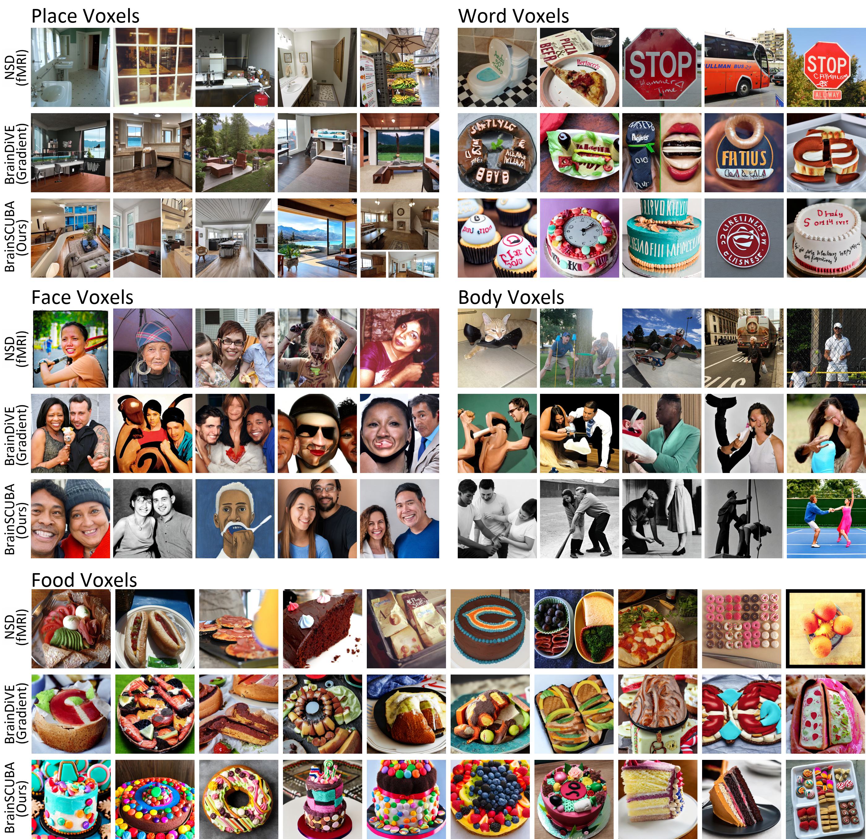
Visualization of the captions can be helpful in highlighting subtle co-occurrence statistics, thereby enriching our understanding of the captions corresponding to images that activate a given region. We utilize a text-to-image diffusion model, and condition the synthesis process on the voxel-wise captions within an ROI (Figure 5).

| \addstackgapFaces | Places | Bodies | Words | Food | Mean | |||||||
|---|---|---|---|---|---|---|---|---|---|---|---|---|
| \addstackgapS2 | S5 | S2 | S5 | S2 | S5 | S2 | S5 | S2 | S5 | S2 | S5 | |
| \addstackgapNSD all stim | 17.1 | 17.5 | 29.4 | 30.7 | 31.5 | 30.3 | 11.0 | 10.1 | 10.9 | 11.4 | 20.0 | 20.0 |
| NSD top-100 | 45.0 | 43.0 | 78.0 | 93.0 | 59.0 | 55.0 | 48.0 | 33.0 | 86.0 | 83.0 | 63.2 | 61.4 |
| \addstackgapBrainDiVE-100 | 68.0 | 64.0 | 100 | 100 | 69.0 | 77.0 | 61.0 | 80.0 | 94.0 | 87.0 | 78.4 | 81.6 |
| \addstackgapBrainSCUBA-100 | 67.0 | 62.0 | 100 | 99.0 | 54.0 | 73.0 | 55.0 | 34.0 | 97.0 | 92.0 | 74.6 | 72.0 |
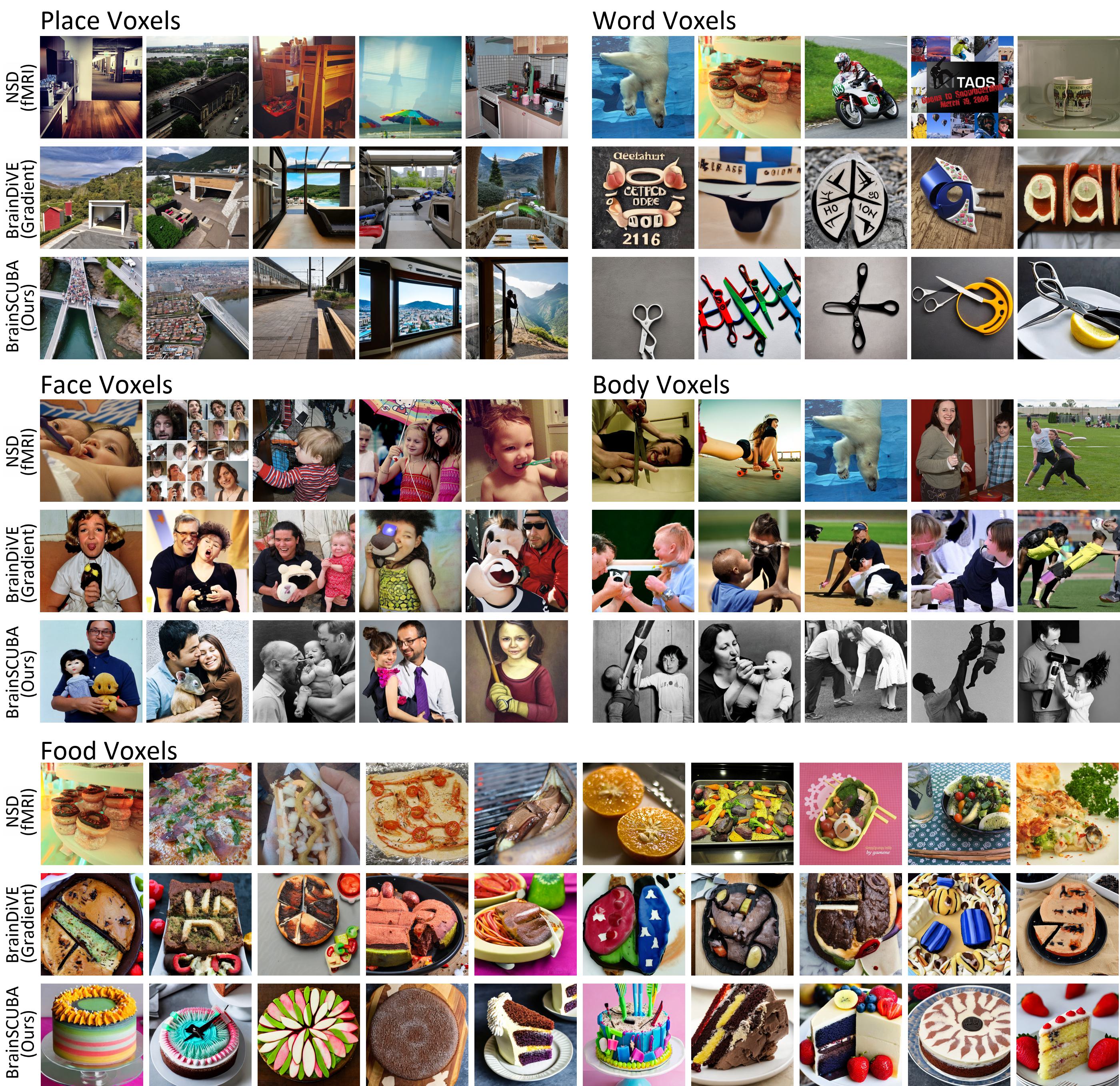
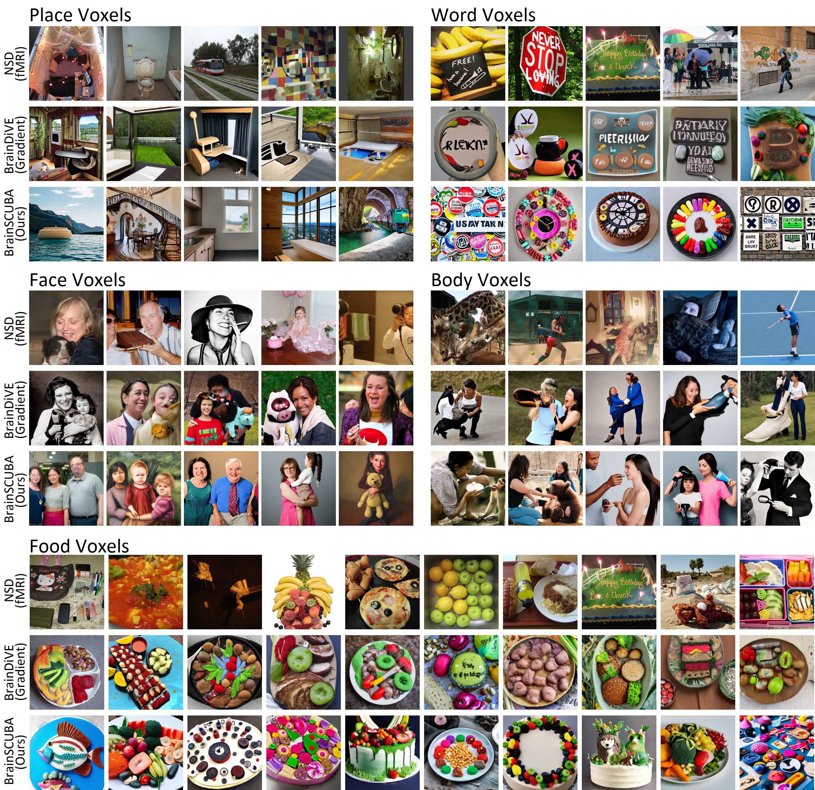
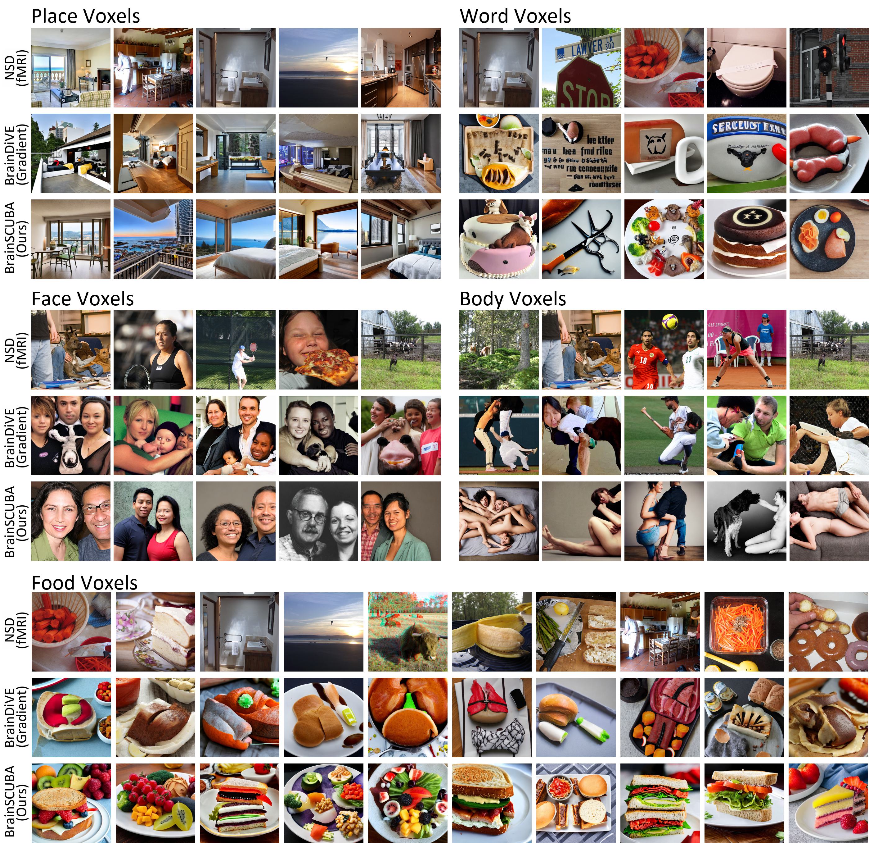
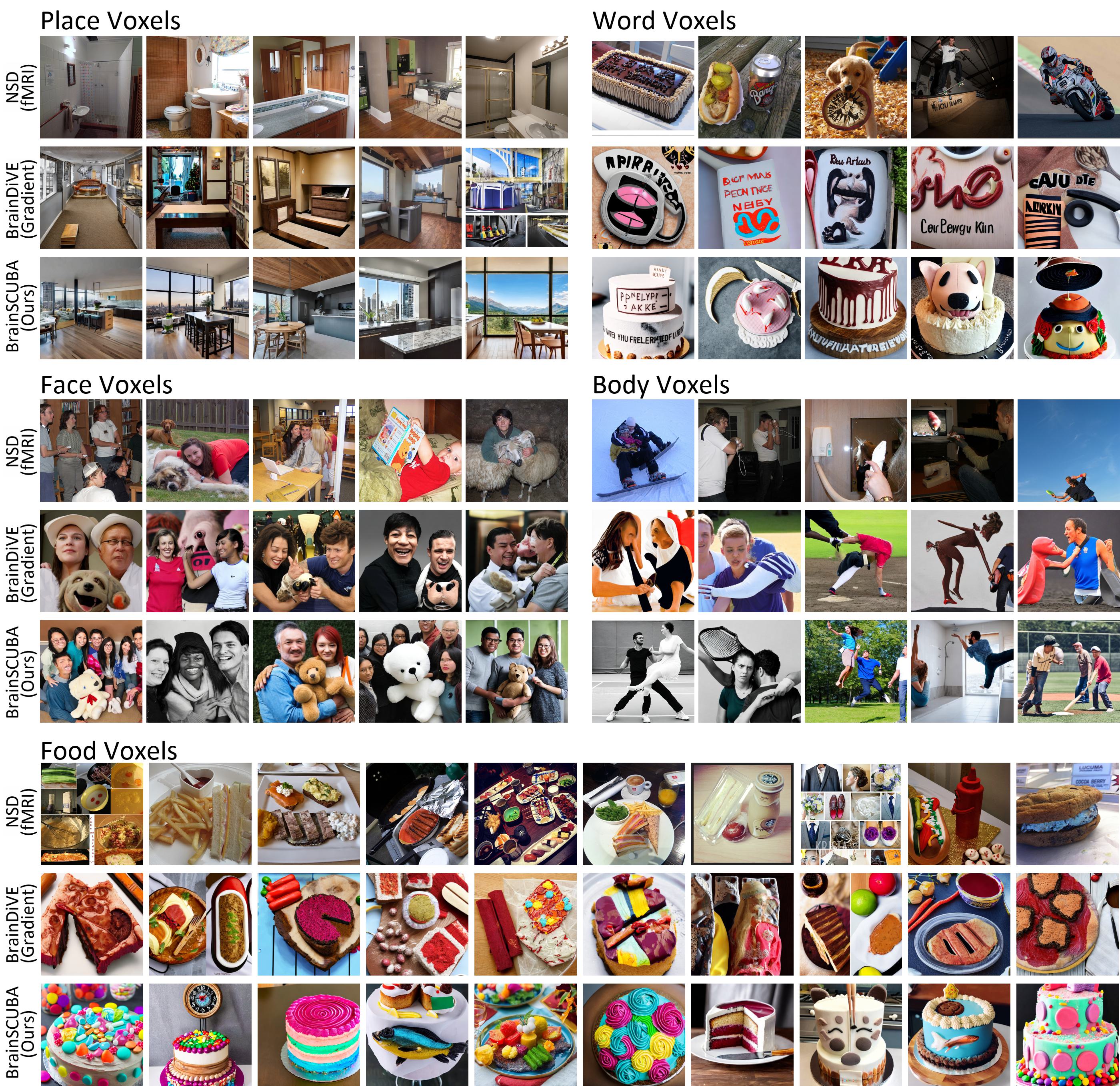
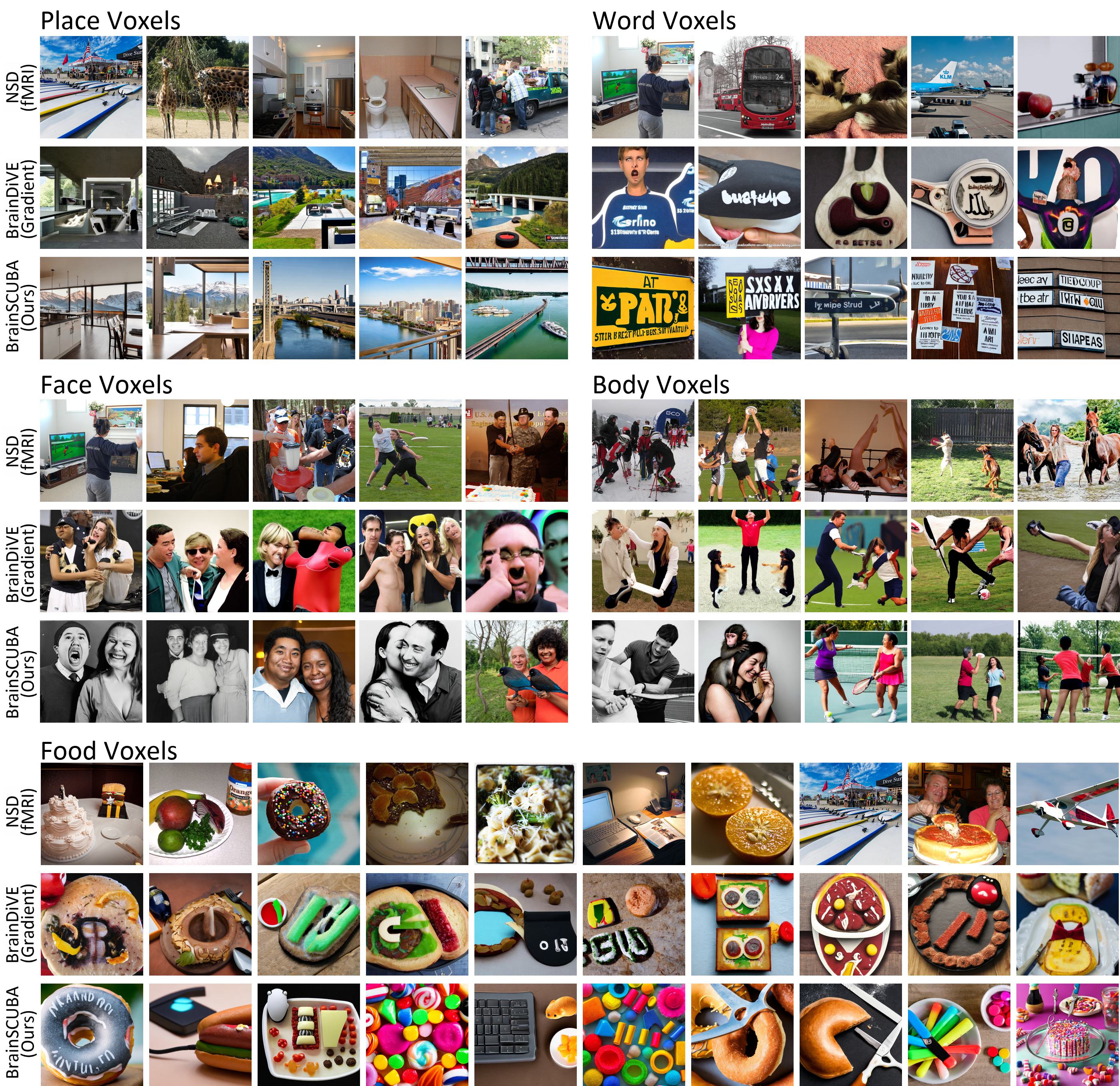
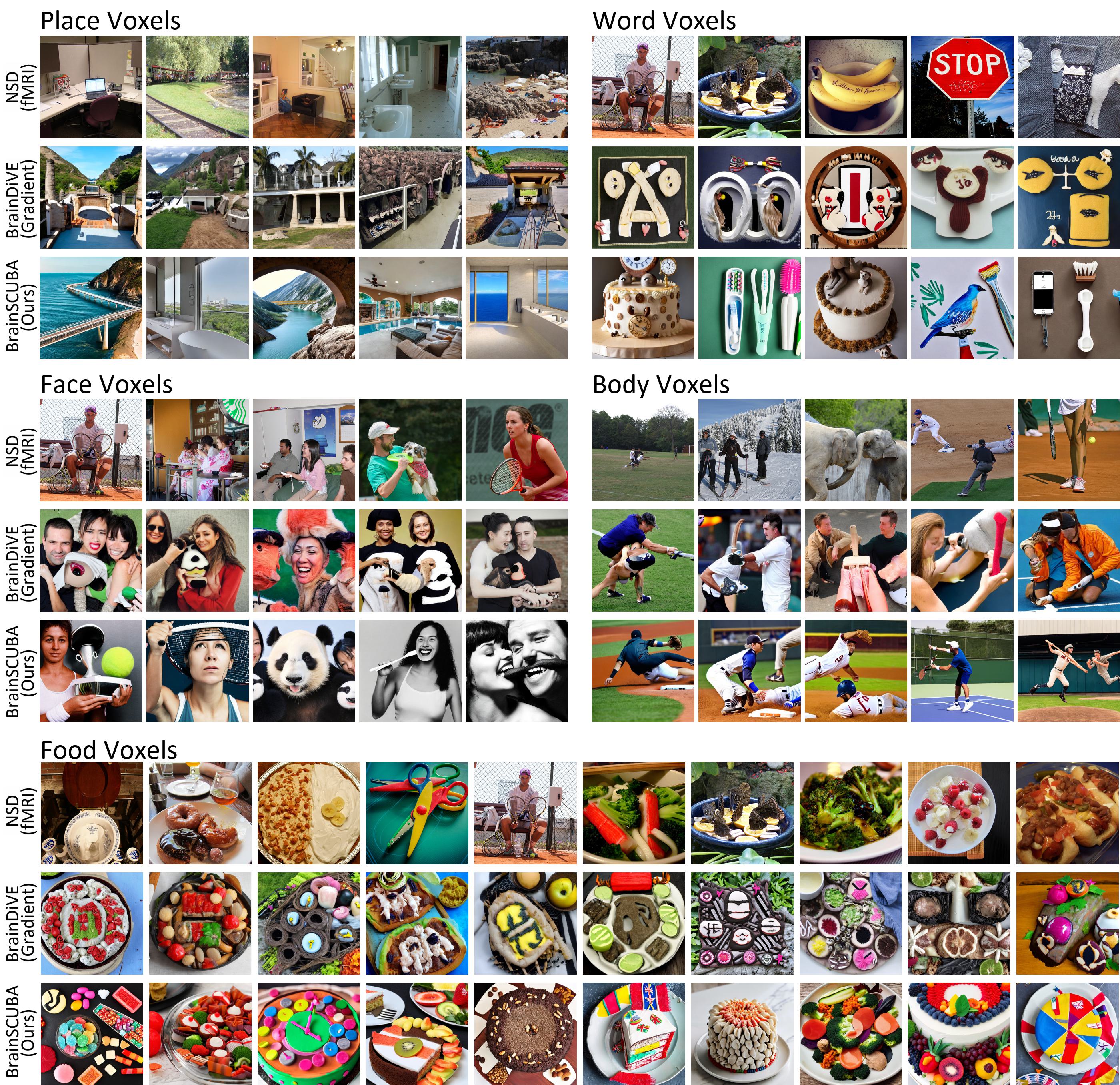
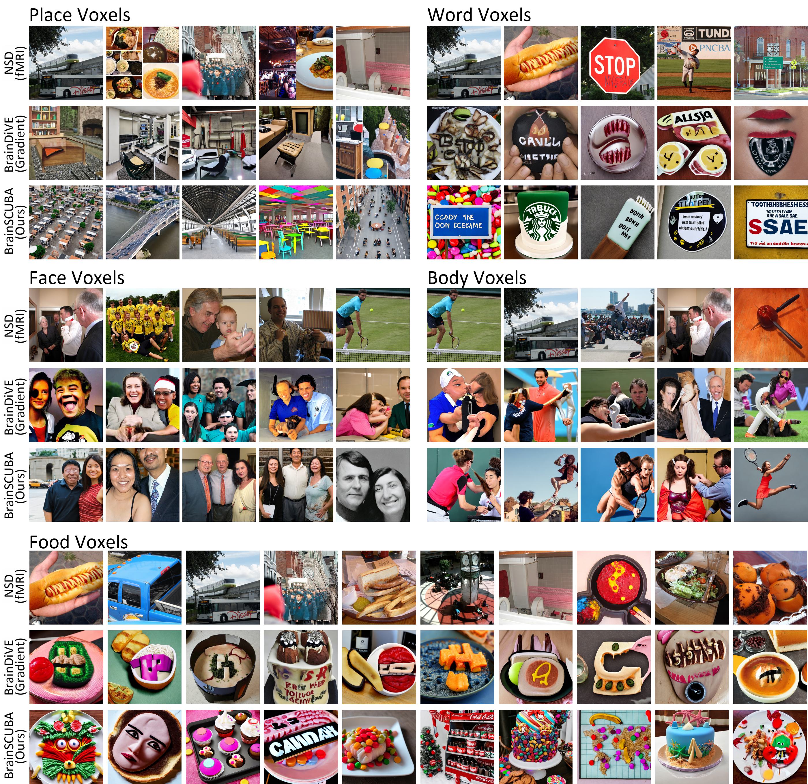
We perform generations per-ROI, subsampling without replacement when the number of voxels/captions in a ROI exceed , and randomly sample the gap when there are fewer than . For face-, place-, word-, body-selective regions, we visualize the top-5 out of images ranked by real average ROI response from the fMRI stimuli (NSD), and the top-5 out of generations ranked by predicted response using the respective BrainDiVE (Luo et al., 2023) and BrainSCUBA encoders. BrainDiVE is used for comparison as it is the state of the art method for synthesizing activating images in the higher visual cortex, and we follow their evaluation procedure. For the food region, we visualize the top-10. Predicted activation is shown in Figure 6, with semantic classification shown in Table 1. Visual inspection suggests our method can generate diverse images semantically aligned with the target category. Our images are generally more visually coherent than those generated by BrainDiVE, and contain more clear text in word voxels, and fewer degraded faces and bodies in the respective regions. This is likely because our images are conditioned on text, while BrainDiVE utilizes the gradient signal alone.
4.4 Investigating the Brain’s Social Network
The intrinsic social nature of humans significantly influences visual perception. This interplay is evident in the heightened visual sensitivity towards social entities such as faces and bodies (Pitcher & Ungerleider, 2021; Kanwisher et al., 1997; Downing et al., 2001). In this section, we explore if BrainSCUBA can provide insights on the finer-grained coding of people in the brain. We use a rule based filter and count the number of captions that contain one of 140 nouns that describe people (person, man, woman, child, boy, girl, family, occupations, and plurals). We visualize the voxels whose captions contain people in Figure 7, and provide a quantitative evaluation in Table 2. We observe that our captions can correctly identify non-person-selective scene, food, and word regions as having lower person content than person-selective ROIs like the FFA or the EBA. Going beyond traditional functional ROIs, we find that the precuneus visual area (PCV) and the temporoparietal junction (TPJ) have a very high density of captions with people. The precuneus has been implicated in third-person mental representations of self (Cavanna & Trimble, 2006; Petrini et al., 2014), while the TPJ has been suggested to be involved in theory of mind and social cognition (Saxe & Kanwisher, 2013). Our results lend support to these hypotheses.

| Non-Person | Person | Other | |||||||
|---|---|---|---|---|---|---|---|---|---|
| RSC | OPA | PPA | Food | Word | EBA | FFA | PCV | TPJ | |
| \addstackgapS1 | 12.9 | 17.3 | 10.6 | 11.5 | 32.0 | 87.2 | 88.5 | 89.7 | 92.1 |
| S2 | 5.58 | 8.15 | 2.70 | 20.0 | 34.8 | 81.4 | 87.2 | 70.8 | 89.1 |
| S5 | 9.31 | 6.43 | 1.95 | 17.8 | 38.4 | 79.5 | 89.4 | 78.5 | 79.9 |
| S7 | 7.14 | 9.87 | 5.99 | 10.7 | 36.9 | 84.3 | 89.5 | 84.2 | 90.3 |
| \addstackgapMean | 8.72 | 10.4 | 5.30 | 15.0 | 35.5 | 83.1 | 88.6 | 80.8 | 87.8 |

| \addstackgapWhich cluster is more… | people per-img | inanimate objs | far away | sports | ||||||||||||
|---|---|---|---|---|---|---|---|---|---|---|---|---|---|---|---|---|
| S1 | S2 | S5 | S7 | S1 | S2 | S5 | S7 | S1 | S2 | S5 | S7 | S1 | S2 | S5 | S7 | |
| \addstackgapEBA-1 (Cluster 1) | 88 | 84 | 91 | 78 | 15 | 11 | 12 | 13 | 62 | 72 | 78 | 63 | 75 | 79 | 85 | 76 |
| EBA-2 (Cluster 2) | 5 | 10 | 4 | 13 | 72 | 80 | 81 | 65 | 21 | 21 | 14 | 25 | 9 | 12 | 6 | 11 |
A close visual examination of Figure 7 suggests a divide within EBA. We perform spherical k-means clustering on joint encoder weights for EBA from S1/S2/S5/S7, and identify two stable clusters. These clusters are visualized in Figure 8. Utilizing the rule parser, we labels the voxels into those that contain a single individual or multiple people, and further visualize the top-nouns within each of these two clusters. While both clusters include general person words like “man” and “woman”, cluster 1 has more nouns that suggest groups of people interacting together (group, game, people), and cluster 2 has words that suggest close-ups of individuals with objects that may be hand-held. To validate our findings, we perform a study where subjects are asked to evaluate the top-100 images from each of the clusters. Results are shown in Table 3. Aligned with the top-nouns, the study suggests that cluster-1 has more groups of people, fewer inanimate objects, and consists of larger scenes. This intriguing novel finding about the fine-grained distinctions in EBA can lead to new hypotheses about its function. This finding also demonstrates the ability of BrainSCUBA to uncover broad functional differences across the visual cortex.
5 Discussion
Limitations and Future Work.
Although our methods can generate semantically faithful descriptions for the broad category selective regions, our approach ultimately relies on a pre-trained captioning model. Due to this, our method reflects the biases of the captioning model. It is further not clear if the most selective object in each region can be perfectly captured by language. Future work could explore the use of more unconstrained captioning models (Tewel et al., 2022) or more powerful language models (Touvron et al., 2023).
Conclusion.
To summarize, in this paper we propose BrainSCUBA, a method which can generate voxel-wise captions to describe each voxel’s semantic selectivity. We explore how the output tiles the higher visual cortex, perform text-conditioned image synthesis with the captions, and apply it to uncover finer-grained patterns of selectivity in the brain within the person class. Our results suggest that BrainSCUBA may be used to facilitate data-driven exploration of the visual cortex.
References
- Allen et al. (2022) Emily J Allen, Ghislain St-Yves, Yihan Wu, Jesse L Breedlove, Jacob S Prince, Logan T Dowdle, Matthias Nau, Brad Caron, Franco Pestilli, Ian Charest, et al. A massive 7t fmri dataset to bridge cognitive neuroscience and artificial intelligence. Nature neuroscience, 25(1):116–126, 2022.
- Bashivan et al. (2019) Pouya Bashivan, Kohitij Kar, and James J DiCarlo. Neural population control via deep image synthesis. Science, 364(6439):eaav9436, 2019.
- Cavanna & Trimble (2006) Andrea E Cavanna and Michael R Trimble. The precuneus: a review of its functional anatomy and behavioural correlates. Brain, 129(3):564–583, 2006.
- Chen et al. (2022) Zijiao Chen, Jiaxin Qing, Tiange Xiang, Wan Lin Yue, and Juan Helen Zhou. Seeing beyond the brain: Conditional diffusion model with sparse masked modeling for vision decoding. arXiv preprint arXiv:2211.06956, 1(2):4, 2022.
- Chen et al. (2023) Zijiao Chen, Jiaxin Qing, Tiange Xiang, Wan Lin Yue, and Juan Helen Zhou. Seeing beyond the brain: Conditional diffusion model with sparse masked modeling for vision decoding. In Proceedings of the IEEE/CVF Conference on Computer Vision and Pattern Recognition, pp. 22710–22720, 2023.
- Cherti et al. (2023) Mehdi Cherti, Romain Beaumont, Ross Wightman, Mitchell Wortsman, Gabriel Ilharco, Cade Gordon, Christoph Schuhmann, Ludwig Schmidt, and Jenia Jitsev. Reproducible scaling laws for contrastive language-image learning. In Proceedings of the IEEE/CVF Conference on Computer Vision and Pattern Recognition, pp. 2818–2829, 2023.
- Cohen et al. (2000) Laurent Cohen, Stanislas Dehaene, Lionel Naccache, Stéphane Lehéricy, Ghislaine Dehaene-Lambertz, Marie-Anne Hénaff, and François Michel. The visual word form area: spatial and temporal characterization of an initial stage of reading in normal subjects and posterior split-brain patients. Brain, 123(2):291–307, 2000.
- Conwell et al. (2023) Colin Conwell, Jacob S. Prince, Kendrick N. Kay, George A. Alvarez, and Talia Konkle. What can 1.8 billion regressions tell us about the pressures shaping high-level visual representation in brains and machines? bioRxiv, 2023. doi: 10.1101/2022.03.28.485868.
- Deniz et al. (2019) Fatma Deniz, Anwar O Nunez-Elizalde, Alexander G Huth, and Jack L Gallant. The representation of semantic information across human cerebral cortex during listening versus reading is invariant to stimulus modality. Journal of Neuroscience, 39(39):7722–7736, 2019.
- Desimone et al. (1984) Robert Desimone, Thomas D Albright, Charles G Gross, and Charles Bruce. Stimulus-selective properties of inferior temporal neurons in the macaque. Journal of Neuroscience, 4(8):2051–2062, 1984.
- Doerig et al. (2022) Adrien Doerig, Tim C Kietzmann, Emily Allen, Yihan Wu, Thomas Naselaris, Kendrick Kay, and Ian Charest. Semantic scene descriptions as an objective of human vision. arXiv preprint arXiv:2209.11737, 2022.
- Downing et al. (2001) Paul E Downing, Yuhong Jiang, Miles Shuman, and Nancy Kanwisher. A cortical area selective for visual processing of the human body. Science, 293(5539):2470–2473, 2001.
- Eickenberg et al. (2017) Michael Eickenberg, Alexandre Gramfort, Gaël Varoquaux, and Bertrand Thirion. Seeing it all: Convolutional network layers map the function of the human visual system. NeuroImage, 152:184–194, 2017.
- Epstein & Kanwisher (1998) Russell Epstein and Nancy Kanwisher. A cortical representation of the local visual environment. Nature, 392(6676):598–601, 1998.
- Felsen & Dan (2005) Gidon Felsen and Yang Dan. A natural approach to studying vision. Nature neuroscience, 8(12):1643–1646, 2005.
- Ferrante et al. (2023) Matteo Ferrante, Furkan Ozcelik, Tommaso Boccato, Rufin VanRullen, and Nicola Toschi. Brain captioning: Decoding human brain activity into images and text. arXiv preprint arXiv:2305.11560, 2023.
- Gallant et al. (1998) Jack L Gallant, Charles E Connor, and David C Van Essen. Neural activity in areas v1, v2 and v4 during free viewing of natural scenes compared to controlled viewing. Neuroreport, 9(7):1673–1678, 1998.
- Gauthier et al. (1999) Isabel Gauthier, Marlene Behrmann, and Michael J Tarr. Can face recognition really be dissociated from object recognition? Journal of cognitive neuroscience, 11(4):349–370, 1999.
- Glasser et al. (2016) Matthew F Glasser, Timothy S Coalson, Emma C Robinson, Carl D Hacker, John Harwell, Essa Yacoub, Kamil Ugurbil, Jesper Andersson, Christian F Beckmann, Mark Jenkinson, et al. A multi-modal parcellation of human cerebral cortex. Nature, 536(7615):171–178, 2016.
- Grill-Spector (2003) Kalanit Grill-Spector. The neural basis of object perception. Current opinion in neurobiology, 13(2):159–166, 2003.
- Gu et al. (2022) Zijin Gu, Keith Wakefield Jamison, Meenakshi Khosla, Emily J Allen, Yihan Wu, Ghislain St-Yves, Thomas Naselaris, Kendrick Kay, Mert R Sabuncu, and Amy Kuceyeski. NeuroGen: activation optimized image synthesis for discovery neuroscience. NeuroImage, 247:118812, 2022.
- Han et al. (2019) Kuan Han, Haiguang Wen, Junxing Shi, Kun-Han Lu, Yizhen Zhang, Di Fu, and Zhongming Liu. Variational autoencoder: An unsupervised model for encoding and decoding fmri activity in visual cortex. NeuroImage, 198:125–136, 2019.
- Huth et al. (2012) Alexander G Huth, Shinji Nishimoto, An T Vu, and Jack L Gallant. A continuous semantic space describes the representation of thousands of object and action categories across the human brain. Neuron, 76(6):1210–1224, 2012.
- Huth et al. (2016) Alexander G Huth, Wendy A De Heer, Thomas L Griffiths, Frédéric E Theunissen, and Jack L Gallant. Natural speech reveals the semantic maps that tile human cerebral cortex. Nature, 532(7600):453–458, 2016.
- Igelström & Graziano (2017) Kajsa M Igelström and Michael SA Graziano. The inferior parietal lobule and temporoparietal junction: a network perspective. Neuropsychologia, 105:70–83, 2017.
- Jain et al. (2023) Nidhi Jain, Aria Wang, Margaret M. Henderson, Ruogu Lin, Jacob S. Prince, Michael J. Tarr, and Leila Wehbe. Selectivity for food in human ventral visual cortex. Communications Biology 2023 6:1, 6:1–14, 2 2023. ISSN 2399-3642. doi: 10.1038/s42003-023-04546-2.
- Kamitani & Tong (2005) Yukiyasu Kamitani and Frank Tong. Decoding the visual and subjective contents of the human brain. Nature neuroscience, 8(5):679–685, 2005.
- Kanwisher et al. (1997) Nancy Kanwisher, Josh McDermott, and Marvin M Chun. The fusiform face area: a module in human extrastriate cortex specialized for face perception. Journal of neuroscience, 17(11):4302–4311, 1997.
- Khosla & Wehbe (2022) Meenakshi Khosla and Leila Wehbe. High-level visual areas act like domain-general filters with strong selectivity and functional specialization. bioRxiv, pp. 2022–03, 2022.
- Khosla et al. (2022) Meenakshi Khosla, N. Apurva Ratan Murty, and Nancy Kanwisher. A highly selective response to food in human visual cortex revealed by hypothesis-free voxel decomposition. Current Biology, 32:1–13, 2022.
- Kubilius et al. (2019) Jonas Kubilius, Martin Schrimpf, Kohitij Kar, Rishi Rajalingham, Ha Hong, Najib Majaj, Elias Issa, Pouya Bashivan, Jonathan Prescott-Roy, Kailyn Schmidt, et al. Brain-like object recognition with high-performing shallow recurrent anns. Advances in neural information processing systems, 32, 2019.
- Kuznetsova et al. (2020) Alina Kuznetsova, Hassan Rom, Neil Alldrin, Jasper Uijlings, Ivan Krasin, Jordi Pont-Tuset, Shahab Kamali, Stefan Popov, Matteo Malloci, Alexander Kolesnikov, et al. The open images dataset v4: Unified image classification, object detection, and visual relationship detection at scale. International Journal of Computer Vision, 128(7):1956–1981, 2020.
- Li et al. (2023a) Junnan Li, Dongxu Li, Silvio Savarese, and Steven Hoi. Blip-2: Bootstrapping language-image pre-training with frozen image encoders and large language models. arXiv preprint arXiv:2301.12597, 2023a.
- Li et al. (2023b) Wei Li, Linchao Zhu, Longyin Wen, and Yi Yang. Decap: Decoding clip latents for zero-shot captioning via text-only training. arXiv preprint arXiv:2303.03032, 2023b.
- Liu et al. (2023) Yulong Liu, Yongqiang Ma, Wei Zhou, Guibo Zhu, and Nanning Zheng. Brainclip: Bridging brain and visual-linguistic representation via clip for generic natural visual stimulus decoding from fmri. arXiv preprint arXiv:2302.12971, 2023.
- Lu et al. (2023) Yizhuo Lu, Changde Du, Dianpeng Wang, and Huiguang He. Minddiffuser: Controlled image reconstruction from human brain activity with semantic and structural diffusion. arXiv preprint arXiv:2303.14139, 2023.
- Luo et al. (2023) Andrew F Luo, Margaret M Henderson, Leila Wehbe, and Michael J Tarr. Brain diffusion for visual exploration: Cortical discovery using large scale generative models. arXiv preprint arXiv:2306.03089, 2023.
- Maguire (2001) Eleanor Maguire. The retrosplenial contribution to human navigation: a review of lesion and neuroimaging findings. Scandinavian journal of psychology, 42(3):225–238, 2001.
- Mai & Zhang (2023) Weijian Mai and Zhijun Zhang. Unibrain: Unify image reconstruction and captioning all in one diffusion model from human brain activity. arXiv preprint arXiv:2308.07428, 2023.
- McCarthy et al. (1997) Gregory McCarthy, Aina Puce, John C Gore, and Truett Allison. Face-specific processing in the human fusiform gyrus. Journal of cognitive neuroscience, 9(5):605–610, 1997.
- McInnes et al. (2018) Leland McInnes, John Healy, and James Melville. Umap: Uniform manifold approximation and projection for dimension reduction. arXiv preprint arXiv:1802.03426, 2018.
- Mei et al. (2010) Leilei Mei, Gui Xue, Chuansheng Chen, Feng Xue, Mingxia Zhang, and Qi Dong. The “visual word form area” is involved in successful memory encoding of both words and faces. Neuroimage, 52(1):371–378, 2010.
- Mokady et al. (2021) Ron Mokady, Amir Hertz, and Amit H Bermano. Clipcap: Clip prefix for image captioning. arXiv preprint arXiv:2111.09734, 2021.
- Naselaris et al. (2011) Thomas Naselaris, Kendrick N Kay, Shinji Nishimoto, and Jack L Gallant. Encoding and decoding in fmri. Neuroimage, 56(2):400–410, 2011.
- Ozcelik & VanRullen (2023) Furkan Ozcelik and Rufin VanRullen. Brain-diffuser: Natural scene reconstruction from fmri signals using generative latent diffusion. arXiv preprint arXiv:2303.05334, 2023.
- Pennock et al. (2023) Ian ML Pennock, Chris Racey, Emily J Allen, Yihan Wu, Thomas Naselaris, Kendrick N Kay, Anna Franklin, and Jenny M Bosten. Color-biased regions in the ventral visual pathway are food selective. Current Biology, 33(1):134–146, 2023.
- Petrini et al. (2014) Karin Petrini, Lukasz Piwek, Frances Crabbe, Frank E Pollick, and Simon Garrod. Look at those two!: The precuneus role in unattended third-person perspective of social interactions. Human Brain Mapping, 35(10):5190–5203, 2014.
- Pitcher & Ungerleider (2021) David Pitcher and Leslie G. Ungerleider. Evidence for a third visual pathway specialized for social perception. Trends in Cognitive Sciences, 25:100–110, 2 2021. ISSN 1364-6613. doi: 10.1016/J.TICS.2020.11.006.
- Ponce et al. (2019) Carlos R Ponce, Will Xiao, Peter F Schade, Till S Hartmann, Gabriel Kreiman, and Margaret S Livingstone. Evolving images for visual neurons using a deep generative network reveals coding principles and neuronal preferences. Cell, 177(4):999–1009, 2019.
- Popham et al. (2021) Sara F Popham, Alexander G Huth, Natalia Y Bilenko, Fatma Deniz, James S Gao, Anwar O Nunez-Elizalde, and Jack L Gallant. Visual and linguistic semantic representations are aligned at the border of human visual cortex. Nature neuroscience, 24(11):1628–1636, 2021.
- Prince et al. (2022) Jacob S Prince, Ian Charest, Jan W Kurzawski, John A Pyles, Michael J Tarr, and Kendrick N Kay. Improving the accuracy of single-trial fmri response estimates using glmsingle. eLife, 11:e77599, nov 2022. ISSN 2050-084X. doi: 10.7554/eLife.77599.
- Puce et al. (1996) Aina Puce, Truett Allison, Maryam Asgari, John C Gore, and Gregory McCarthy. Differential sensitivity of human visual cortex to faces, letterstrings, and textures: a functional magnetic resonance imaging study. Journal of neuroscience, 16(16):5205–5215, 1996.
- Radford et al. (2021) Alec Radford, Jong Wook Kim, Chris Hallacy, A. Ramesh, Gabriel Goh, Sandhini Agarwal, Girish Sastry, Amanda Askell, Pamela Mishkin, Jack Clark, Gretchen Krueger, and Ilya Sutskever. Learning transferable visual models from natural language supervision. In ICML, 2021.
- Ratan Murty et al. (2021) N Apurva Ratan Murty, Pouya Bashivan, Alex Abate, James J DiCarlo, and Nancy Kanwisher. Computational models of category-selective brain regions enable high-throughput tests of selectivity. Nature communications, 12(1):5540, 2021.
- Ren et al. (2021) Ziqi Ren, Jie Li, Xuetong Xue, Xin Li, Fan Yang, Zhicheng Jiao, and Xinbo Gao. Reconstructing seen image from brain activity by visually-guided cognitive representation and adversarial learning. NeuroImage, 228:117602, 2021.
- Saxe & Kanwisher (2013) Rebecca Saxe and Nancy Kanwisher. People thinking about thinking people: the role of the temporo-parietal junction in “theory of mind”. In Social neuroscience, pp. 171–182. Psychology Press, 2013.
- Schuhmann et al. (2022) Christoph Schuhmann, Romain Beaumont, Richard Vencu, Cade Gordon, Ross Wightman, Mehdi Cherti, Theo Coombes, Aarush Katta, Clayton Mullis, Mitchell Wortsman, et al. Laion-5b: An open large-scale dataset for training next generation image-text models. arXiv preprint arXiv:2210.08402, 2022.
- Seeliger et al. (2018) Katja Seeliger, Umut Güçlü, Luca Ambrogioni, Yagmur Güçlütürk, and Marcel AJ van Gerven. Generative adversarial networks for reconstructing natural images from brain activity. NeuroImage, 181:775–785, 2018.
- Shen et al. (2019) Guohua Shen, Tomoyasu Horikawa, Kei Majima, and Yukiyasu Kamitani. Deep image reconstruction from human brain activity. PLoS computational biology, 15(1):e1006633, 2019.
- Shen et al. (2021) Sheng Shen, Liunian Harold Li, Hao Tan, Mohit Bansal, Anna Rohrbach, Kai-Wei Chang, Zhewei Yao, and Kurt Keutzer. How much can clip benefit vision-and-language tasks? arXiv preprint arXiv:2107.06383, 2021.
- Sun et al. (2023) Quan Sun, Yuxin Fang, Ledell Wu, Xinlong Wang, and Yue Cao. Eva-clip: Improved training techniques for clip at scale. arXiv preprint arXiv:2303.15389, 2023.
- Takagi & Nishimoto (2022) Yu Takagi and Shinji Nishimoto. High-resolution image reconstruction with latent diffusion models from human brain activity. bioRxiv, pp. 2022–11, 2022.
- Tewel et al. (2022) Yoad Tewel, Yoav Shalev, Idan Schwartz, and Lior Wolf. Zerocap: Zero-shot image-to-text generation for visual-semantic arithmetic. In Proceedings of the IEEE/CVF Conference on Computer Vision and Pattern Recognition, pp. 17918–17928, 2022.
- Touvron et al. (2023) Hugo Touvron, Louis Martin, Kevin Stone, Peter Albert, Amjad Almahairi, Yasmine Babaei, Nikolay Bashlykov, Soumya Batra, Prajjwal Bhargava, Shruti Bhosale, et al. Llama 2: Open foundation and fine-tuned chat models. arXiv preprint arXiv:2307.09288, 2023.
- Vaswani et al. (2017) Ashish Vaswani, Noam Shazeer, Niki Parmar, Jakob Uszkoreit, Llion Jones, Aidan N Gomez, Łukasz Kaiser, and Illia Polosukhin. Attention is all you need. Advances in neural information processing systems, 30, 2017.
- Walker et al. (2019) Edgar Y Walker, Fabian H Sinz, Erick Cobos, Taliah Muhammad, Emmanouil Froudarakis, Paul G Fahey, Alexander S Ecker, Jacob Reimer, Xaq Pitkow, and Andreas S Tolias. Inception loops discover what excites neurons most using deep predictive models. Nature neuroscience, 22(12):2060–2065, 2019.
- Wang et al. (2022) Aria Yuan Wang, Kendrick Kay, Thomas Naselaris, Michael J Tarr, and Leila Wehbe. Incorporating natural language into vision models improves prediction and understanding of higher visual cortex. BioRxiv, pp. 2022–09, 2022.
- Wen et al. (2018) Haiguang Wen, Junxing Shi, Wei Chen, and Zhongming Liu. Deep residual network predicts cortical representation and organization of visual features for rapid categorization. Scientific reports, 8(1):3752, 2018.
- Yamins et al. (2014) Daniel LK Yamins, Ha Hong, Charles F Cadieu, Ethan A Solomon, Darren Seibert, and James J DiCarlo. Performance-optimized hierarchical models predict neural responses in higher visual cortex. Proceedings of the national academy of sciences, 111(23):8619–8624, 2014.
Appendix A Appendix
Sections
-
1.
Visualization of each subject’s top-nouns for category selective voxels (A.1)
-
2.
Visualization of UMAPs for all subjects (A.2)
-
3.
Novel image generation for all subjects (A.3)
-
4.
Distribution of “person” representations across the brain for all subjects (A.4)
-
5.
Additional extrastriate body area (EBA) clustering results (A.5)
-
6.
Human study details (A.6)
-
7.
Training and inference details (A.7)
A.1 Visualization of each subject’s top-nouns for category selective voxels

A.2 Visualization of UMAPs for all subjects


A.3 Novel image generation for all subjects

A.4 Distribution of “person” representations across the brain for all subjects

| \addstackgapNon-Person | Person | Other | |||||||
|---|---|---|---|---|---|---|---|---|---|
| RSC | OPA | PPA | Food | Word | EBA | FFA | PCV | TPJ | |
| \addstackgapS1 | 12.9 | 17.3 | 10.6 | 11.5 | 32.0 | 87.2 | 88.5 | 89.7 | 92.1 |
| S2 | 5.58 | 8.15 | 2.70 | 20.0 | 34.8 | 81.4 | 87.2 | 70.8 | 89.1 |
| S3 | 6.57 | 16.9 | 4.49 | 24.4 | 33.9 | 84.7 | 90.3 | 75.3 | 83.2 |
| S4 | 4.40 | 14.7 | 4.47 | 20.0 | 37.8 | 78.9 | 90.3 | 66.5 | 88.9 |
| S5 | 9.31 | 6.43 | 1.95 | 17.8 | 38.4 | 79.5 | 89.4 | 78.5 | 79.9 |
| S6 | 16.7 | 28.2 | 6.93 | 27.1 | 48.8 | 91.8 | 97.8 | 75.4 | 79.1 |
| S7 | 7.14 | 9.87 | 5.99 | 10.7 | 36.9 | 84.3 | 89.5 | 84.2 | 90.3 |
| S8 | 15.7 | 30.9 | 9.84 | 42.6 | 57.5 | 86.7 | 96.2 | 71.2 | 89.7 |
| \addstackgapMean | 9.78 | 16.5 | 5.86 | 21.8 | 40.0 | 84.3 | 91.2 | 76.4 | 86.5 |
A.5 Additional extrastriate body area (EBA) clustering results

| \addstackgapSingle | Multiple | |||
|---|---|---|---|---|
| EBA-1 | EBA-2 | EBA-1 | EBA-2 | |
| \addstackgapS1 | 21.8 | 68.5 | 78.2 | 31.5 |
| S2 | 31.5 | 69.5 | 68.6 | 30.5 |
| S5 | 28.8 | 75.2 | 71.2 | 24.8 |
| S7 | 29.0 | 63.8 | 71.0 | 36.2 |
| \addstackgapMean | 27.8 | 69.3 | 72.3 | 30.8 |
A.6 Human study details
Ten subjects were recruited via prolific.co. These subjects are aged ; asian, black, white; men, women. For each NSD subject (S1/S2/S5/S7), we select the top- images for each cluster as ranked by the real average fMRI response. Each of the images were randomly split into non-overlapping subgroups.
Questions were posed in two formats. In the first format, subjects were simultaneously presented with images from the two clusters, and select the set where an attribute was more prominent, possible answers include cluster-1/cluster-2/same. The second format asked subjects to evaluate a set of image from a single cluster, and answer yes/no on if an attribute/object-type was present in most of the images.
For each question, a human evaluator would perform comparisons, from splits and the NSD subjects; with human evaluators per question. For the human study results in section 4.4, we collected total responses for the four questions.
Due to space constraints, we present the single set attribute evaluation (second format described above) results here in the appendix. We divide the results into two tables for presentation purposes.
| \addstackgapAre most images… | social | sports | large-scale scene | animals | ||||||||||||
|---|---|---|---|---|---|---|---|---|---|---|---|---|---|---|---|---|
| S1 | S2 | S5 | S7 | S1 | S2 | S5 | S7 | S1 | S2 | S5 | S7 | S1 | S2 | S5 | S7 | |
| \addstackgapEBA-1 | 88 | 80 | 85 | 85 | 90 | 85 | 88 | 100 | 80 | 85 | 83 | 85 | 20 | 20 | 18 | 23 |
| EBA-2 | 28 | 23 | 35 | 45 | 28 | 25 | 30 | 50 | 38 | 28 | 33 | 60 | 30 | 30 | 30 | 28 |
| \addstackgapAre most images… | artificial objs | body parts | human faces | multi person | ||||||||||||
|---|---|---|---|---|---|---|---|---|---|---|---|---|---|---|---|---|
| S1 | S2 | S5 | S7 | S1 | S2 | S5 | S7 | S1 | S2 | S5 | S7 | S1 | S2 | S5 | S7 | |
| \addstackgapEBA-1 | 78 | 78 | 80 | 75 | 73 | 80 | 78 | 83 | 85 | 78 | 75 | 75 | 100 | 60 | 100 | 85 |
| EBA-2 | 85 | 75 | 83 | 80 | 35 | 30 | 40 | 55 | 28 | 20 | 15 | 45 | 23 | 8 | 18 | 25 |
A.7 Training and inference details
We perform our experiments on a mixture of Nvidia V100 (16GB and 32GB variants), 4090, and 2080 Ti cards. Network training code was implemented using pytorch. Generating one caption for every voxel in higher visual cortex ( voxels) in a single subject can be completed in less than an hour on a 4090. Compared to brainDiVE on the same V100 GPU type, caption based image synthesis with 50 diffusion steps can be done in seconds, compared to their gradient based approach of seconds.
For the encoder training, we use the Adam optimizer with decoupled weight decay set to . Initial learning rate is set to and decays exponentially to over the 100 training epochs. We train each subject independently. The CLIP ViT-B/32 backbone is executed in half-precision (fp16) mode.
During training, we resize the image to . Images are augmented by randomly scaling the pixel values between , followed by normalization using CLIP image mean and variance. Prior to input to the network, the image is randomly offset by up to pixels along either axis, with the empty pixels filled in with edge padding. A small amount of normal noise with is independely added to each pixel.
During softmax projection, we set the temperature parameter to . We observe higher cosine similarity between pre- and post- projection vectors with lower temperatures, but going even lower causes numerical issues. Captions are generated using beam search with a beam width of . A set of 2 million images are used for the projection, and we repeat this with 5 sets. We select the best out of by measuing the CLIP similarity between the caption and the fMRI weights using the original encoder. Sentences are converted to lower case, and further stripped of leading and trailing spaces for analysis.