CauDR: A Causality-inspired Domain Generalization Framework for Fundus-based Diabetic Retinopathy Grading
Abstract
Diabetic retinopathy (DR) is the most common diabetic complication, which usually leads to retinal damage, vision loss, and even blindness. A computer-aided DR grading system has a significant impact on helping ophthalmologists with rapid screening and diagnosis. Recent advances in fundus photography have precipitated the development of novel retinal imaging cameras and their subsequent implementation in clinical practice. However, most deep learning-based algorithms for DR grading demonstrate limited generalization across domains. This inferior performance stems from variance in imaging protocols and devices inducing domain shifts. We posit that declining model performance between domains arises from learning spurious correlations in the data. Incorporating do-operations from causality analysis into model architectures may mitigate this issue and improve generalizability. Specifically, a novel universal structural causal model (SCM) was proposed to analyze spurious correlations in fundus imaging. Building on this, a causality-inspired diabetic retinopathy grading framework named CauDR was developed to eliminate spurious correlations and achieve more generalizable DR diagnostics. Furthermore, existing datasets were reorganized into 4DR benchmark for DG scenario. Results demonstrate the effectiveness and the state-of-the-art (SOTA) performance of CauDR.
1 introduction
.
Diabetic retinopathy (DR), the main complication of diabetes, leads to the visual impairment of patients through gradually damaging the retinal blood vessels and diminishing the retinal fundus light-sensitive inner coating [43]. In 2022, more than 536.6 million people suffered from diabetes, and more patients are projected in the future [40]. If left untreated, DR would affect the patient’s vision and cause severe visual impairment, even blindness. Thus, early screening and diagnosis are critical for the effective prevention and treatment of DR. Fundus photography is the most commonly used medical imaging technique to examine the typical pathologies of DR, including hemorrhages, exudates, microaneurysms, and retinal neovascularization [17]. The development of optics and semiconductor technology has promoted the iterative update of cameras rapidly [24, 49, 28, 16]. Fundus images from innovated cameras typically exhibit various representations [3], while the pathological manifestations therein are often minuscule, requiring careful inspections with significant efforts of ophthalmologists. Accompanied the number of patients continues to escalate and the burden on the healthcare system becomes increasingly heavy, advanced technologies in imaging and analysis have been developed to facilitate the early detection and optimal management of DR patients. Therefore, a generalized method based on fundus imaging can improve the efficacy of the current time-consuming and labor-intensive DR screening and diagnosis [10].
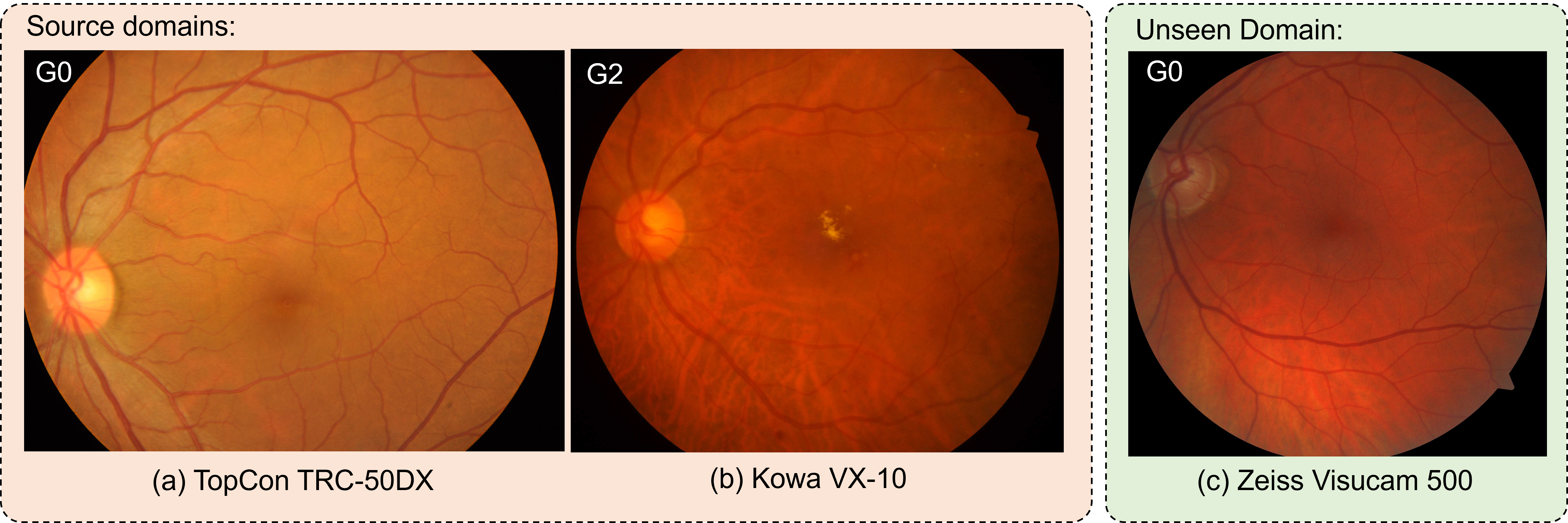
Recently, deep learning has been successful in tackling various complex computer vision tasks, which attracts researchers to develop deep learning-based DR grading methods [11, 9]. Generally, deep learning models are trained to learn the correlations between input and target labels. However, such correlations usually do not imply causality due to the existence of confounder[27]. As shown in Fig.1, a DR grading model trained on source domains from imaging devices (a) and (b) may recognize the fundus image from (c) as instead of based on its high-saturation appearance, causing spurious correlations with DR grades. In this case, the imaging process of different devices can be regarded as the confounder[27], statistically (or spuriously) but not causally correlates with two grades, i.e., and . These spurious correlations often degrade the performance on unseen domains due to the lack of these confounders. The discrepancies between the source and unseen domains are termed as domain shifts[9]. To mitigate this issue, domain generalization (DG)[50] is introduced to train a robust model that can perform well on the data sampled from unseen (target) domains.
During training, DG methods only leverage images from source domains, without the accessibility to target domains. In related publications, most works aim to learn invariant features across domains using meta-learning frameworks[18], data augmentations[51], or new regularization terms[2]. From the perspective of causality[27], learning invariant features is equivalent to perform cutting off or so called do-operation on spurious correlations. This usually requires interventions, such as fixing the variable of objects and varying other irrelevant variables[29]. For example, a straightforward intervention is to take more fundus images with all possible imaging devices for a fixed cohort of patients to amplify source domains. However, this is an unrealistic and impractical way in light of ethical issues and privacy concerns.
In this work, we propose a causality-inspired framework, i.e., CauDR, to implement virtual interventions on the fundus images to cut off spurious correlations between imaging devices and DR grades. Specifically, to analyze the involved variables and their correlations in fundus imaging, we propose a novel structural causal model (SCM)[27] to reveal the possible spurious correlations. Then, we identify the spuriously correlated variables in frequency space through discrete cosine transform (DCT)[18]. Finally, the do-operation is implemented by exchanging the domain variant frequencies while keeping domain invariant ones. In summary, we make the following contributions:
-
•
From a causal view, we investigate the DG paradigm for the DR grading task and then propose a novel and effective causality-inspired framework (CauDR) to achieve generalizable performance on the unseen domain.
-
•
We propose a novel SCM to help us analyze the involved variables in the imaging process and their correlations. This SCM can also lay the groundwork for the development of new equipment for fundus photography.
-
•
To carry out the virtual intervention, we propose 1) a spectrum decomposition step to convert the fundus image into frequency space to simplify the image processing; 2) a novel frequency channel identification strategy to identify spuriously correlated variables; 3) an exchanging-based do-operation to cut off spurious correlations. These three important steps are the cores of our proposed framework.
-
•
To evaluate the generalizable performance, we re-organize the existing four DR grading datasets based on imaging devices to build a DR grading benchmark, called 4DR, in the DG scenario, offering opportunities for future works to explore new DG methods.
-
•
The proposed CauDR framework attains state-of-the-art performance on the 4DR benchmark, surpassing other domain generalization baseline methods developed for both natural and medical image analysis. Moreover, the in-depth examination of causality and its integration into DR grading conducted in this work may spur the research community to devote greater attention to domain generalization and causality-based approaches.
2 Related works
2.1 Grading of Diabetic Retinopathy
The widely used disease severity classification system for DR [44] includes five grades: normal, mild, moderate, severe non-proliferative, and proliferative, corresponding to to , respectively. Early works on automated DR grading rely on hand-crafted features and utilize machine learning algorithms to classify these features[35]. Recently, deep learning-based DR grading models have been proposed, most of which are based on off-the-shelf models developed for natural image processing or their variants of more effective network structure designs. For example, Gulshan et al.[14] trained an Inception-v3 network on fundus images for the DR grading task. Inspired by this idea, Alantary et al.[1] introduced a multi-scale network to learn more detailed features. To encourage models to focus on informative regions, attention mechanisms were considered and incorporated into DR grading methods. Xiao et.al[45] combined an enhanced Inception module with a squeeze-and-excitation (SE) module and achieved an improved DR grading performance. Later, HA-Net[36] was proposed to utilize multiple attention stages to learn global features of DR. However, the common assumption of independent and identically distributed (I.I.D.) variables among training and testing data in these methods is hard to hold in real-world scenarios[4], leading to a degraded performance on unseen data, which compromises the potential clinic values.
2.2 Domain Generalization
DG aims to learn invariant features across multiple source domains for a well-generalizable capability on unseen target domains[7, 50]. To achieve this aim, some works assumed exact formats of discrepancies between domains and then designed strategies to reduce these differences. For example, Xu et.al [48] treated image appearances as the main discrepancies between different domains and then proposed domain MixUp to linearly interpolates image-label pairs between domains to reduce the possible spurious correlations between domain and appearance. Later, Nam et.al[26] proposed to disentangle image style and contents to reduce style bias among domains, assuming image styles are sensitive to domain shifts.
In addition, encouraging a model to learn domain-invariant representations across multiple domains is another strategy for handling DG problems. In this regard, Sun et.al[39] proposed a new loss, termed CORAL loss, to minimize discrepancies between the feature covariance across multiple domains in the training set, which explicitly aligns the learned feature distributions to learn domain-invariant representations. Based on a similar strategy, Li et.al[19] designed a novel framework to match learned feature distributions across source domains by minimizing the Maximum Mean Discrepancy (MMD) and then aligned the matched representations to a prior distribution by adversarial learning to learn universal representations. In addition to the above-mentioned methods taht align features, several recent works have explored aligning gradients between domains to constrain the model’s learning. For example, Shi et.al[38] and Rame et.al [34] incorporated new measures to align inter-domain gradients during the training process, encouraging the model to learn invariant features among domains.
On the other hand, DG has attracted increasing attention in the field of medical image analysis, where domain shifts are often related to variations in clinical centers, imaging protocols, and imaging devices[22] [12]. Li et al.[20] proposed to learn the invariant features by restricting the distribution of encoded features to follow a predefined Gaussian distribution, while Wang et al. [42] developed the DoFE framework for generalizable fundus image segmentation by enriching image features with domain prior knowledge learned from multiple source domains. Based on Fishr [34], Atwany et al. [4] recently adopted the stochastic weight averaging densely (SWAD) technique to find flat minima during DR grading training for better generalization on fundus images from unseen datasets.
2.3 Causality-inspired DG
Causality[27] is a branch of research that explores the connections between various causes and their corresponding effects, with the primary goal to comprehend the underlying mechanisms and patterns behind the occurrence of events. Theoretically, the relationship of should be the same across domains. For example, an object is considered as a cat because it has the cat’s characteristics, which are consistent in different domains. Therefore, the causality-inspired model is a feasible method to tackle DG problems. By considering causality, Arjovsky et. al[2] proposed a new regularization term to constrain the optimal classifier (learn ) in each domain to be the same by minimizing the invariant risk, called Invariant Risk Minimization (IRM). Similarly, Chevalley et.al [8] developed a framework CauIRL to minimize the distributional distance between intervened batches in latent space by using MMD or CORAL technique, encouraging the learning of invariant representations across domains. However, these works mainly focus on natural image processing instead of medical image analysis. More recently, Ouyang et.al[29] proposed to incorporate causality into data augmentation strategy to synthesize domain-shifted training examples in an organ segmentation task, which extends the distributions of training datasets to cover the potentially unseen data distributions. As for our task, the fundus image grading is relatively more challenging due to the complicated factors associated with DR severity.
3 Methodology
This section provides a detailed description of our causality-inspired framework. We first formulate the DG problem, followed by a novel causality-based perspective on the image generation process. The proposed methodology is then elaborated.
3.1 Problem Formulation
A training image set is composed of image pairs and its label , which are sampled from a joint distribution . The test image pairs are sampled from to form the set . In a regular learning setting, the model is trained on to learn the mapping from image space to label space by minimizing the empirical risk , where denotes the loss function used in the training phase. The trained model is expected to perform well on the test set if , which is often difficult to be satisfied considering the complexity of real-world scenarios. For instance, fundus imaging in different vendors would produce images of diverse appearances.
Current study extends the regular learning paradigm by considering the existence of mismatching between the joint distributions of in training and testing sets, where different are treated as different domains. More specifically, we characterize the training set formed by domains: . The goal of domain generalization is to learn a predictor on that can perform well at the unseen test set .
Due to the condition of , the domain generalization problem[13] holds a strong assumption that there exist invariant features or information across multiple domains, which is expected to be learned by the predictor so that it can generalize well to unseen domains. Extracting features that are invariant from the training set is the key to solve the generalization problem.
It should be noted that domain generalization differs from domain adaptation on accessing unseen domains, where the images from unseen domain are unavailable during the training process in the former but available in the latter. As shown in Table 1, we demonstrate different learning paradigms to highlight the characteristics of DG settings[13].
| Learning Paradigm | Training set | Test set |
|---|---|---|
| Supervised Learning | ||
| Semi-Supervised Learning | ||
| Domain Adaptation | ||
| Domain Generalization |
3.2 Structural Causal Model
To understand and analyze the relationships between involved factors in the fundus imaging process, we propose the SCM shown in Fig.2(a), including the observed image , the context factor , the imaging device factor , and the image label , where the direct links between nodes denote causalities, i.e., cause effect. During the imaging process, the context factor and the imaging device factor are logically combined together to generate an observed image by the internal underlying mechanism (the yellow dotted box in Fig.2(a)). To illustrate this mechanism, two mediation nodes, including (the domain-invariant features) and (the domain-variant features), are introduced to logically connect , , and . It is worth noting that this SCM can also be applied to other medical modalities if they have a similar imaging process as the fundus. Specifically, we detail each connection in SCM as follows:
1) : the context factor , such as the retinal vessels or specific lesions, directly determines the imaged contents in the image through the mediation of the variable , which denotes the feature representations of these contents (logically captured by the device ). Ideally, only involves the contents specific to patients, without other irrelevant effects from the device (domain-invariant). denotes the materialization of features into concrete image contents in (from feature space to spatial space). means the lesions in the retina determine its image-level label, e.g. DR grades in our task.
2) : During the imaging process, the device inevitably introduces irrelevant contents into the final image . In our assumptions, these contents contain not only noise, such as Gaussian noise and speckle noise, but also device-related imaging biases. For example, fundus images of the same patient acquired using different devices may have different hues and saturation due to different hardware and software settings. In this SCM, represents the features of these irrelevant contents, which are device/domain-variant and independent of the context factor . In addition, means the materialization process, resulting in noise or color bias in the fundus image.
3) : An image can be regarded as the combination of domain-invariant features and domain-variant features , i.e., , where denotes the underlying combination and materialization process to generate the observed image . The path belongs to the fork structure in causality[30], where should be is dependent on and is the confounder. This dependent relationship brings the spurious correlation between two variables, compromising the generalization ability of the trained model on the images from another unseen imaging device . It should be noted that the underlying mechanism of in an imaging device is complex and even unknown. The aforementioned process only provides the direction of information flow instead of detailed mechanisms.
4) : Our classification task aims to train a model to learn a function mapping from an image and its image-level label : . Considering the irrelevant contents (introduced by ) in , the ideal model should only take as input: , in order to gain better generalization ability than that of . Our method aims to reduce the gap between and to improve generalization ability through the do-operation.
In our study, we assume and are inaccessible: ophthalmologists usually choose different imaging devices based on their availability, which means there are no correlations between device and patients, corresponding to the context factor by being the captured retina. Therefore, our method implements virtual interventions through do-operation to cut off the path to make and become independent, reducing the influence induced by spurious correlations between and . The increased independence between and improves the generalization capability of the model has been proved as well. In the following section, we describe the implementations of virtual interventions, do-operations, and the structure of the proposed model.

3.3 Virtual Interventions
To cut off spurious correlations between domains, an intuitive but impractical strategy is to take fundus images of patients using all possible imaging devices. However, it is impractical . Fortunately, virtual interventions through do-operation can also achieve similar effects. The deep learning-based model in the regular training paradigm is trained to learn . After introducing virtual interventions, our goal becomes to learn a new classifier: , where operator is the mathematical representation of intervention. Specifically, the do-operation can be implemented by stratifying the confounder, i.e., device factor :
| (1) |
where denotes the number of all available devices. Considering the dependent relationship between and : , we have: . Furthermore, we adopt the Normalized Weighted Geometric Mean [46] to move the outer sum into the inner to achieve the batch approximation of Eq.(1):
| (2) |
However, it is still hard to calculate this equation due to the unknown function. If we treat this process as a feature fusion procedure, it usually can be implemented as [15]. In this study, we adopt the implementation of to simplify the complex imaging process and also for better computability. Based on this simplification, we can only change the part for any input image to achieve Eq.(2) (detailed in subsection E), if we can split the image into (the domain-invariant features) and (the domain-variant features) parts (detailed in subsection D).
3.4 Spectrum Decomposition
Splitting a fundus image into and is a vital step in our task. For example, considering an image of a cat on lawn, and are used to represent the pixel set of the foreground (cat) and background (lawn), corresponding to and in the high-dimensional feature space, respectively. Then, an ideal classifier is equivalent to considering is independent of its label . Theoretically, we should use only for model training to directly cut off the spurious correlations and achieve an optimal performance. Based on this strategy, Wei et al. [32] proposed a classification framework by first segmenting the foreground through a segmentation network and then taking current and randomized as the input to improve the generalization performance on ImageNet[11].
However, it is difficult to figure out which pixel belongs to or in a fundus image because the DR severity and grades are jointly determined by multiple factors [17]. Xu et al. [47] found that an image can be decomposed into informative and non-informative parts relative to subsequent vision tasks, where the former can be considered as the foreground and the latter belongs to . Inspired by this work, we introduce a spectrum decomposition step to convert a fundus image into its frequency space, where the complex task of splitting fore and background can be implemented as a simple task to identify informative and non-informative frequencies.
For an image , Discrete Cosine Transform (DCT) is first used to convert it from the spatial image space into the frequency space. Then, band-pass filters are employed to decompose the frequency signals into 64 channels:
| (3) |
where denotes the band-pass filter executed on channel of frequency signals. In practice, the DCT coefficients in the JPEG format images are treated as the frequency representations, where different blocks of coefficients approximately implement the filtering process, i.e., for the input image . Then, the frequency channels can be decomposed into the subsets of and . In this case, denotes , where . The method to identify frequencies associated with and is detailed in the following section.
3.5 Frequency Channel Identification
Based on our hypothesis, different channels (frequencies) in carry diverse information for the downstream tasks. To split them into two parts, the informative (salient channels, ) and non-informative (trivial ones, ) parts, we introduce a channel attention-based gate module by optimizing an external regularization in the training objectives.
For the sample, , in a batched input, this gate module estimates whether its channels belong to or by reaching the trade-off between fewer channels in and higher performance. During implementation, there are four strategies to achieve channel identification: original estimation from the gate module (OE), batched original estimation (BOE) with continuous values, batched average estimation (BAE) with threshold, and the pre-defined indexes (FE) belonging to in natural images [47], as shown in Fig.3. Based on the ablation study about these four choices, we adopt the average estimation in a batch as the final estimation of a single sample:

| (4) |
where is an indicator function to output if the input condition is satisfied. For the sample in batch inputs (batch size is B), denotes its batch averaged channel estimation. is a threshold hyper-parameter to control the estimation sensitivity, where a larger value leads to a smaller set of . The selection of is discussed in the Ablation Analysis section.
3.6 Do-operation
After the identification of frequency channels, we now consider the implementation of the do-operation in Eq.2, i.e., iterating different while keeping unchanged. Generally, there are two strategies, as shown in Fig. 4: 1) directly exchange the channels of between two arbitrary input samples, and 2) exchange the statistical characteristics of frequency channels between two input samples, such as the mean and standard deviation. We hypothesize that the co-occurrence of and in one image will lead to spurious correlations during training. Therefore, the first method is able to cut off this co-occurrence relation directly by randomizing , which is also validated in subsequent experiments.
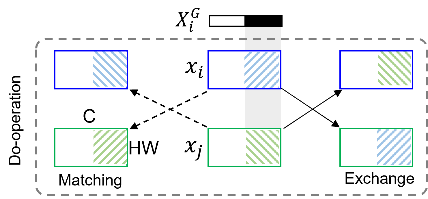
Based on the experiments, our framework adopts the exchaning-based strategy to implement do-operation during the training phase, as shown in the right part of Fig.4. For each input, and , and corresponding frequency representation and , the channels belonging to (predicted as 0 in the gate module) are exchanged between them to form new samples, which are inputs of the subsequent networks. By constantly and randomly sampling inputs, spurious correlations between devices and fundus images are disordered to cut off ,
3.7 Network Structures
The structure of the task model in our framework is shown in Fig.5, which is based on the ResNet50. In our implementation, we remove the input layers ( convolution), the dashed arrow, to adapt to the DCT coefficients after do-operation. During training, the gate module is constrained by the regularization term: , where denotes the channel in . This term would select fewer input channels for (the remaining channels belong to ). There is an adversarial training schema between and classification performance: lower means fewer , which would degrade the accuracy ( is more informative than ).

In addition, do-operation requires the combination of both and channels. During training, however, the first convolutional layer may block the trivial channels by zero weights. To avoid this situation, we introduce a new regulation term to constrain it to select at least half of the channels: , where denotes the normalization of the first layer.
Finally, the standard cross-entropy loss is used to optimize the classification performance. By incorporating the above-mentioned regularization terms, the objective function of our model is shown below:
| (5) |
where indicates the relative weight of the and is empirically set to 0.18.
4 Results
4.1 Dataset and DR Grading
In order to validate the effectiveness of the proposed framework, we collected 4 public datasets containing subjects from India [31], China [23, 21] and Paraguay [5]. Then, we curated them into 4 domains based on imaging devices to build a DR grading benchmark, termed 4DR. The 4DR benchmark contains a total of 5082 images and five DR grades ( to ), as shown in Table. 2. Other classes irrelevant to DR grades to in each dataset were discarded to focus on the current task of DR grading.
| Dataset | Domain No. | Device Types | G0 | G1 | G2 | G3 | G4 | Total | Origin |
|---|---|---|---|---|---|---|---|---|---|
| IDRiD[31] | Domain 1 | Kowa VX-10 | 168 | 25 | 157 | 86 | 58 | 494 | India |
| DeepDRiD[23] | Domain 2 | Topcon TRC-NW300 | 918 | 222 | 396 | 352 | 112 | 2000 | China |
| Sustech-SYSU [21] | Domain 3 | Topcon TRC-50DX | 631 | 24 | 365 | 73 | 58 | 1151 | China |
| DR2021 Version 0.3[5] | Domain 4 | Zeiss Visucam 500 | 711 | 6 | 110 | 349 | 261 | 1437 | Paraguay |
4.2 Implementation Details
The proposed framework was implemented using Pytorch. During training, the pre-trained weight on the ImageNet dataset was first loaded. Then, we utilized the SGD optimizer to train the model in steps with a learning rate of and weight decay of . The learning rate was decayed by multiplying after reaching and of the total steps. To accelerate the forward process, we adopted the offline DCT transformation by first reading DCT coefficients from the JPEG images and then saving the DCT results into the local disk for fast online loading during training, where the size of the JPEG image and DCT coefficients are and , respectively. Only random horizontal flip is used as the basic data augmentation strategy in the pre-processing step. All experiments were conducted on one NVIDIA RTX 3060 12GB GPU with a batch size of 96. Each experiment was repeated three times by using different random seeds.
In addition, we implemented most of the comparable methods by using the code in the DomainBed[13] benchmark. For the algorithms not in this benchmark, we utilize their official implementation to ensure fair comparisons.
4.3 Experimental Results on DR Grading
4.3.1 Comparison With Baseline Models
We trained the commonly used ResNet50 [15] on all source domains in a regular learning manner (I.I.D assumption) as the baseline models. As shown in Table 3, our proposed CauDR outperforms the baseline by significant margins (average accuracy increase of ), which clearly demonstrates the effectiveness and better generalization ability of our method.
In addition, the baseline under-performs all the DG-optimized methods, demonstrating the necessity of study in domain generalization issues.
4.3.2 Comparison with Appearance-based Methods
In MixUp[48] and SagNet[26], authors assumed the image appearance differences result in discrepancies across domains. Therefore, they designed strategies to randomize or remove appearance while keeping the contents fixed. As shown in Table 3, both MixUp and SagNet achieve better results than ResNet50. However, those assumptions are limited as they only consider the image appearance in spatial space. In contrast, our proposed frequency-based operation covers more types of discrepancies and produces performance improvements of and when compared with MixUp and SagNet, respectively.
4.3.3 Comparison with Representation-based Methods
Instead of directly manipulating the input data, designing strategies in feature or gradients space to encourage learning domain-invariant representations across domains is also one of the main directions to solve DG problems. We implemented features alignment-based (CORAL[39], MMD[19]) and gradients alignment-based (Fish[38], Fishr[34], DRGen[4]) methods on our task to evaluate their performance. As shown in Table 3, MMD achieves in average accuracy while CORAL performs poorly but still slightly outperforms ResNet50.
On the other hand, Fish and Fishr adopted new measures to align gradients across domains to promote the learning of domain-invariant features, while DRGen incorporated the SWAD technique into Fishr to search for the flat minima during training. As shown in the Table 3, Fish, Fishr, and DRGen outperform the ResNet50 but fail to surpass our method. This may reveal the challenges to learn invariant representations by enforcing gradient directions on the small and class-imbalanced fundus datasets.
4.3.4 Comparison With Causality-based Methods
Currently, there are only limited studies focused on the causality for the DG problems in image classification tasks, such as DR grading. IRM[2] assumed the underlying causal relationship (observable features interested variable ) in each domain is constant and then proposed a regularization term to constrain the optimal classifier (learn ) in each domain to be the same. Similarly, CauIRL[8] was developed to minimize the distributional distances (measured by MMD or CORAL, termed Cau_MMD and Cau_CORAL, respectively) between batches intervened by the confounder in the latent space to encourage the learning of invariant representations under the pre-defined SCM. As shown in Table 3, the performance of IRM slightly outperforms CauIRLs, i.e. Cau_MMD and Cau_CORAL. However, IRM performs worse than other DG methods and its performance is inferior to our method by a considerable margin of . The complicated invariant and variant features in our task may result in this performance gap, where optimizing the invariance of the classifier alone is not sufficient to learn the domain-invariant features. Likewise, the regularization of encoded features in CauIRLs may not be effective in handling current complicated scenarios. In addition, the implementation of intervention is important and the results demonstrate that our exchange-based intervention outperforms the distance constrain-based intervention in CauIRLs. More experiments on the implementations of intervention are detailed in the Ablation Analysis section.
4.4 Ablation Analysis
| Method | Domain 1 | Domain 2 | Domain 3 | Domain 4 | Average |
|---|---|---|---|---|---|
| w/o Gate | 61.05 | ||||
| w/o Fuse | 61.04 | ||||
| Ours | 61.82 |
4.4.1 Effectiveness of Loss Terms in Eq.5
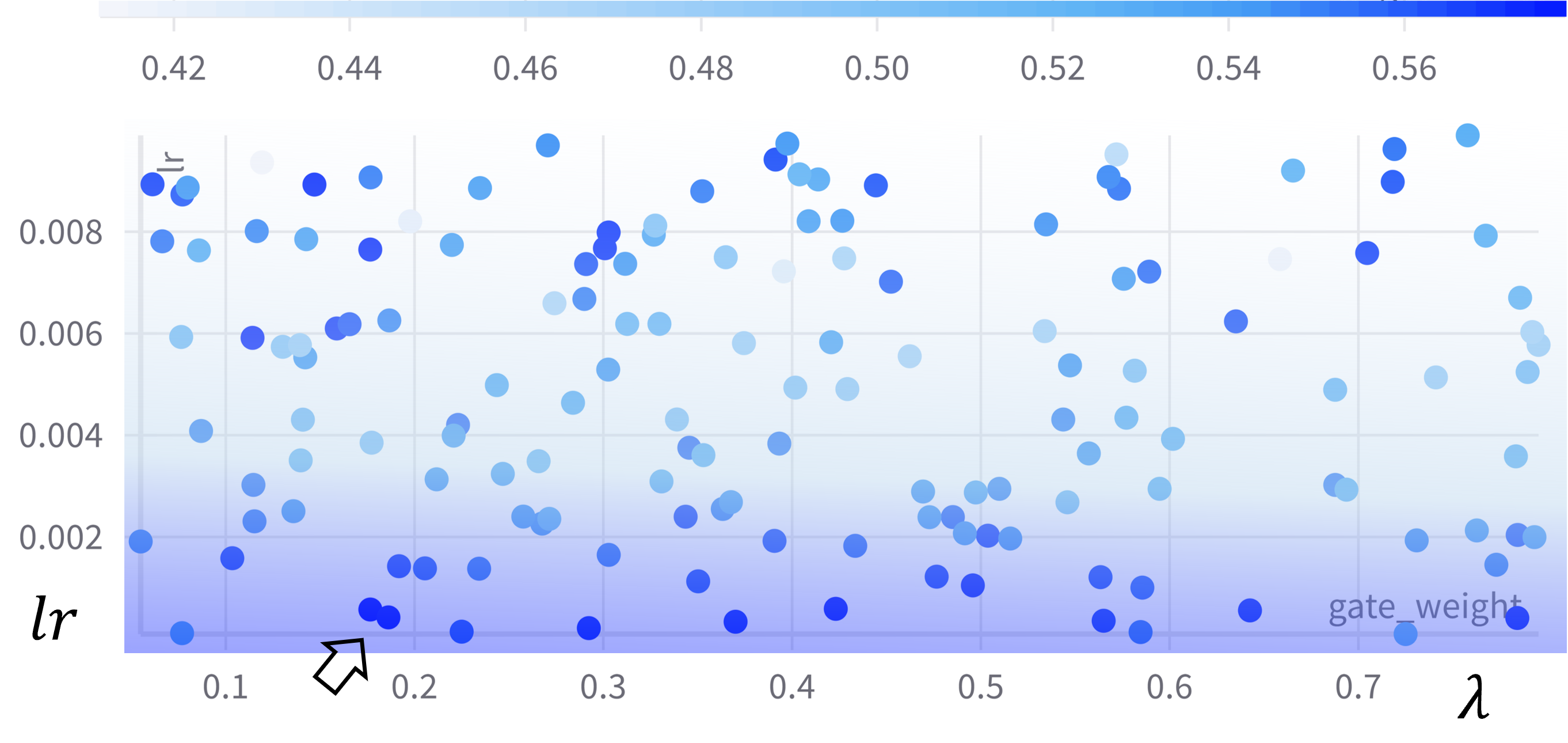
Firstly, we utilize the hyper-parameters searching tool in wandb[6] to find the optimal and the learning rate. From Fig.6, we determine the acceptable parameters: and . Then, we evaluate the effectiveness of gate and fuse regularization terms by removing one of them. The average performance drop (about ) can be observed from Table 4 after removing the gate or fuse term. Removing the gate loss term may degrade the channel identification accuracy, further reducing the generalization ability of the model. Similarly, the removal of the fuse loss term also reduces the performance gain of the proposed virtual interventions.
4.4.2 Influence of in Eq.4
We further explored the influence of in Eq.4 by separately setting its value to 0.1, 0.2, 0.4, 0.6, 0.8, and 0.9. From the results in Table 5, it can be observed that the average accuracy increases first and then decreases along with the increase of . Therefore, we chose in Eq.4 for the optimal performance.
| Domain 1 | Domain 2 | Domain 3 | Domain 4 | Average | |
|---|---|---|---|---|---|
| 0.1 | 61.22 | ||||
| 0.2 | 61.13 | ||||
| 0.4 | 61.82 | ||||
| 0.6 | 61.20 | ||||
| 0.8 | 61.21 | ||||
| 0.9 | 61.13 |
4.4.3 Channel Identification and Do-operation
In section 3.5 and 3.6, we introduce four strategies to implement the channel identification, as shown in Fig.3, and two ways for the do-operation in Fig.4. Therefore, eight methods can be achieved by randomly combining strategies of channel identification and do-operation implementation, as shown in Table 6.
| Do | Domain 1 | Domain 2 | Domain 3 | Domain 4 | Average |
|---|---|---|---|---|---|
| Ex+OE | 61.39 | ||||
| Ex+BOE | 61.37 | ||||
| Ex+BAE | 61.82 | ||||
| Ex+FE | 61.79 | ||||
| Ma+OE | 58.96 | ||||
| Ma+BOE | 58.91 | ||||
| Ma+BAE | 58.97 | ||||
| Ma+FE | 58.76 |
From the results, the exchange-based methods outperform the match-based methods by a margin of circa 2.5 on the average accuracy, indicating the effectiveness of the exchanging mechanism, although the matching-based variants perform better than other DG-related algorithms. Furthermore, we found that different methods in either exchange-based or match-based group achieve similar performance, signifying the pre-trained weights in natural images still provide correct estimations on and in fundus images. Eventually, we adopted BAE strategy in our method for its optimal performance.
4.4.4 Visualizations of Do-operation
For an arbitrary image , we first visualize its DCT coefficients ( channels) in Fig.7 with three maps, corresponding to RGB channels in the spatial space, where each map consists of channels (small images). In each map, the frequency gradually increases from the upper left to the bottom right by a shape path[47].
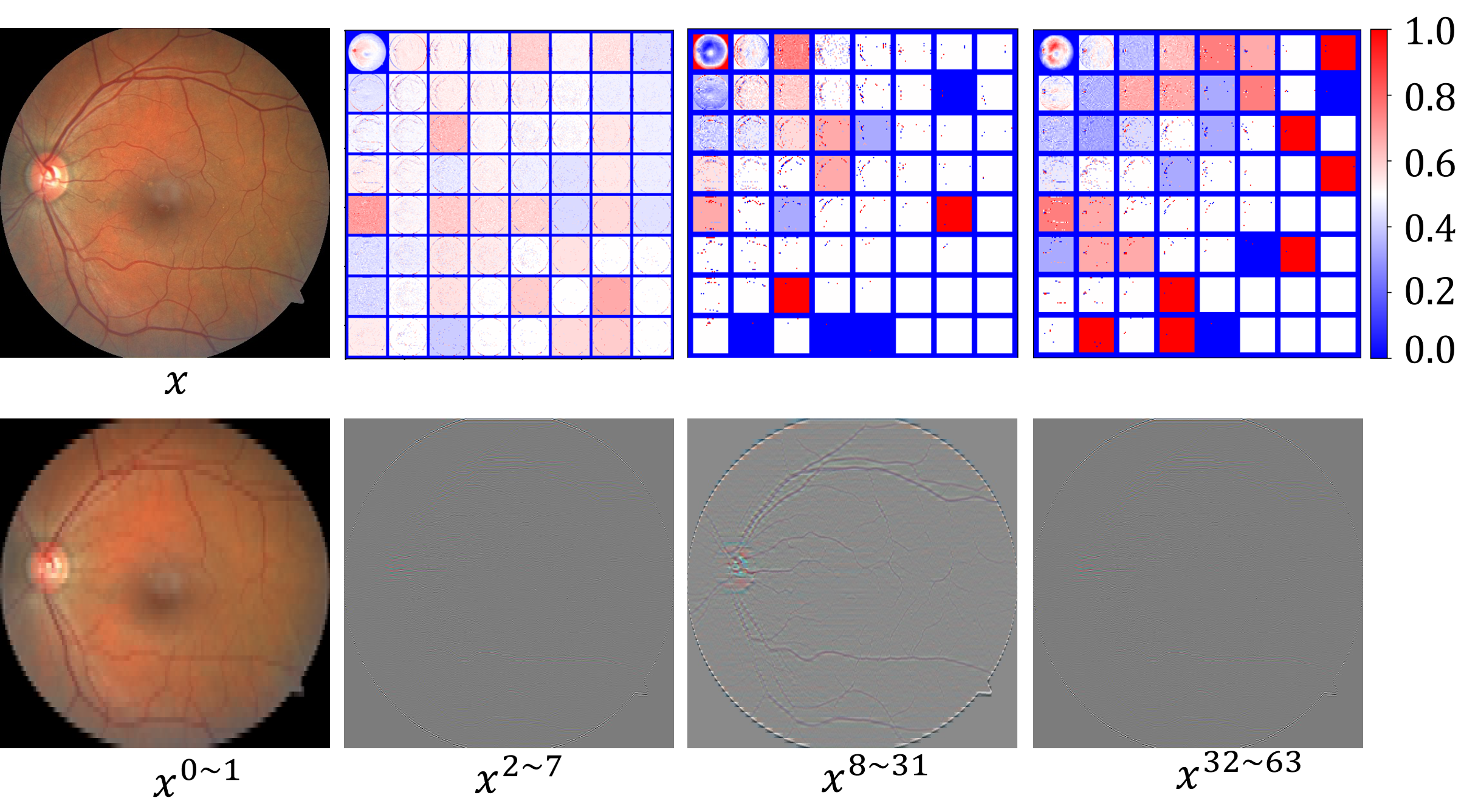
From Fig. 7, we find that the low frequencies usually contain more contour information, such as the vessels and the optic disk. As the frequency increases, more detailed local information becomes available. We also observe this phenomenon in the reconstructed images in the second row by exchanging the subset of frequencies between two images.
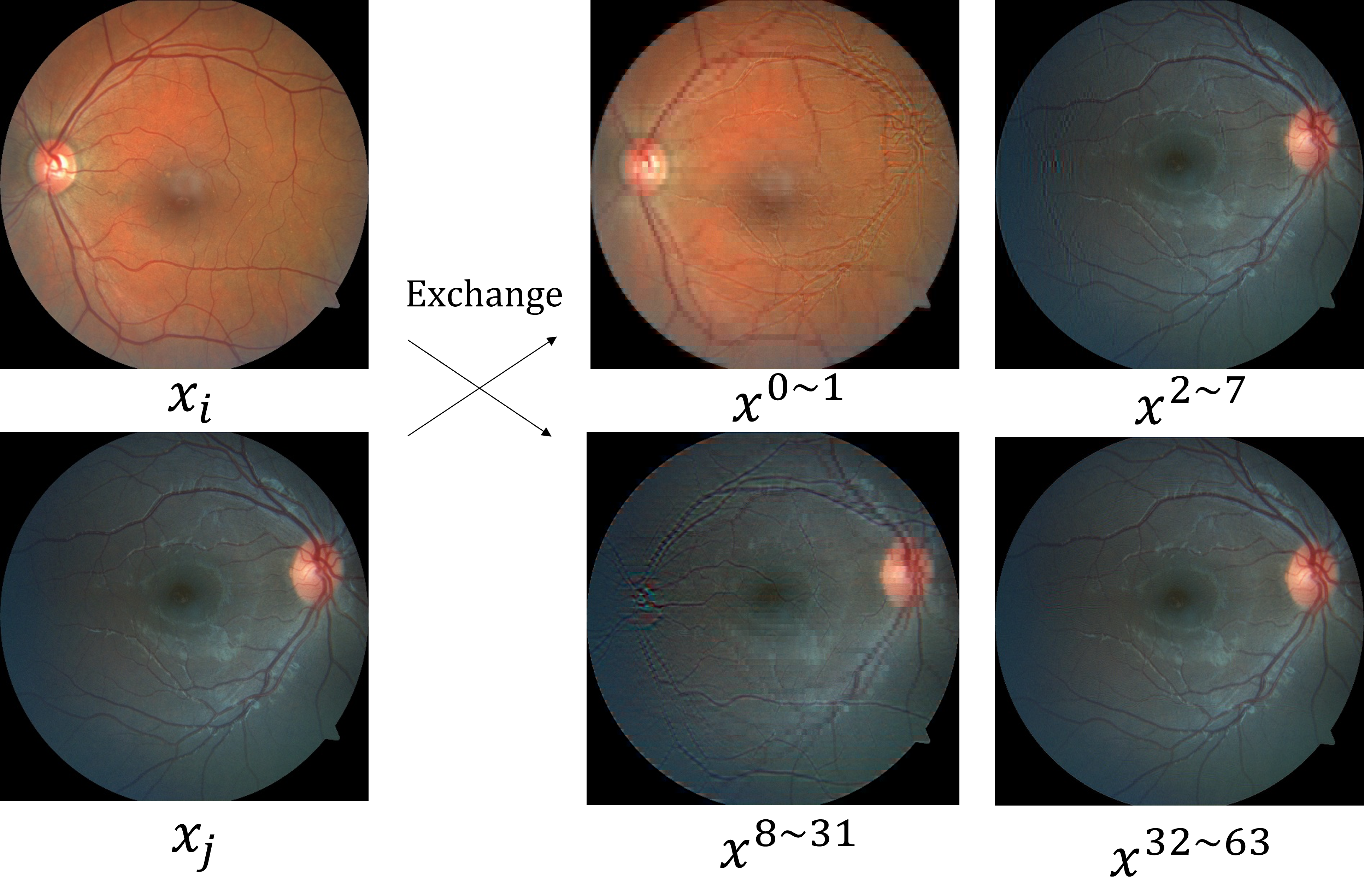
Then, we randomly sample images and to visualize the effects of do-operation: exchanging the subset of frequencies between them. As shown in Fig.8, the intervented images simultaneously include the contents from two images in different frequencies, where the domain-related information is expected to be disordered by this exchange.
4.4.5 Visualizations for Feature Representations
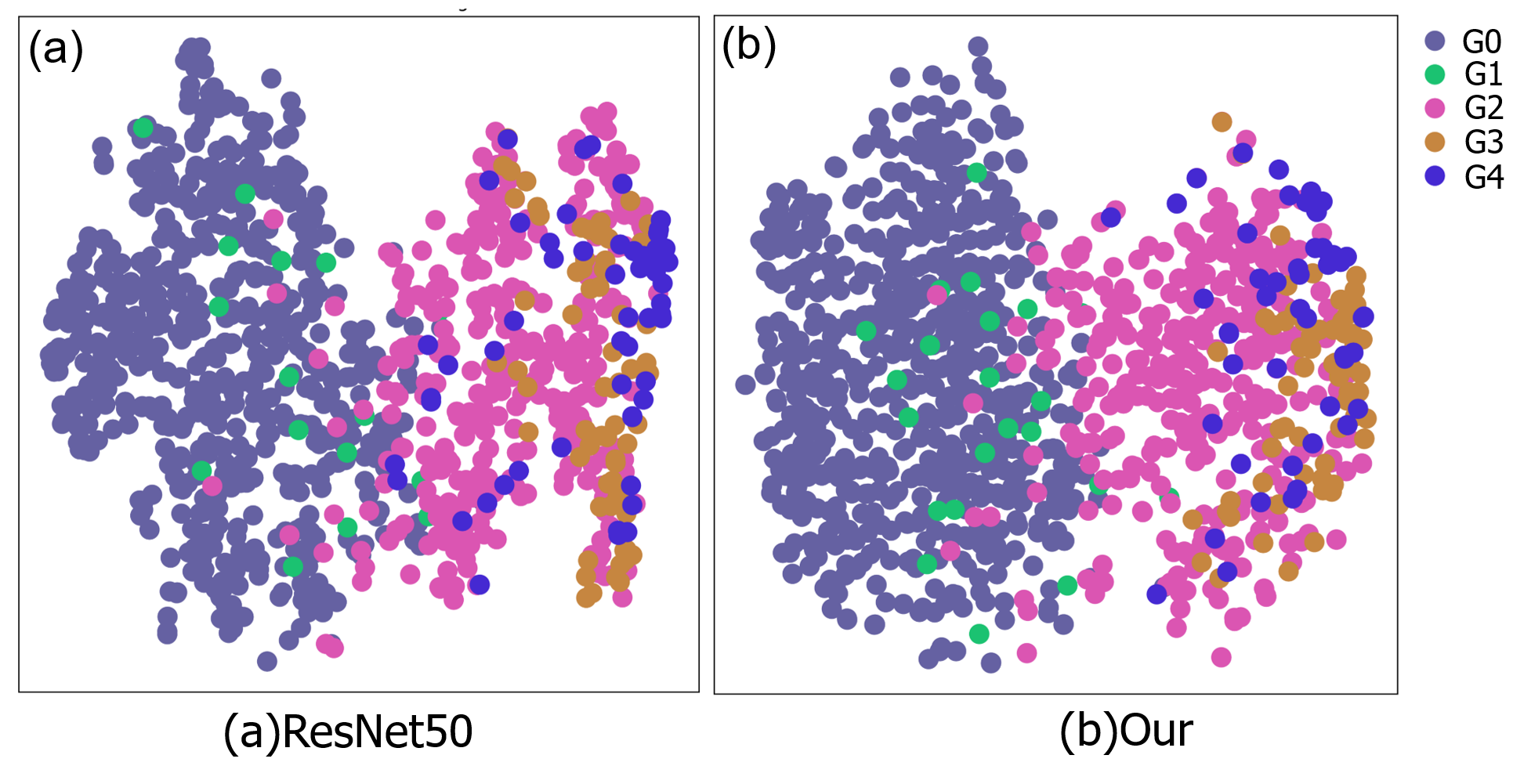
We utilize the t-SNE[41] technique to visualize the learned feature representations extracted from the Domain 2 dataset using the baseline ResNet50 and our method. As shown in Fig.9, the class representations extracted using our method are more sparsely separated than those extracted using the baseline method. For example, the representations of are mixed with those of and in Fig.9(a), while this mixing effect is not obvious in Fig.9(b). This indicates that the features extracted by our method have a clear decision boundary, which is beneficial for the classification task.
5 Discussion
Our proposed method demonstrates better generalizable performance on DR grading compared with other DG methods. Nevertheless, our method still has limitations. First, the limited source domains in the training set hinder the learning of invariant features across domains due to the lack of enough invariability. Collecting more images from different devices would alleviate this issue and further improve the overall generalizable performance on unseen datasets. However, in practice, collecting and annotating more fundus images is troublesome and expensive when considering the involved privacy and ethical matters and the clinical experience of annotators. One practical and potential solution is to employ semi-supervised learning(SSL) to take advantage of the large amount of unlabeled data, which also contains the desired invariability across domains. The second limitation is that only one single modality image, the color fundus image, was involved in our experiments. Conducting experiments on other modality images would provide evidence for the effectiveness of our proposed method. In addition, involving other tasks, such as optic cup/disk and retinal vessel segmentation, would further extend our method for validating the causality in more applications. Thirdly, we only apply do-operation on the frequency space instead of the high-dimensional feature space generated by the network, which may be another suitable representation to disentangle and by carefully designing proxy tasks. Extending the current implementation of do-operation into feature space worth further investigating. Finally, during experiments, we ignore the class imbalance issue in each source domain to only focus on improving the average accuracy of five classes. Different class ratios in source and target domains may also affect the generalizable performance.
Future work would explore the incorporation of causality and SSL paradigm to involve more unlabeled data. The regular SSL setting assumes that each sample has an equal opportunity to be the unlabeled one, which may not be a valid assumption in some cases. For example, if considering the practical cost, the annotator usually tends to label the representative images, which contain more apparent key features related to the downstream task, such as the retina hemorrhage regions (the key metrics to grade DR) in our task. In this case, the representative ones have less chance of being the unlabeled set. Therefore, we intend to mitigate this selection bias through the causal perspective to encourage the model to learn invariant features across domains when utilizing unlabeled images in training. Furthermore, the causality would also be helpful in large visual models [33, 37] recently emerging with massive parameters and datasets to potentially enhance in-context learning on huge datasets. In medical scenarios, leveraging causal correlations between medical concepts and clinical findings is critical and important for generalist medical artificial intelligence (GMAI)[25] to carry out diverse tasks. Therefore, exploring causal learning in the mentioned settings will also be in our future research plans.
6 Conclusions
In this work, we present a generalizable DR grading system by incorporating causality into the training pipeline to learn invariant features across multiple domains. Specifically, a structure causal model (SCM) is first proposed to model the fundus imaging process by analyzing the involved factors and their relationships. Then, we determine the features associated with spurious correlations and propose virtual interventions implemented by do-operation to cut off these correlations for better performance on images from unseen domains. To evaluate the performance, we collect four public fundus datasets associated with DR grading and reorganize them into 4 non-overlapping domains based on imaging devices to build a benchmark, i.e., 4DR. Finally, we conduct comprehensive experiments on this benchmark to demonstrate the effectiveness of our proposed CauDR framework. In the future, we will further extend this causality-inspired DG paradigm to more modalities and tasks.
Declaration of competing interest
The authors declare that they have no conflict of interest.
References
- [1] Mohammad T Al-Antary and Yasmine Arafa. Multi-scale attention network for diabetic retinopathy classification. IEEE Access, 9:54190–54200, 2021.
- [2] Martin Arjovsky, Léon Bottou, Ishaan Gulrajani, and David Lopez-Paz. Invariant risk minimization. arXiv preprint arXiv:1907.02893, 2019.
- [3] Ryo Asaoka, Masaki Tanito, Naoto Shibata, Keita Mitsuhashi, Kenichi Nakahara, Yuri Fujino, Masato Matsuura, Hiroshi Murata, Kana Tokumo, and Yoshiaki Kiuchi. Validation of a deep learning model to screen for glaucoma using images from different fundus cameras and data augmentation. Ophthalmology Glaucoma, 2(4):224–231, 2019.
- [4] Mohammad Atwany and Mohammad Yaqub. Drgen: Domain generalization in diabetic retinopathy classification. In Medical Image Computing and Computer Assisted Intervention–MICCAI 2022: 25th International Conference, Singapore, September 18–22, 2022, Proceedings, Part II, pages 635–644. Springer, 2022.
- [5] Veronica Elisa Castillo Benítez, Ingrid Castro Matto, Julio César Mello Román, José Luis Vázquez Noguera, Miguel García-Torres, Jordan Ayala, Diego P Pinto-Roa, Pedro E Gardel-Sotomayor, Jacques Facon, and Sebastian Alberto Grillo. Dataset from fundus images for the study of diabetic retinopathy. Data in brief, 36:107068, 2021.
- [6] Lukas Biewald. Experiment tracking with weights and biases, 2020. Software available from wandb.com.
- [7] Gilles Blanchard, Gyemin Lee, and Clayton Scott. Generalizing from several related classification tasks to a new unlabeled sample. Advances in neural information processing systems, 24, 2011.
- [8] Mathieu Chevalley, Charlotte Bunne, Andreas Krause, and Stefan Bauer. Invariant causal mechanisms through distribution matching. arXiv preprint arXiv:2206.11646, 2022.
- [9] Dolly Das, Saroj Kr Biswas, and Sivaji Bandyopadhyay. A critical review on diagnosis of diabetic retinopathy using machine learning and deep learning. Multimedia Tools and Applications, 81(18):25613–25655, 2022.
- [10] Taraprasad Das, Brijesh Takkar, Sobha Sivaprasad, Thamarangsi Thanksphon, Hugh Taylor, Peter Wiedemann, Janos Nemeth, Patanjali D Nayar, Padmaja Kumari Rani, and Rajiv Khandekar. Recently updated global diabetic retinopathy screening guidelines: commonalities, differences, and future possibilities. Eye, 35(10):2685–2698, 2021.
- [11] Jia Deng, Wei Dong, Richard Socher, Li-Jia Li, Kai Li, and Li Fei-Fei. Imagenet: A large-scale hierarchical image database. In 2009 IEEE conference on computer vision and pattern recognition, pages 248–255. Ieee, 2009.
- [12] Qi Dou, Daniel Coelho de Castro, Konstantinos Kamnitsas, and Ben Glocker. Domain generalization via model-agnostic learning of semantic features. Advances in Neural Information Processing Systems, 32, 2019.
- [13] Ishaan Gulrajani and David Lopez-Paz. In search of lost domain generalization. arXiv preprint arXiv:2007.01434, 2020.
- [14] Varun Gulshan, Lily Peng, Marc Coram, Martin C Stumpe, Derek Wu, Arunachalam Narayanaswamy, Subhashini Venugopalan, Kasumi Widner, Tom Madams, Jorge Cuadros, et al. Development and validation of a deep learning algorithm for detection of diabetic retinopathy in retinal fundus photographs. Jama, 316(22):2402–2410, 2016.
- [15] Kaiming He, Xiangyu Zhang, Shaoqing Ren, and Jian Sun. Deep residual learning for image recognition. In Proceedings of the IEEE conference on computer vision and pattern recognition, pages 770–778, 2016.
- [16] Meng Huang, Shiyu Sun, Than S Saini, Qiang Fu, Lin Xu, Dong Wu, Haonan Ren, Li Shen, Thomas W Hawkins, John Ballato, et al. Raman amplification at 2.2 m in silicon core fibers with prospects for extended mid-infrared source generation. Light: Science and Applications, 2023.
- [17] Samuel C Lee, Elisa T Lee, Yiming Wang, Ronald Klein, Ronald M Kingsley, and Ann Warn. Computer classification of nonproliferative diabetic retinopathy. Archives of ophthalmology, 123(6):759–764, 2005.
- [18] Da Li, Yongxin Yang, Yi-Zhe Song, and Timothy Hospedales. Learning to generalize: Meta-learning for domain generalization. In Proceedings of the AAAI conference on artificial intelligence, volume 32, 2018.
- [19] Haoliang Li, Sinno Jialin Pan, Shiqi Wang, and Alex C Kot. Domain generalization with adversarial feature learning. In Proceedings of the IEEE conference on computer vision and pattern recognition, pages 5400–5409, 2018.
- [20] Haoliang Li, YuFei Wang, Renjie Wan, Shiqi Wang, Tie-Qiang Li, and Alex Kot. Domain generalization for medical imaging classification with linear-dependency regularization. Advances in Neural Information Processing Systems, 33:3118–3129, 2020.
- [21] Li Lin, Meng Li, Yijin Huang, Pujin Cheng, Honghui Xia, Kai Wang, Jin Yuan, and Xiaoying Tang. The sustech-sysu dataset for automated exudate detection and diabetic retinopathy grading. Scientific Data, 7(1):409, 2020.
- [22] Quande Liu, Qi Dou, and Pheng-Ann Heng. Shape-aware meta-learning for generalizing prostate mri segmentation to unseen domains. In International Conference on Medical Image Computing and Computer-Assisted Intervention, pages 475–485. Springer, 2020.
- [23] Ruhan Liu, Xiangning Wang, Qiang Wu, Ling Dai, Xi Fang, Tao Yan, Jaemin Son, Shiqi Tang, Jiang Li, Zijian Gao, et al. Deepdrid: Diabetic retinopathy—grading and image quality estimation challenge. Patterns, page 100512, 2022.
- [24] Zhuolin Liu, Furu Zhang, Kelvy Zucca, Anant Agrawal, and Daniel X Hammer. Ultrahigh-speed multimodal adaptive optics system for microscopic structural and functional imaging of the human retina. Biomedical Optics Express, 13(11):5860–5878, 2022.
- [25] Michael Moor, Oishi Banerjee, Zahra Shakeri Hossein Abad, Harlan M Krumholz, Jure Leskovec, Eric J Topol, and Pranav Rajpurkar. Foundation models for generalist medical artificial intelligence. Nature, 616(7956):259–265, 2023.
- [26] Hyeonseob Nam, HyunJae Lee, Jongchan Park, Wonjun Yoon, and Donggeun Yoo. Reducing domain gap by reducing style bias. In Proceedings of the IEEE/CVF Conference on Computer Vision and Pattern Recognition, pages 8690–8699, 2021.
- [27] Leland Gerson Neuberg. Causality: models, reasoning, and inference, by judea pearl, cambridge university press, 2000. Econometric Theory, 19(4):675–685, 2003.
- [28] Giuseppe Ortolano, Alberto Paniate, Pauline Boucher, Carmine Napoli, Sarika Soman, Silvania F Pereira, Ivano Ruo-Berchera, and Marco Genovese. Quantum enhanced non-interferometric quantitative phase imaging. Light: Science & Applications, 12(1):171, 2023.
- [29] Cheng Ouyang, Chen Chen, Surui Li, Zeju Li, Chen Qin, Wenjia Bai, and Daniel Rueckert. Causality-inspired single-source domain generalization for medical image segmentation. IEEE Transactions on Medical Imaging, 2022.
- [30] Judea Pearl, Madelyn Glymour, and Nicholas P Jewell. Causal inference in statistics: A primer. 2016. Google Ascholar there is no corresponding record for this reference, 2016.
- [31] Prasanna Porwal, Samiksha Pachade, Ravi Kamble, Manesh Kokare, Girish Deshmukh, Vivek Sahasrabuddhe, and Fabrice Meriaudeau. Indian diabetic retinopathy image dataset (idrid): a database for diabetic retinopathy screening research. Data, 3(3):25, 2018.
- [32] Wei Qin, Hanwang Zhang, Richang Hong, Ee-Peng Lim, and Qianru Sun. Causal interventional training for image recognition. IEEE Transactions on Multimedia, 2021.
- [33] Jianing Qiu, Lin Li, Jiankai Sun, Jiachuan Peng, Peilun Shi, Ruiyang Zhang, Yinzhao Dong, Kyle Lam, Frank P-W Lo, Bo Xiao, et al. Large ai models in health informatics: Applications, challenges, and the future. IEEE Journal of Biomedical and Health Informatics, 2023.
- [34] Alexandre Rame, Corentin Dancette, and Matthieu Cord. Fishr: Invariant gradient variances for out-of-distribution generalization. In International Conference on Machine Learning, pages 18347–18377. PMLR, 2022.
- [35] Sohini Roychowdhury, Dara D Koozekanani, and Keshab K Parhi. Dream: diabetic retinopathy analysis using machine learning. IEEE journal of biomedical and health informatics, 18(5):1717–1728, 2013.
- [36] Nagur Shareef Shaik and Teja Krishna Cherukuri. Hinge attention network: A joint model for diabetic retinopathy severity grading. Applied Intelligence, 52(13):15105–15121, 2022.
- [37] Peilun Shi, Jianing Qiu, Sai Mu Dalike Abaxi, Hao Wei, Frank P-W Lo, and Wu Yuan. Generalist vision foundation models for medical imaging: A case study of segment anything model on zero-shot medical segmentation. Diagnostics, 13(11):1947, 2023.
- [38] Yuge Shi, Jeffrey Seely, Philip HS Torr, N Siddharth, Awni Hannun, Nicolas Usunier, and Gabriel Synnaeve. Gradient matching for domain generalization. arXiv preprint arXiv:2104.09937, 2021.
- [39] Baochen Sun and Kate Saenko. Deep coral: Correlation alignment for deep domain adaptation. In European conference on computer vision, pages 443–450. Springer, 2016.
- [40] Hong Sun, Pouya Saeedi, Suvi Karuranga, Moritz Pinkepank, Katherine Ogurtsova, Bruce B Duncan, Caroline Stein, Abdul Basit, Juliana CN Chan, Jean Claude Mbanya, et al. Idf diabetes atlas: Global, regional and country-level diabetes prevalence estimates for 2021 and projections for 2045. Diabetes research and clinical practice, 183:109119, 2022.
- [41] Laurens Van der Maaten and Geoffrey Hinton. Visualizing data using t-sne. Journal of machine learning research, 9(11), 2008.
- [42] Shujun Wang, Lequan Yu, Kang Li, Xin Yang, Chi-Wing Fu, and Pheng-Ann Heng. Dofe: Domain-oriented feature embedding for generalizable fundus image segmentation on unseen datasets. IEEE Transactions on Medical Imaging, 39(12):4237–4248, 2020.
- [43] Wei Wang and Amy CY Lo. Diabetic retinopathy: pathophysiology and treatments. International journal of molecular sciences, 19(6):1816, 2018.
- [44] Charles P Wilkinson, Frederick L Ferris III, Ronald E Klein, Paul P Lee, Carl David Agardh, Matthew Davis, Diana Dills, Anselm Kampik, R Pararajasegaram, Juan T Verdaguer, et al. Proposed international clinical diabetic retinopathy and diabetic macular edema disease severity scales. Ophthalmology, 110(9):1677–1682, 2003.
- [45] Zhitao Xiao, Yaxin Zhang, Jun Wu, and Xinxin Zhang. Se-midnet based on deep learning for diabetic retinopathy classification. In 2021 7th International Conference on Computing and Artificial Intelligence, pages 92–98, 2021.
- [46] Kelvin Xu, Jimmy Ba, Ryan Kiros, Kyunghyun Cho, Aaron Courville, Ruslan Salakhudinov, Rich Zemel, and Yoshua Bengio. Show, attend and tell: Neural image caption generation with visual attention. In International conference on machine learning, pages 2048–2057. PMLR, 2015.
- [47] Kai Xu, Minghai Qin, Fei Sun, Yuhao Wang, Yen-Kuang Chen, and Fengbo Ren. Learning in the frequency domain. In Proceedings of the IEEE/CVF Conference on Computer Vision and Pattern Recognition, pages 1740–1749, 2020.
- [48] Minghao Xu, Jian Zhang, Bingbing Ni, Teng Li, Chengjie Wang, Qi Tian, and Wenjun Zhang. Adversarial domain adaptation with domain mixup. In Proceedings of the AAAI conference on artificial intelligence, volume 34, pages 6502–6509, 2020.
- [49] Xincheng Yao, Taeyoon Son, and Jiechao Ma. Developing portable widefield fundus camera for teleophthalmology: Technical challenges and potential solutions. Experimental Biology and Medicine, 247(4):289–299, 2022.
- [50] Kaiyang Zhou, Ziwei Liu, Yu Qiao, Tao Xiang, and Chen Change Loy. Domain generalization: A survey. IEEE Transactions on Pattern Analysis and Machine Intelligence, 2022.
- [51] Kaiyang Zhou, Yongxin Yang, Yu Qiao, and Tao Xiang. Mixstyle neural networks for domain generalization and adaptation. arXiv preprint arXiv:2107.02053, 2021.