[1]This work was supported in part by the National Natural Science Foundation of China under Grant 62172444 and U22A2041, in part by the Shenzhen Science and Technology Program under Grant KQTD20200820113106007, in part by the Natural Science Foundation of Hunan Province under Grant 2022JJ30753, in part by the Central South University Innovation-Driven Research Pro-gramme under Grant 2023CXQD018, and in part by the High Performance Computing Center of Central South University.
[prefix = Junbin] [prefix = Jin] \cormark[1] [prefix= Hanhe]
[prefix= Hulin] [prefix= Shirui] [prefix= Yi] \cormark[1]
[cor1]Corresponding author
Multi-modal Multi-kernel Graph Learning for Autism Prediction and Biomarker Discovery
Abstract
Due to its complexity, graph learning-based multi-modal integration and classification is one of the most challenging obstacles for disease prediction. To effectively offset the negative impact between modalities in the process of multi-modal integration and extract heterogeneous information from graphs, we propose a novel method called MMKGL (Multi-modal Multi-Kernel Graph Learning). For the problem of negative impact between modalities, we propose a multi-modal graph embedding module to construct a multi-modal graph. Different from conventional methods that manually construct static graphs for all modalities, each modality generates a separate graph by adaptive learning, where a function graph and a supervision graph are introduced for optimization during the multi-graph fusion embedding process. We then propose a multi-kernel graph learning module to extract heterogeneous information from the multi-modal graph. The information in the multi-modal graph at different levels is aggregated by convolutional kernels with different receptive field sizes, followed by generating a cross-kernel discovery tensor for disease prediction. Our method is evaluated on the benchmark Autism Brain Imaging Data Exchange (ABIDE) dataset and outperforms the state-of-the-art methods. In addition, discriminative brain regions associated with autism are identified by our model, providing guidance for the study of autism pathology.
keywords:
Multi-modal integration \sepGraph learning \sepMulti-kernel learning\sepAutism prediction \sepBiomarker discovery1 Introduction
The development of medical devices has facilitated the acquisition of multi-modal clinical data. It has been found that multi-modal data could benefit disease prediction as it contains more information than single-modal data (Mohammadi-Nejad et al., 2017) by taking advantage of the joint and supplementary information. Recently, a few approaches that aim to make use of multi-modal data for disease prediction have been proposed, including multi-modal fusion methods based on attention mechanisms (Zhu et al., 2022; Zheng et al., 2022), multi-task learning (Adeli et al., 2019; Jie et al., 2015), and nonnegative matrix decomposition (Zhou et al., 2019; Peng et al., 2022). However, these approaches rely on feature learning and do not adequately explore the structure and cross-modal relationships between modalities. As a result, how to fully exploit the knowledge of multi-modal is still a long-standing research topic.
Owing to the inherent properties of graphs (Su et al., 2020) such as information aggregation and relationship modeling, graph models provides an efficient way for integrating multi-modal information. Graph neural networks (GNNs) have been proposed in (Welling and Kipf, 2016). Due to their advantages of joint population learning and information mining, a growing number of GNN-based studies have been proposed for disease prediction. PopGCN (Parisot et al., 2017) is the first application of graph convolutional networks to Autism and Alzheimer’s disease. It combines brain imaging information and phenotypic information jointly to construct disease population graphs and perform disease prediction to distinguish normal individuals from patients. InceptionGCN (Kazi et al., 2019a) explored the impacts of the size of convolutional filters on disease prediction based on PopGCN, and tried to find the optimal convolutional filter size. MMGL (Zheng et al., 2022) used a self-attentive mechanism to learn complementary information between modalities and is used to construct disease population graphs.
Although the above studies have achieved promising results for disease prediction, there are some limitations. One of the most challenging problems is that the domain distributions of different modalities vary significantly. It leads to the fact that when multi-modal data are directly integrated, the less expressive modalities may suppress the expression of other modal data. This phenomenon is known as the inter-modal negative impact. Another major issue is that most of the existing graph convolutional network-based disease prediction methods use a size-fixed convolutional filter, which cannot well extract the heterogeneous information over the graph.
To overcome these problems and to effectively utilize multimodal data, we propose a novel framework entitled Multi-modal Multi-Kernel Graph Learning (MMKGL). The contributions of our work are summarized as follows:
-
•
We propose a multi-modal graph embedding module that generates multiple graphs adaptively, where each graph corresponds to one modal data. A function graph and a supervision graph are introduced to optimize the multi-graph fusion embedding process, which effectively mitigates the negative impacts between modalities.
-
•
We propose a multi-kernel graph learning network that extracts heterogeneous information by using convolutional kernels with different receptive field sizes. To minimize the graph noise, a relational attention mechanism (RAM) is deployed to adaptively tune the graph structure in the training process.
-
•
The proposed framework is evaluated on the Autism Brain Imaging Data Exchange (ABIDE) dataset. The results verify the validity of our proposed framework and demonstrate its advantages over state-of-the-art methods. The code of MMKGL will be made available at the time of publication.
-
•
Discriminative brain regions and subnetworks with discriminatory properties are discovered, giving guidance for the study of autism pathology.
2 Related Work
In this section, we mainly review automatic disease diagnosis works from multi-modal analysis and graph neural networks.
2.1 Multi-modal Analysis for Disease Prediction
Multi-modal analysis for disease prediction aims to explore complementary and specific information of modalities in a specific way and use them for disease prediction. Traditional multi-modal approaches usually concatenate multi-modal features in the original space to perform disease prediction (Ritter et al., 2015). However, such approaches cannot take full advantage of the complementary information presented in multi-modal data.
An increasing number of researchers have started to explore more intricate multi-modal approaches (Peng et al., 2022; Zhou et al., 2019; Zheng et al., 2022; Song et al., 2022; Zhang et al., 2022b; Zhu et al., 2022). Peng et al. (2022) utilized a joint non-negative matrix decomposition algorithm (GJNMFO) to identify abnormal brain regions associated with diseases by projecting three modal data into different coefficient matrices. Zhou et al. (2019) learned a latent space for multi-modal data that retains modality-specific information, and then projected the features of the latent space into the label space for prediction. Song et al. (2022) constructed a fused brain functional connectivity networks by applying different strength penalty terms for different modalities. To combine heterogeneous structural information between multi-modal states, Zhang et al. (2022b) proposed a fusion algorithm based on graph popular regularization. Zhu et al. (2022) proposed a triple network to explore higher-order discriminative information in multi-modal while extracting complementary information from fMRI and DTI (Diffusion Tensor Imaging) using cross-attention. Zheng et al. (2022) exploited attention-based pattern-aware learning to mine complementary information between multi-modal data. Unlike the fusion at the feature level in (Zheng et al., 2022), our framework transforms multi-modal data into graphs for fusion.
2.2 Graph Neural Networks in Disease Prediction
Graph neural networks (GNNs) (Welling and Kipf, 2016) provide a practical solution for exploiting potential information from different modalities. Most early studies on GNNs-based disease prediction rely on either single modal data (Kazi et al., 2019a) or manually constructed static graphs (Parisot et al., 2017). For the former, it lacks rich multi-modal information. For the latter, it brings a lot of noise to the graph.
Some GNNs methods that use multi-modal data and adaptive graph learning have been proposed in recent years (Huang and Chung, 2022; Cosmo et al., 2020; Zheng et al., 2022; Ji and Zhang, 2022; Li et al., 2022; Zhang et al., 2022a). Huang and Chung (2022) constructed a pairwise encoder between subjects from phenotypic information to learn the adjacency matrix weights. Cosmo et al. (2020) learned optimal graphs for downstream classification of diseases based on dynamic and local graph pruning. Ji and Zhang (2022) learned graph hash functions based on phenotypic information and then used them to transform deep features into hash codes to predict the classes of brain networks. Li et al. (2022) proposed an edge-based graph path convolution algorithm that aggregates information from different paths and the algorithm is suitable for dense graphs like brain function networks. Gürbüz and Rekik (2021) proposed the multi-view graph normalizer network (MGN-Net) to normalize and integrate a set of multi-view biological networks into a single connectional template. Chaari et al. (2022) introduce a multi-graph integration and classifier network (MICNet) and applies to gender fingerprinting. Zhang et al. (2022a) designed a local-to-global graph neural network to explore the global relationships between individuals and the local connections between brain regions. Zheng et al. (2022) used the complementary and shared information between multi-modalities to learn the structure of graphs through an adaptive learning mechanism of graphs. Different from the two-layer graph convolutional network used in (Zheng et al., 2022), our multi-kernel graph learning network contains different convolutional kernels that can efficiently extract the heterogeneous information of a graph.

3 METHODOLOGY
3.1 Overview
For multi-modal data, the inputs of the m-th modality consists of features of N subjects. For the multi-modal features of each subject , there is an associated label . The task is defined on the dataset with X as the input and Y as the prediction target. Given a graph , is a set of nodes and is a set of edges (e.g. if and only if the distance between and is below a threshold), where denotes the -th node which represents a subject in autism prediction. describes the relationship between subject and . In addition, the adjacency matrix is derived from the set of edges , where corresponds to the weights of .
As shown in Fig. 1, our proposed framework mainly consists of two modules, i.e., Multi-modal Graph Embedding and Multi-Kernel Graph Learning.
Multi-modal Graph Embedding constructs a separate graph for each modality by adaptive graph construction. Then, multi-graph fusion is performed under the supervision of a function graph and a supervision graph . Finally, phenotypic information is embedded into the fused graph to generate a multi-modal graph .
Multi-Kernel Graph Learning acquires heterogeneous information of the multi-modal graph through convolutional kernels of different sizes. Among them, relational attention mechanism is proposed to adjust the weights of individual relations in the multi-modal graph with the self-adaptation. Finally, a cross-kernel discovery tensor is generated by fusing the heterogeneous information and used for autism prediction.
3.2 Data acquisition and Preprocessing
We validate our framework on the publicly available Autism Brain Imaging Data Exchange (ABIDE) (Di Martino et al., 2014) dataset. It collects data from 17 international sites and contains neuroimaging and phenotypic data of 1,112 subjects. In this study, followed by the same imaging criteria in (Abraham et al., 2017), 871 subjects were selected, where 403 are with ASD and 468 are with typical control (TC). To ensure equitability and reproducibility, the fMRI data was processed by using the Configurable Pipeline for the Analysis of Connectomes (CPAC) from the Preprocessed Connectomes Project (PCP) (Craddock et al., 2013). The preprocessed functional images were registered to a standard anatomical space (MNI152). Subsequently, the average time series of brain regions were extracted according to the automatic anatomical atlas (AAL).
Four modalities were selected for this study. 1) Brain functional connectivity (FC): Calculated from time series of brain regions based on Pearson’s correlation coefficient. 2) Phenotype information (PHE): Including age, gender, site, and scale information. 3) Automated anatomical quality assessment metrics (Anat): Reflecting indicators such as smoothness of Voxels, percentage of Artifact Voxels, and signal to noise ratio. 4) Automated functional quality assessment metrics (Func): Reflecting indicators such as entopy focus criterion, standardized DVARS, and mean distance to median volume. Anat and Func are proven to be effective in disease prediction (Zheng et al., 2022).
3.3 Multi-modal Graph Embedding
3.3.1 Graph Construction
We divide the four modalities into two categories based on their properties: 1) Continuous Data : FC, Anat, and Func. 2) Discrete Data : PHE.
For continuous features , we calculate cosine similarity between pairs of subjects and maps onto the [0, 1] interval by rescaling to construct an adjacency matrix of -th modality, where includes FC, Anat, and Func. The similarity between subject and subject is defined as:
| (1) |
where is the feature of -th modality of -th subject. In order to allow the features of each modality to learn adaptively during the composition, the projection transformation of the subspace is applied to the features before estimating the cosine similarity. is the projection matrix (Implemented by a full connectivity layer) of the -th modal.
For discrete features , i.e., PHE, we calculate the correlation between pairs of subjects using attribute matching. We construct an adjacency matrix for -th modality. The similarity between subject and subject is estimated as:
| (2) |
where denotes the attribute matching function used in discrete features for variables of type , is the discrete feature of its corresponding type. For example, if is gender with a binary type feature, we let to be . Similarly, when is age data, we let to be .
In addition, we introduce a supervision graph and a function graph for optimization during the multi-graph fusion embedding, defined as:
| (3) |
| (4) |
where is Gaussian kernel, and are FC features of subject and . The reason for choosing is that it has the best representational ability.
3.3.2 Graph Integration
In order to learn towards the optimal graph for , we design a loss function to optimize the learning of graph structure. The reason for constructing is that GNNs are able to classify unknown nodes quite simply if given a fully labeled graph (optimal graph). The loss is defined as follows:
| (5) |
where is the supervision graph. We supervise the construction of the modal graph by with the expectation that the transformation ability of the projection matrix of will have good generalization ability.
Meanwhile, to prevent the overfitting problem caused by the supervision graph , we introduce a function graph constructed by the modal feature with the best expressiveness. Function graph supervise the generation of validation and test set nodes as a way to enhance the generalization of the model, defined as follows:
| (6) |
Finally, the objective function of Multi-modal Graph Embedding can be expressed as the following equation:
| (7) |
Using the loss function , we can optimize the generation and fusion embedding of multiple modal graphs well to obtain a multi-modal graph that is close to the optimal graph and use it for autism prediction.
The adjacency matrix A of the multi-modal graph can be obtained from the adjacency matrices and , which is given by:
| (8) |
where and are the number of continuous and discrete modalities, respectively. is matrix Hadamard product. In our experiment, and are 3 and 1, respectively.
Input: Multi-modal data X, continuous-type modal , discrete-type modal , prediction target Y.
MMGE:
MKGL:
3.4 Multi-Kernel Graph Learning
The multi-modal graph consists of edges (relationships) and nodes (subjects). To characterize the connections between nodes and nodes in the multi-modal graph , a better choice is the regularized Laplacian matrix: , where A is the multi-modal graph adjacency matrix, is the degree matrix of the nodes, and is a identity matrix. Since is a real symmetric positive semdefinite matrix, we can use the matrix decomposition as , where is a matrix composed of eigenvectors and is a matrix composed of eigenvalues. For the node features X ( is replaced by X for simplicity of writing) and the adjacency matrix A, we can obtain the representation of the graph convolution on the multi-modal graph :
| (9) |
where . To reduce calculation costs , Chebyshev graph convolution uses Chebyshev polynomials to approximate the spectral graph convolution. In the field of polynomial function approximation, Chebyshev polynomials are usually preferred due to their numerical stability and computational efficiency. Introducing the polynomial, let , so that the following equation is obtained:
| (10) |
Then by shifting the eigenvector right into the summation equation and passing the equation , we can obtain the following equation:
| (11) |
where is the order of the Chebyshev polynomial. is the Chebyshev polynomial defined recursively with and . Bringing it into Eq. (11) shows that:
| (12) |
where , is the rescaled graph Laplace operator. Similar to convolutional neural networks, the polynomial is a -order domain aggregator that integrates the information of neighboring nodes at steps from the central node.
According to the above derivation, the forward propagation form of the Chebyshev convolutional layer can be obtained:
| (13) |
where represents the Chebyshev convolution of order on the graph . , where is the feature dimension of input X. , where is the output dimension of the fully connected layer .
3.4.1 Cross-kernel Discovery Tensor
With the information aggregation of multiple Chebyshev graph convolutional networks of different orders, we obtain the heterogeneous information of the input features X on the graph . Before X is input to the feature fusion module, we train each Chebyshev network by the cross-entropy loss function so that its output has a better representation before fusion. The loss function is shown as follows:
| (14) |
where is the output of a Chebyshev convolutional network of order . Next, we use the fusion module to fuse the outputs of multiple Chebyshev convolutional networks. Fusion module is designed to learn the cross-correlation of higher-level intra-view and heterogeneous graph information in the label space. For the predicting probability distribution ( ) of the -th sample from the output of different Chebyshev convolutional networks , we construct a Cross-Kernel Discovery Tensor (CKDT) , where is the number of classes. The formula is defined as follows:
| (15) |
is then flattened to a 1-dimensional vector and the final prediction is made using the fully connected network , where the loss function is written as:
| (16) |
The objective function of Multi-Kernel Graph Learning can be expressed as the following equation:
| (17) |
Eventually we optimize our model using and until convergence, and the total loss function can be expressed as follows:
| (18) |
where and are the weight parameters of the corresponding loss functions, respectively. Algorithm 1 details the procedure of our proposed MMKGL framework.
3.4.2 Relational Attention Mechanism
To reduce the noise of the multi-modal graph . we propose a Relational Attention Mechanism (RAM) to learn specific information between subjects. Specifically, we first filter the more valuable individual relationships from the multi-modal graph by threshold. A less noisy adjacency matrix is generated by the learnable parameters , i.e., edges of the same class are weighted more and those across class are weighted less. The RAM can be expressed as:
| (19) |
where are the subject’s FC feature embedding, and is the learned relational attention score, which represents the informational relational reference importance of subject to subject . Learned parameter , where is the dimension of feature and is the dimension of the hidden unit, is the relational attention operator, To make the relational attention scores easily comparable across subjects, we normalize all choices for subject using the Softmax function:
| (20) |
where is the normalize relational attention weight and is the neighboring node with which subject is associated.
In this study, the relational attention operator is a single-layer feedforward neural network. The information relationship between subjects can be expressed as:
| (21) |
where denotes the transpose, denotes the concatenate operation. is the activation function, in our experiment, we use LeakyRelu and the negative input slope ( = 0.2) of the nonlinear activation function . To ensure the stability of the relationship between pairs of subjects, we extend the RAM to be multi-head, which can be expressed as:
| (22) |
where is the number of heads in the multi-head RAM, is the relational attention operator of the -th head, and is the learnable weight parameter of the -th head.
4 EXPERIMENTAL RESULTS AND ANALYSIS
4.1 Experimental Settings
| Method | ACC (%) | AUC (%) | SEN (%) | SPE (%) | Modal Type | Graph Type |
| PopGCN (Parisot et al., 2017) | 69.80 3.35 | 70.32 3.90 | 73.35 7.74 | 80.27 6.48 | Single | Static |
| MultiGCN (Kazi et al., 2019c) | 69.24 5.90 | 70.04 4.22 | 70.93 4.68 | 74.33 6.07 | Multiple | Static |
| InceptionGCN (Kazi et al., 2019a) | 72.69 2.37 | 72.81 1.94 | 80.29 5.10 | 74.41 6.22 | Single | Static |
| LSTMGCN (Kazi et al., 2019b) | 74.92 7.74 | 74.71 7.92 | 78.57 11.6 | 78.87 7.79 | Multiple | Static |
| LG-GNN (Zhang et al., 2022a) | 81.75 1.10 | 85.22 1.01 | 83.22 1.84 | 82.96 0.94 | Multiple | Static |
| EVGCN (Huang and Chung, 2022) | 85.90 4.47 | 84.72 4.27 | 88.23 7.18 | 79.90 7.37 | Multiple | Dynamic |
| LGL (Cosmo et al., 2020) | 86.40 1.63 | 85.88 1.75 | 86.31 4.52 | 88.42 3.04 | Multiple | Dynamic |
| MMGL (Zheng et al., 2022) | 89.77 2.72 | 89.81 2.56 | 90.32 4.21 | 89.30 6.04 | Multiple | Dynamic |
| MMKGL | 91.08 0.59 | 91.01 0.63 | 91.97 0.64 | 90.05 1.37 | Multiple | Dynamic |
For a fair comparison, we performed a K-fold (K=5) cross-validation experiment on the ABIDE dataset. To be more specific, the dataset was split into 5 non-overlapping subsets. Each time we left one for test, and the rest for training and validation. In the training process, the model that performs best on the validation set was taken and evaluated on the test set. We repeated it 10 times, and the average performance was reported.
Four widely used metrics were applied to evaluate the performance, including accuracy (ACC), area under the curve (AUC), sensitivity (SEN), and specificity (SPE). In our experiment, hyper-parameters and were set to 1 empirically.
4.2 Comparison with previous work
We compare our approach with state-of-the-art (SOTA) methods, including PopGCN (Parisot et al., 2017), InceptionGCN (Kazi et al., 2019a), MultiGCN (Kazi et al., 2019c), LSTMGCN (Kazi et al., 2019b), LG-GNN (Zhang et al., 2022a), EVGCN (Huang and Chung, 2022), LGL (Cosmo et al., 2020), and MMGL (Zheng et al., 2022). Among them, both PopGCN and InceptionGCN are early disease prediction studies based on single modal data and manual construction of static graphs. MultiGCN, LSTMGCN, LG-GNN, EVGCN, LGL, and MMGL are SOTA works that make use of multi-modal data for disease prediction. Moreover, EVGCN, LGL, and MMGL are dynamic in constructing graphs.
The experimental results are shown in Table 1. As can be seen, approaches that use multi-modal generally outperform those use single modal. The average performance of the multi-modal approaches (82.72%) is 11.48% higher than the average performance of the single modal approach (71.24%). Furthermore, the performance of the methods that use dynamic graphs, i.e., EVGCN, LGL, and MMGL, has a large performance improvement over the methods that use static graphs. This confirms the advantage of dynamic graphs over static graphs, i.e., sacrificing a negligible amount of training time in exchange for model performance as well as learnability.
Among the approaches that construct graphs dynamically, the MMGL method outperforms the EVGCN and LGL methods. One possible reason is that MMGL employs cross-modal attention mechanism, which helps to capture valuable multi-modal complementary information. In contrast, our method outperforms MMGL in all four metrics. Compared to the traditional dynamic graph construction methods, our method uses multi-modal graph embedding to construct dynamic graphs, which alleviates the problem of negative effects between modal fusion. The supervision graph and function graph are also used to optimize the graph fusion process. In light of this, our dynamic graph method outperforms the SOTA methods.
4.3 Ablation Study
4.3.1 Effect of MMGE on MMKGL
| Method | Modal | MKGL | ACC (%) | AUC (%) | SEN (%) | SPE (%) | |||
| PHE | Anat | Func | FC | ||||||
| Backbone (Single Modal) | 52.99 0.96 | 51.86 0.97 | 67.13 2.35 | 36.58 2.62 | |||||
| 53.75 1.61 | 52.18 1.64 | 73.49 3.41 | 31.07 4.11 | ||||||
| 54.17 0.64 | 52.08 0.74 | 79.91 3.80 | 24.25 4.51 | ||||||
| 75.07 0.87 | 74.75 0.88 | 78.98 1.47 | 70.51 1.78 | ||||||
| MMKGL (Multi-modal) | 77.76 2.13 | 77.55 2.15 | 81.15 1.60 | 73.95 3.31 | |||||
| 81.81 1.52 | 81.41 1.49 | 86.30 2.47 | 76.52 2.38 | ||||||
| 87.87 0.63 | 87.58 0.72 | 90.67 0.91 | 84.48 2.06 | ||||||
| 88.47 0.96 | 88.36 0.93 | 90.86 1.91 | 85.85 1.28 | ||||||
| 91.08 0.59 | 91.01 0.63 | 91.97 0.64 | 90.05 1.37 | ||||||
To evaluate the effectiveness of different modalities, we separately input four modalities FC, Anat, Func, and PHE into the two-layer graph convolutional network (backbone). As shown in Table 2, all four modalities demonstrate their effectiveness for autism prediction, with FC exhibiting the strongest representation ability and achieving an accuracy of 75.07%, the highest among the four modalities.
Next, we evaluate the performance of modal combinations on the basis of MMKGL. We replace the backbone with MKGL and add other modalities incrementally, with FC as the primary modality. As shown in Table 2, the accuracy of MKGL+FC is 77.76%, which represents a 2.69% improvement over the backbone’s accuracy (75.07%). Considering that Anat and Func are imaging information, while PHE belongs to clinical phenotype information, we first incorporate PHE into MKGL+FC, resulting in a performance of 81.81%. This indicates that PHE can effectively complement FC and improve the prediction performance of the model. We subsequently add Anat and Func to MKGL+FC+PHE, and the performance of MKGL+FC+PHE+Anat and MKGL+FC+PHE+Func reaches 88.47% and 87.87%, respectively. This suggests that Anat and Func can synergize well with the FC+PHE modality. The four modalities (FC+Func+Anat+PHE) demonstrate a higher accuracy of 91.08% than the three modalities, indicating that FC, Func, Anat, and PHE can be effectively integrated through MMGE.
4.3.2 Effect of MKGL on MMKGL
To validate the effectiveness of Multi-Kernel Graph Learning (MKGL) module, we fix the MMGE module and investigate the contribution of different components of MKGL.
-
[(1)]
-
1.
GCN: It uses the graph convolution network only.
-
2.
GCN + CKDT: It adds the CKDT to GCN.
-
3.
GCN + RAM: It integrates the RAM to GCN.
-
4.
MKGL: it combines both RAM and CKDT with GCN, i.e., GCN+RAM+CKDT.
| Method | MMGE | MKGL | ACC (%) | AUC (%) | SEN (%) | SPE (%) | ||
| GCN | RAM | CKDT | ||||||
| MMKGL | 80.61 1.24 | 80.66 1.23 | 79.94 2.12 | 81.39 1.99 | ||||
| 81.13 1.12 | 81.15 1.17 | 80.79 1.90 | 81.51 2.73 | |||||
| 89.45 0.79 | 89.37 0.78 | 90.47 1.32 | 88.26 1.16 | |||||
| 91.08 0.59 | 91.01 0.63 | 91.97 0.64 | 90.05 1.37 | |||||
The experimental results are shown in Table 3. It can be observed that the accuracy of using GCN alone is 80.61%. By separately adding CKDT and RAM to GCN, the accuracy of GCN+CKDT and GCN+RAM improve to 81.13% and 89.45%, respectively. It indicates that RAM and CKDT effectively utilize the multi-modal graph generated by MMGE to extract discriminative information for autism prediction. The accuracy of GCN+RAM+CKDT (MKGL) is 91.08%, which validates the effectiveness of MKGL. Using MKGL, our model is improved by 10.47%, which shows the promising ability to combine GCN, RAM, and CKDT.
4.3.3 Effect of Feature Fusion Method
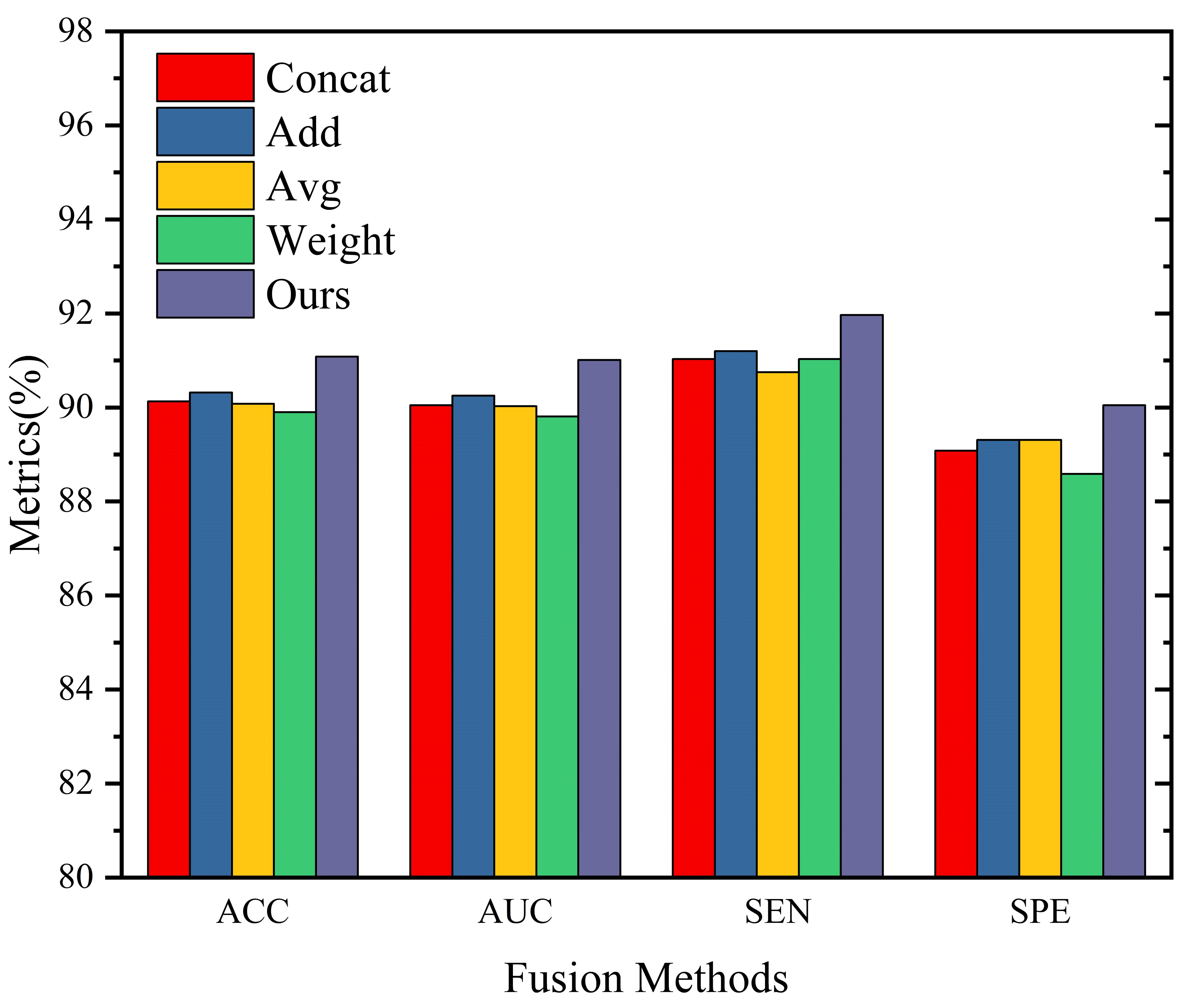
To verify the effectiveness of the feature fusion method in CKDT, we replaced it with feature concatenation (Concat), feature addition (Add), feature weighting (Weight), and feature averaging (Avg). For feature weighting, we obtain the optimal expression by assigning different weights to the multi-modal features.
The experimental results are shown in Fig. 2. In terms of accuracy ranking, the performance of the fusion methods from high to low is Ours, Add, Concat, Avg, and Weight. Unexpectedly, the performance of the feature weighting fusion method is the worst, even lower than that of Avg. This may be due to the model learning inappropriate modality weights, which leads to relatively poor performance. According to previous research, the feature weighting fusion method generally performs well in these traditional feature fusion methods. It is worth noting that our fusion module outperforms traditional methods on all evaluation metrics. This indicates that the fusion method in CKDT can effectively integrate heterogeneous information on the graph.

4.3.4 Multiple Convolutional Kernel Combinations Analysis
To investigate the influence of convolutional kernel size on the model, we evaluate the performance of single convolutional kernels separately using a single graph convolutional network (MMGE+RAM). is the convolution kernel receptive field size, in our experiment, . Subsequently, we test a combination (e.g., 1+2+3, 2+3+4,…) of three randomly selected convolutional kernels from the convolutional kernels.

The experimental results are shown in Fig. 3(a). As can be seen from, the performance of the model shows a trend of increasing and then decreasing with the increase of the receptive field . The model performance reaches the maximum when =3. Too small convolutional kernel size, i.e., =1, cannot effectively extract information from the multi-modal graph , and too large convolutional kernels, e.g., =5, causes an oversmoothing effect. As can be seen from Fig. 3(b), model achieves the best performance among all combinations when the convolutional kernel combination is =2+3+4. This also confirms the conclusion obtained in Fig. 3(a), i.e., the closer the convolution kernel size is to 3, the better the performance.
4.4 Train Set Ratio Analysis
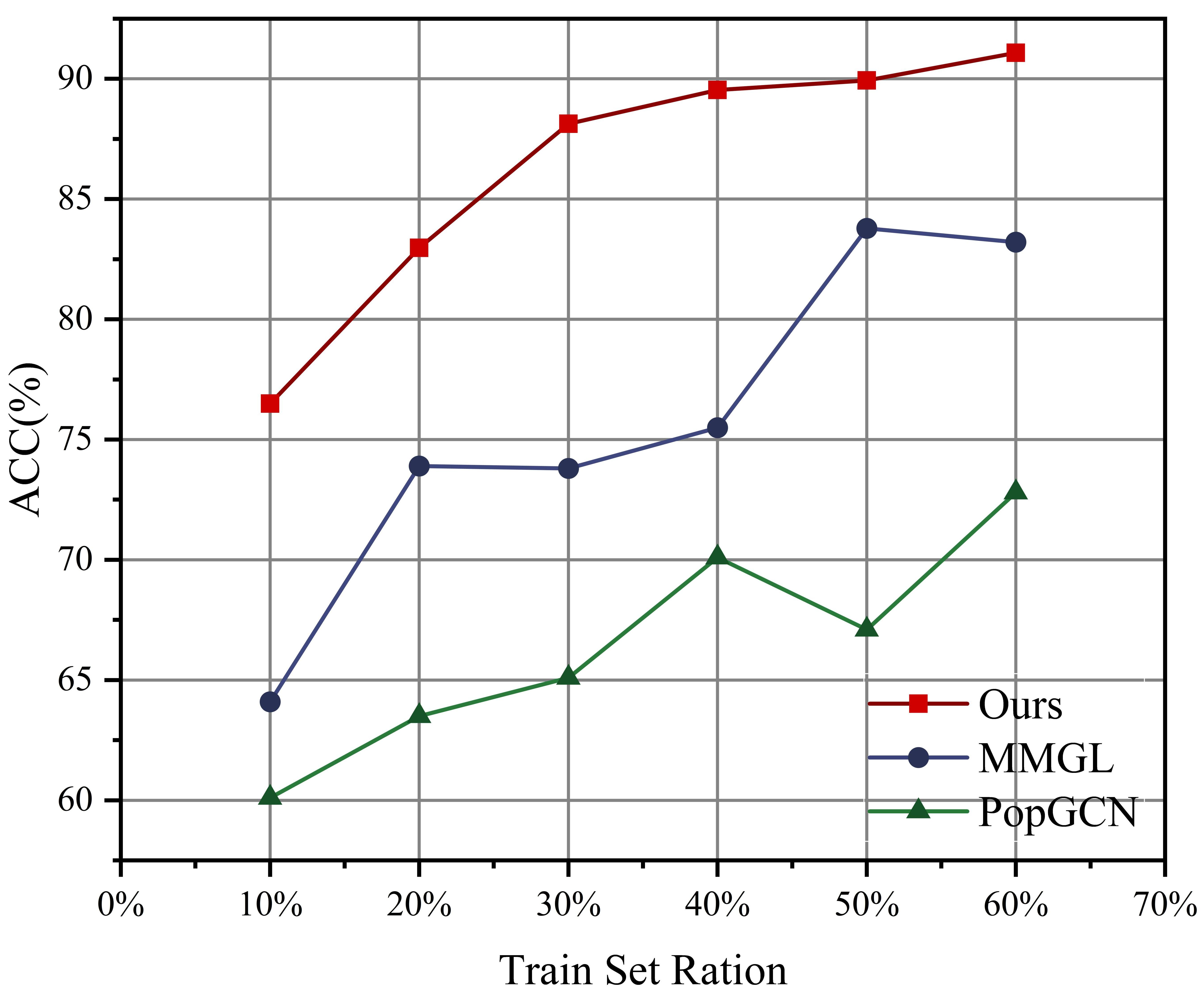
It is well known that one challenge in the field of deep learning in medicine is the lack of training data. In this context, we explore the performance of the model with a smaller data set. In the normal case, we usually use the traditional data partitioning approach, i.e., we divide the dataset into training, validation, and test sets in the ratio of 60%, 20%, and 20%, respectively.
As shown in Fig. 4, we set the training set ratio from 10% to 60%, and compare our method with MMGL and PopGCN with the same training set ratio, it is obvious that our method performs much better than the other two methods in the same ratio. Compared with MMGL and PopGCN that fluctuate largely when training set ratio increases from 30% to 60%, our method shows a smooth and stable increase. It is worth mentioning that in the lowest 10% percentage, our method works much better than MMGL and PopGCN, which indicates that our method is well suited for the domain with small data volume.

5 Discussions
In this section, we analyze the brain regions and their constituent subnetworks that are of significant discriminatory for autism diagnosis in our model. Specifically, we use a gradient-based approach (Selvaraju et al., 2017) to present FC self-attentive weighting coefficients from the model for all subjects and to obtain the average attentional level of these FCs. Notably, we used the automatic anatomical labeling (AAL) atlas and select the top K functional connections that are most discriminatory, respectively. The top 10 and top 20 most discriminating functional connections, standardized by their weight scores, are shown in Fig. 5.

5.1 Discriminative Brain Regions
As shown in Table 4, the discriminative brain areas that distinguish autism from healthy controls included the following: Frontal Area) FSO.R, FRM.L, FIOP.R, FMOR.R. Motorium) CAU.L. Sensorium) PCEN.R. Cerebellum) VER.1.2, VER.4.5, VER.8, VER.9, CC2.L. There are also some important brain areas that have a relatively small weighting, but still contribute to the diagnosis of autism, such as FIOR.L, SMA.R, HIP.R, PAI.R, PRCE.L, PRCE.R. Below we discuss the role of the discriminative brain regions that we find in the pathogenesis pattern of autism of previous studies.

In a study of related literature, it was found that in the frontal orbit (FSO.R, FIOR.L), patients with asd respond to mildly aversive visual and auditory stimuli (Green et al., 2015). There are abnormalities in the morphological structure of the ossicle (FIOP) in patients with autism and normal people, and there is a correlation with social barriers. ASD’s palpebral activity is relatively calm (Yamasaki et al., 2010). Frontal middle gyrus (FRM.L) gene expression in ASD is different from normal (Crider et al., 2014). The volume of the caudate nucleus is enlarged in medication-naive subjects with autism (Langen et al., 2007). The strength of connectivity within and between different functional subregions of the precentral gyrus (PRCE) is associated with the diagnosis of ASD and the severity of ASD features (Samson et al., 2012). The postcentral gyrus (PCEN) is responsible for somatosensory sensation. The postcentral gyrus cortical thickness and gray matter concentration are reduced in autistic subjects, and research has determined that the postcentral gyrus is a key brain area for ASD (Fatemi et al., 2018). Cerebellar worms have been implicated in the regulation of limbic functions, including mood, sensory reactivity, and salience detection. Association study between posterior earthworm and mesocerebellar cortex suggests cerebellum plays key role in autism (Fatemi et al., 2012).
| ID | ROI | Anatomical Region | FC-10 Weight | FC-20 Weight |
| 1 | PRCE.L | Precentral_L | - | 1.76% |
| 2 | PRCE.R | Precentral_R | - | 1.53% |
| 3 | FSU.L | Frontal_Sup_L | - | 1.06% |
| 6 | FSO.R | Frontal_Sup_Orb_R | 19.26% | 14.51% |
| 7 | FRM.L | Frontal_Mid_L | 12.72% | 9.58% |
| 11 | FIOP.L | Frontal_Inf_Oper_L | 12.09% | 10.32% |
| 12 | FIOP.R | Frontal_Inf_Oper_R | 5.93% | 8.99% |
| 15 | FIOR.L | Frontal_Inf_Orb_L | 2.71% | 2.04% |
| 17 | ROP.L | Rolandic_Oper_L | - | 1.40% |
| 20 | SMA.R | Supp_Motor_Area_R | 3.22% | 2.43% |
| 22 | OLF.R | Olfactory_R | - | 0.96% |
| 26 | FMOR.R | Frontal_Mid_Orb_R | - | 3.31% |
| 38 | HIP.R | Hippocampus_R | 2.49% | 1.87% |
| 58 | PCEN.R | Postcentral_R | 11.84% | 8.92% |
| 62 | PAI.R | Parietal_Inf_R | - | 1.76% |
| 68 | PREC.R | Precuneus_R | - | 1.28% |
| 71 | CAU.L | Caudate_L | 7.42% | 6.69% |
| 74 | PUTA.R | Putamen_R | - | 1.02% |
| 76 | PALL.R | Pallidum_R | - | 1.01% |
| 82 | TES.R | Temporal_Sup_R | - | 1.53% |
| 93 | CC2.L | Cerebelum_Crus2_L | 3.20% | 2.41% |
| 109 | VER.1.2 | Vermis_1_2 | 7.03% | 5.30% |
| 111 | VER.4.5 | Vermis_4_5 | 2.98% | 2.25% |
| 113 | VER.7 | Vermis_7 | - | 1.21% |
| 114 | VER.8 | Vermis_8 | 4.74% | 3.57% |
| 115 | VER.9 | Vermis_9 | 4.37% | 3.29% |
5.2 Discriminative subnetworks
We construct 2 subnetworks, as shown in Fig. 6(b). They are the subnetwork FPCF and FCVH. FPCF connects PCEN.R, CAU.L, and FIOP.R with the brain area FSO.R as the core. According to the research of Green et al. (2015), the blood oxygen signals of the amygdala and the frontal eye (FSO.R) in adolescents with ASD showed significant correlation changes under sensory stimulation. The amygdala and caudate nucleus (CAU.L) are directly connected in the brain. In the subnetwork we found in FPCF, CAU.L and FSO.R are connected. This may suggest that CAU.L provides a bridge for the connection between the amygdala and FSO.R. According to Glerean et al. (2016), CAU.L is clearly connected to PCEN.R in the Ventro-temporal limbic (VTL) subnetwork that differs most between the ASD and normal groups. At the same time, in VTL, CAU.L also has a certain connection with the amygdala.
The subnetwork FCVH connects CC2.L, VER.1.2, and HIP.R with the brain region FRM.L as the core. Numerous studies from neurocognitive and neuroimaging have shown that FRM is associated with the pathophysiology of ASD (Barendse et al., 2013). In addition, there is bisexual dimorphism in the middle frontal gyrus (Goldstein et al., 2001), which may be why men are four times more likely to develop autism than women. According to research by Fatemi et al. (2012), neuropathological abnormalities in autism were found in the cerebellum (CC2.L). In the subnetwork FCVH, both FRM.L and HIP.R are related top memory, and FRM.L is mainly responsible for short-term memory.
According to Table 4, there is an abnormal phenomenon, that is, the weight of the brain region FIOP.R has increased by 3.06%. This shows that on the basis of FC-10, most of the newly added functional connections in FC-20 are related to FIOP.R. In this regard, we use FIOP.R as the core to visualize the brain regions (FSU.L, ROP.L, SMA.R, OLR.R, HIP.R, CAU.L, FSO.R) connected to it, The results are shown in Fig. 6(a). According to the study by Yamasaki et al. (2010), impaired social skills in autistic patients are associated with reduced gray matter volume at the FIOP site. ROP.L has also been shown to be associated with autism in the subnetwork AUD discovered by Glerean et al. (2016). (Carper and Courchesne, 2005) found that the FSU.L region of autism patients was significantly enlarged compared with controls. Enticott et al. (2009). identified SMA.R as a possible source of motor dysfunction in autism by examining motor-related potentials (MRPs).

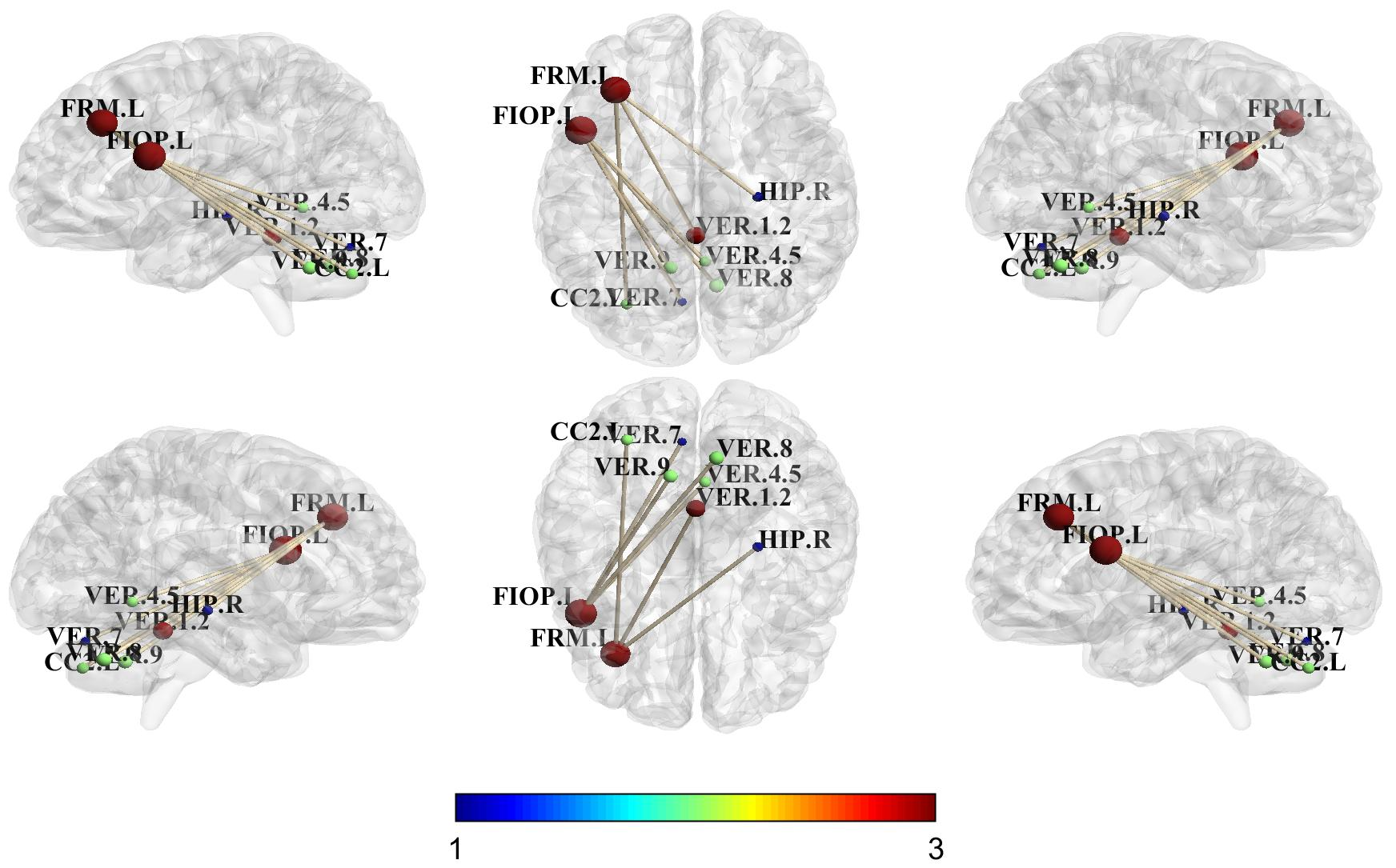
As shown in Fig. 5(a), the brain regions related to the cerebellum account for a larger proportion. We visualize the brain regions of the cerebellum (CC2.L, VER.1.2, VER.4.5, VER.7, VER.8, VER.9) and their connected FIOP.L, FRM.L. The results are as follows shown in Fig. 7. As the central part of the cerebellum, vermis (VER) has the functions of regulating muscle tone, maintaining the balance of the body, and coordinating movements. Stanfield et al. (2008) concluded that the area of lobules I-V and VI-VII of the vermis are reduced in individuals with autism compared to controls. Loss of Purkinje cells in the posterior vermis and cerebellar intermediate cortex is the most consistent neuropathology in post-mortem dissection studies of the brains of individuals with autism (Webb et al., 2009). For more information on the pathological mechanism of the cerebellum in autism, please refer to (Fatemi et al., 2012).
6 Conclusion
In this study, we propose multi-modal multi-kernel graph learning for autism prediction and biomarker discovery. Our proposed multi-modal graph embedding is well suited to alleviate the problem of negative effects between modalities. In addition, our proposed multi-kernel graph learning network is capable of extracting heterogeneous information from multi-modal graphs for autism prediction and biomarker discovery. Finally, we find some important brain regions and subnetworks with important discriminatory properties for autism by a gradient-based approach. These findings provide important guidance for the study of autism pathological mechanisms.
References
- Abraham et al. (2017) Abraham, A., Milham, M.P., Di Martino, A., Craddock, R.C., Samaras, D., Thirion, B., Varoquaux, G., 2017. Deriving reproducible biomarkers from multi-site resting-state data: An autism-based example. NeuroImage 147, 736–745.
- Adeli et al. (2019) Adeli, E., Meng, Y., Li, G., Lin, W., Shen, D., 2019. Multi-task prediction of infant cognitive scores from longitudinal incomplete neuroimaging data. NeuroImage 185, 783–792.
- Barendse et al. (2013) Barendse, E.M., Hendriks, M.P., Jansen, J.F., Backes, W.H., Hofman, P.A., Thoonen, G., Kessels, R.P., Aldenkamp, A.P., 2013. Working memory deficits in high-functioning adolescents with autism spectrum disorders: neuropsychological and neuroimaging correlates. Journal of Neurodevelopmental Disorders 5, 1–11.
- Carper and Courchesne (2005) Carper, R.A., Courchesne, E., 2005. Localized enlargement of the frontal cortex in early autism. Biological Psychiatry 57, 126–133.
- Chaari et al. (2022) Chaari, N., Gharsallaoui, M.A., Akdağ, H.C., Rekik, I., 2022. Multigraph classification using learnable integration network with application to gender fingerprinting. Neural Networks 151, 250–263.
- Cosmo et al. (2020) Cosmo, L., Kazi, A., Ahmadi, S.A., Navab, N., Bronstein, M., 2020. Latent-graph learning for disease prediction, in: International Conference on Medical Image Computing and Computer-assisted Intervention, Springer. pp. 643–653.
- Craddock et al. (2013) Craddock, C., Benhajali, Y., Chu, C., Chouinard, F., Evans, A., Jakab, A., Khundrakpam, B.S., Lewis, J.D., Li, Q., Milham, M., et al., 2013. The neuro bureau preprocessing initiative: open sharing of preprocessed neuroimaging data and derivatives. Frontiers in Neuroinformatics 7, 27.
- Crider et al. (2014) Crider, A., Thakkar, R., Ahmed, A.O., Pillai, A., 2014. Dysregulation of estrogen receptor beta (er), aromatase (cyp19a1), and er co-activators in the middle frontal gyrus of autism spectrum disorder subjects. Molecular Autism 5, 1–10.
- Di Martino et al. (2014) Di Martino, A., Yan, C.G., Li, Q., Denio, E., Castellanos, F.X., Alaerts, K., Anderson, J.S., Assaf, M., Bookheimer, S.Y., Dapretto, M., et al., 2014. The autism brain imaging data exchange: towards a large-scale evaluation of the intrinsic brain architecture in autism. Molecular Psychiatry 19, 659–667.
- Enticott et al. (2009) Enticott, P.G., Bradshaw, J.L., Iansek, R., Tonge, B.J., Rinehart, N.J., 2009. Electrophysiological signs of supplementary-motor-area deficits in high-functioning autism but not asperger syndrome: an examination of internally cued movement-related potentials. Developmental Medicine & Child Neurology 51, 787–791.
- Fatemi et al. (2012) Fatemi, S.H., Aldinger, K.A., Ashwood, P., Bauman, M.L., Blaha, C.D., Blatt, G.J., Chauhan, A., Chauhan, V., Dager, S.R., Dickson, P.E., et al., 2012. Consensus paper: pathological role of the cerebellum in autism. The Cerebellum 11, 777–807.
- Fatemi et al. (2018) Fatemi, S.H., Wong, D.F., Brašić, J.R., Kuwabara, H., Mathur, A., Folsom, T.D., Jacob, S., Realmuto, G.M., Pardo, J.V., Lee, S., 2018. Metabotropic glutamate receptor 5 tracer [18f]-fpeb displays increased binding potential in postcentral gyrus and cerebellum of male individuals with autism: A pilot pet study. Cerebellum & Ataxias 5, 1–8.
- Glerean et al. (2016) Glerean, E., Pan, R.K., Salmi, J., Kujala, R., Lahnakoski, J.M., Roine, U., Nummenmaa, L., Leppämäki, S., Nieminen-von Wendt, T., Tani, P., et al., 2016. Reorganization of functionally connected brain subnetworks in high-functioning autism. Human Brain Mapping 37, 1066–1079.
- Goldstein et al. (2001) Goldstein, J.M., Seidman, L.J., Horton, N.J., Makris, N., Kennedy, D.N., Caviness Jr, V.S., Faraone, S.V., Tsuang, M.T., 2001. Normal sexual dimorphism of the adult human brain assessed by in vivo magnetic resonance imaging. Cerebral Cortex 11, 490–497.
- Green et al. (2015) Green, S.A., Hernandez, L., Tottenham, N., Krasileva, K., Bookheimer, S.Y., Dapretto, M., 2015. Neurobiology of sensory overresponsivity in youth with autism spectrum disorders. JAMA Psychiatry 72, 778–786.
- Gürbüz and Rekik (2021) Gürbüz, M.B., Rekik, I., 2021. Mgn-net: a multi-view graph normalizer for integrating heterogeneous biological network populations. Medical Image Analysis 71, 102059.
- Huang and Chung (2022) Huang, Y., Chung, A.C., 2022. Disease prediction with edge-variational graph convolutional networks. Medical Image Analysis 77, 102375.
- Ji and Zhang (2022) Ji, J., Zhang, Y., 2022. Functional brain network classification based on deep graph hashing learning. IEEE Transactions on Medical Imaging 41, 2891–2902. doi:10.1109/TMI.2022.3173428.
- Jie et al. (2015) Jie, B., Zhang, D., Cheng, B., Shen, D., Initiative, A.D.N., 2015. Manifold regularized multitask feature learning for multimodality disease classification. Human Brain Mapping 36, 489–507.
- Kazi et al. (2019a) Kazi, A., Shekarforoush, S., Arvind Krishna, S., Burwinkel, H., Vivar, G., Kortüm, K., Ahmadi, S.A., Albarqouni, S., Navab, N., 2019a. Inceptiongcn: receptive field aware graph convolutional network for disease prediction, in: International Conference on Medical Image Computing and Computer-assisted Intervention, Springer. pp. 73–85.
- Kazi et al. (2019b) Kazi, A., Shekarforoush, S., Arvind Krishna, S., Burwinkel, H., Vivar, G., Wiestler, B., Kortüm, K., Ahmadi, S.A., Albarqouni, S., Navab, N., 2019b. Graph convolution based attention model for personalized disease prediction, in: International Conference on Medical Image Computing and Computer-assisted Intervention, Springer. pp. 122–130.
- Kazi et al. (2019c) Kazi, A., Shekarforoush, S., Kortuem, K., Albarqouni, S., Navab, N., et al., 2019c. Self-attention equipped graph convolutions for disease prediction, in: 2019 IEEE 16th International Symposium on Biomedical Imaging, IEEE. pp. 1896–1899.
- Langen et al. (2007) Langen, M., Durston, S., Staal, W.G., Palmen, S.J., 2007. Caudate nucleus is enlarged in high-functioning medication-naive subjects with autism. Biological Psychiatry 62, 262–266.
- Li et al. (2022) Li, Y., Zhang, X., Nie, J., Zhang, G., Fang, R., Xu, X., Wu, Z., Hu, D., Wang, L., Zhang, H., Lin, W., Li, G., 2022. Brain connectivity based graph convolutional networks and its application to infant age prediction. IEEE Transactions on Medical Imaging 41, 2764–2776. doi:10.1109/TMI.2022.3171778.
- Mohammadi-Nejad et al. (2017) Mohammadi-Nejad, A.R., Hossein-Zadeh, G.A., Soltanian-Zadeh, H., 2017. Structured and sparse canonical correlation analysis as a brain-wide multi-modal data fusion approach. IEEE Transactions on Medical Imaging 36, 1438–1448.
- Parisot et al. (2017) Parisot, S., Ktena, S.I., Ferrante, E., Lee, M., Moreno, R.G., Glocker, B., Rueckert, D., 2017. Spectral graph convolutions for population-based disease prediction, in: International Conference on Medical Image Computing and Computer-assisted Intervention, Springer. pp. 177–185.
- Peng et al. (2022) Peng, P., Zhang, Y., Ju, Y., Wang, K., Li, G., Calhoun, V.D., Wang, Y.P., 2022. Group sparse joint non-negative matrix factorization on orthogonal subspace for multi-modal imaging genetics data analysis. IEEE/ACM Transactions on Computational Biology and Bioinformatics 19, 479–490. doi:10.1109/TCBB.2020.2999397.
- Ritter et al. (2015) Ritter, K., Schumacher, J., Weygandt, M., Buchert, R., Allefeld, C., Haynes, J.D., 2015. Multimodal prediction of conversion to alzheimer’s disease based on incomplete biomarkers∗. Alzheimer’s & Dementia: Diagnosis, Assessment & Disease Monitoring 1, 206–215.
- Samson et al. (2012) Samson, F., Mottron, L., Soulières, I., Zeffiro, T.A., 2012. Enhanced visual functioning in autism: An ale meta-analysis. Human Brain Mapping 33, 1553–1581.
- Selvaraju et al. (2017) Selvaraju, R.R., Cogswell, M., Das, A., Vedantam, R., Parikh, D., Batra, D., 2017. Grad-cam: Visual explanations from deep networks via gradient-based localization, in: Proceedings of the IEEE International Conference on Computer Vision, pp. 618–626.
- Song et al. (2022) Song, X., Zhou, F., Frangi, A.F., Cao, J., Xiao, X., Lei, Y., Wang, T., Lei, B., 2022. Multi-center and multi-channel pooling gcn for early ad diagnosis based on dual-modality fused brain network. IEEE Transactions on Medical Imaging .
- Stanfield et al. (2008) Stanfield, A.C., McIntosh, A.M., Spencer, M.D., Philip, R., Gaur, S., Lawrie, S.M., 2008. Towards a neuroanatomy of autism: a systematic review and meta-analysis of structural magnetic resonance imaging studies. European Psychiatry 23, 289–299.
- Su et al. (2020) Su, C., Tong, J., Zhu, Y., Cui, P., Wang, F., 2020. Network embedding in biomedical data science. Briefings in bioinformatics 21, 182–197.
- Webb et al. (2009) Webb, S.J., Sparks, B.F., Friedman, S.D., Shaw, D.W., Giedd, J., Dawson, G., Dager, S.R., 2009. Cerebellar vermal volumes and behavioral correlates in children with autism spectrum disorder. Psychiatry Research: Neuroimaging 172, 61–67.
- Welling and Kipf (2016) Welling, M., Kipf, T.N., 2016. Semi-supervised classification with graph convolutional networks, in: International Conference on Learning Representations.
- Yamasaki et al. (2010) Yamasaki, S., Yamasue, H., Abe, O., Suga, M., Yamada, H., Inoue, H., Kuwabara, H., Kawakubo, Y., Yahata, N., Aoki, S., et al., 2010. Reduced gray matter volume of pars opercularis is associated with impaired social communication in high-functioning autism spectrum disorders. Biological Psychiatry 68, 1141–1147.
- Zhang et al. (2022a) Zhang, H., Song, R., Wang, L., Zhang, L., Wang, D., Wang, C., Zhang, W., 2022a. Classification of brain disorders in rs-fmri via local-to-global graph neural networks. IEEE Transactions on Medical Imaging .
- Zhang et al. (2022b) Zhang, Y., Zhang, H., Xiao, L., Bai, Y., Calhoun, V.D., Wang, Y.P., 2022b. Multi-modal imaging genetics data fusion via a hypergraph-based manifold regularization: Application to schizophrenia study. IEEE Transactions on Medical Imaging 41, 2263–2272. doi:10.1109/TMI.2022.3161828.
- Zheng et al. (2022) Zheng, S., Zhu, Z., Liu, Z., Guo, Z., Liu, Y., Yang, Y., Zhao, Y., 2022. Multi-modal graph learning for disease prediction. IEEE Transactions on Medical Imaging 41, 2207–2216. doi:10.1109/TMI.2022.3159264.
- Zhou et al. (2019) Zhou, T., Liu, M., Thung, K.H., Shen, D., 2019. Latent representation learning for alzheimer’s disease diagnosis with incomplete multi-modality neuroimaging and genetic data. IEEE Transactions on Medical Imaging 38, 2411–2422.
- Zhu et al. (2022) Zhu, Q., Wang, H., Xu, B., Zhang, Z., Shao, W., Zhang, D., 2022. Multi-modal triplet attention network for brain disease diagnosis. IEEE Transactions on Medical Imaging doi:10.1109/TMI.2022.3199032.