Electronic Properties of Single-Layer CoO2/Au(111)
Abstract
We report direct measurements via angle-resolved photoemission spectroscopy (ARPES) of the electronic dispersion of single-layer CoO2. The Fermi contour consists of a large hole pocket centered at the point. To interpret the ARPES results, we use density functional theory (DFT) in combination with the multi-orbital Gutzwiller Approximation (DFT+GA), basing our calculations on crystalline structure parameters derived from x-ray photoelectron diffraction and low-energy electron diffraction. Our calculations are in good agreement with the measured dispersion. We conclude that the material is a moderately correlated metal. We also discuss substrate effects, and the influence of hydroxylation on the CoO2 single-layer electronic structure.
pacs:
68.90.+g,73.22.-f,73.20.At,79.60.-iI I. INTRODUCTION
Layered bulk crystals based on hexagonal CoO2 Takada et al. (2003); Sakurai et al. (2015); Raveau and Seikh (2015) display intriguing electronic and phononic properties that arise from the quasi-two-dimensional (quasi-2D) nature of the atomic layers. For example, when the CoO2 layers are interleaved with H2O, Na+, and H3O+, superconductivity is observed at transition temperatures TC of approximately 4–5 K Takada et al. (2003); Sakurai et al. (2015), and the nearly 2D character of the CoO2 layers is understood to be a key aspect of these superconducting properties Takada et al. (2003); Lorenz et al. (2003); Milne et al. (2004); Wang et al. (2005). Similarities to the high- copper oxides are also notable. In both cases, a strong anisotropy between the in-plane and out-of-plane directions is key to the materials’ electronic properties Nakamura and Uchida (1993); Terasaki et al. (1997); Basov et al. (1999); Sugiura et al. (2009). Atomic layers that intervene between oxide layers play, in both cases, a complex role that goes beyond simple doping to determine the special many-body physics of the whole system Yang et al. (2005); Raghu et al. (2012); Takahata et al. (2000) (although the superconducting phases certainly also have a critical dependence on doping Schaak et al. (2003); Foo et al. (2004); Rybicki et al. (2016)).
Given that the complex electronic properties of these bulk systems arise as quasi-2D physics in weakly interacting, atomically thin oxide layers, it is natural to ask whether any of the interesting electronic properties of the bulk persist in the single-layer (SL) limit. The electronic properties of a SL material can differ in important ways from those of layered bulk parent compounds. For example, among the transition metal dichalcogenides, the band dispersion Kuc et al. (2011); Zhu et al. (2011) and electronic correlations Feng et al. (2018); Xi et al. (2015) can be significantly modified in the SL limit.
A method has recently been developed for epitaxially fabricating rotationally aligned SL CoO2 islands on Au(111), Pt(111), and Ag(111) substrates Walton et al. (2015); Fester et al. (2017a). So far, the SL system has been studied in the context of applications to catalysis Fester et al. (2018a). Here we investigate the electronic properties of the SL, which—to the best of our knowledge—have not yet been studied experimentally, although a recent theory paper has predicted that the SL might manifest 2D ferromagnetism and undergo a superconducting transition at = 25–28 K Nguyen et al. (2019). Besides laying the groundwork for new directions in the very active field of 2D-materials research, our study aims to clarify our understanding of layered CoO2-based compounds.
II II. METHODS
Our growth procedure is based on the synthesis method that has been previously described by Walton, et al. Walton et al. (2015). It consists of two steps, performed in-situ in a vacuum chamber with a base pressure of low- mbar. First, we evaporate elemental Co while simultaneously exposing the sample to O2 at a chamber pressure of mbar. In this step, the sample temperature is ca. 380 K. This forms SL CoO on the Au(111) substrate. We use a growth rate of approximately one monolayer CoO per hour. We then stop depositing Co and increase the local O2 pressure, using a moveable O2 doser that we bring to within a few mm of the sample face: with this we further oxidize the CoO for two hours, to form CoO2. The sample temperature is in this second step is ca. 325 K and the chamber pressure is mbar, but the local pressure at the sample face is presumably much higher (likely as much as two orders of magnitude). The full procedure results in SL CoO2 islands Walton et al. (2015).
Electronic structure measurements were made by angle-resolved photoemission spectroscopy (ARPES) at the SGM3 beamline of the ASTRID2 Synchrotron Light Source in Denmark Hoffmann et al. (2004). Incident light is linearly polarized in the direction parallel to the scattering plane; the angle between analyzer axis and incident light axis is 50°. Sample quality and coverage were assessed in situ via a combination of x-ray photoelectron spectroscopy (XPS), low-energy electron diffraction (LEED), and scanning-tunneling microscopy (STM). The sample was at room temperature during STM characterisation Sup , while it was at 30(5) K during XPS, LEED and ARPES measurements. The width of the Fermi edge in the ARPES measurements (fitted from projected bulk continuum states of the Au substrate) was approximately 80 meV.
In order to interpret the ARPES measurements, a precise knowledge of structural parameters is crucial. Previous studies of SL CoO2 on noble metal (111) surfaces Walton et al. (2015); Fester et al. (2017a, b) have used STM and XPS to obtain structural information about the SL. However, XPS is a rather indirect method of assessing structure. STM does provide structural information, but mainly only about the top layer of atoms, and it is limited in its scope to local measurements, so it is not an optimal probe of average atomic structure over macroscopic areas. Here we use x-ray photoelectron diffraction (XPD) as a direct, high-resolution probe of the local geometric structure around each chemically distinct type of emitter, averaged over the area of the beam spot. Measurements were made at the SuperESCA beamline at Elettra, the synchrotron radiation facility in Trieste, Italy Baraldi et al. (2003). Here the incident light is linearly polarized in the horizontal plane, where also the electron energy analyser lies, at an angle of 70° with respect to the photon beam. The overall energy resolution was below 100 meV and 200 meV in the energy range from 650 eV to 1150 eV photon energy, respectively. The high resolution spectra of O 1s and Co 2p core levels were measured in normal emission conditions and the binding energy was normalized to the Fermi level of the Au substrate. Sample cleanliness, layer quality and order where checked with XPS and LEED prior to the XPD measurements.
XPD patterns are constructed by collecting XPS spectra across a range of polar () angles, from 70°(normal incidence in the configuration of SuperESCA) to normal emission, and azimuthal angles () across a range of 130°, with an angular resolution in the order of 1°. More details can be found in Bana et al. (2018) and Bignardi et al. (2019). For the measurements in this study, the sample was at room temperature.
In the peak fitting analysis of the resulting data, the photoemission intensity (, ) of each component is extracted from every XPS spectrum by the use of a Doniach-Sunjic fitting function Doniach and Sunjic (1970) with Shirley background subtraction. From this we obtain the modulation function , defined as
| (1) |
where is the average intensity for an azimuthal scan at polar angle . The XPD pattern is a projection of the modulation function for a particular peak. We compare measured XPD patterns to multiple scattering simulations for trial structures that are generated using the program package Electron Diffraction in Atomic Clusters (EDAC) García de Abajo et al. (2001). The agreement between measured and simulated XPD patterns is then quantified, via calculation of a reliability factor that is defined as the sum of the normalised mean-square deviation of the experimental () and theoretical () modulation functions,
| (2) |
for each emission angle . By this definition, an R-factor of 0 corresponds to perfect agreement, while an R-factor of 1 indicates uncorrelated data Woodruff (2007). (Anti-correlated data give an R-factor of 2.) Values of R less than 0.3 are generally taken to indicate relatively good agreement (e.g., Bana et al. (2018)). We take the confidence interval of the minimized value to be Bignardi et al. (2019); Bana et al. (2018), with being the ratio of the solid angle of the measurements to the solid-angular resolution.
III III. RESULTS
Electronic structure determination.
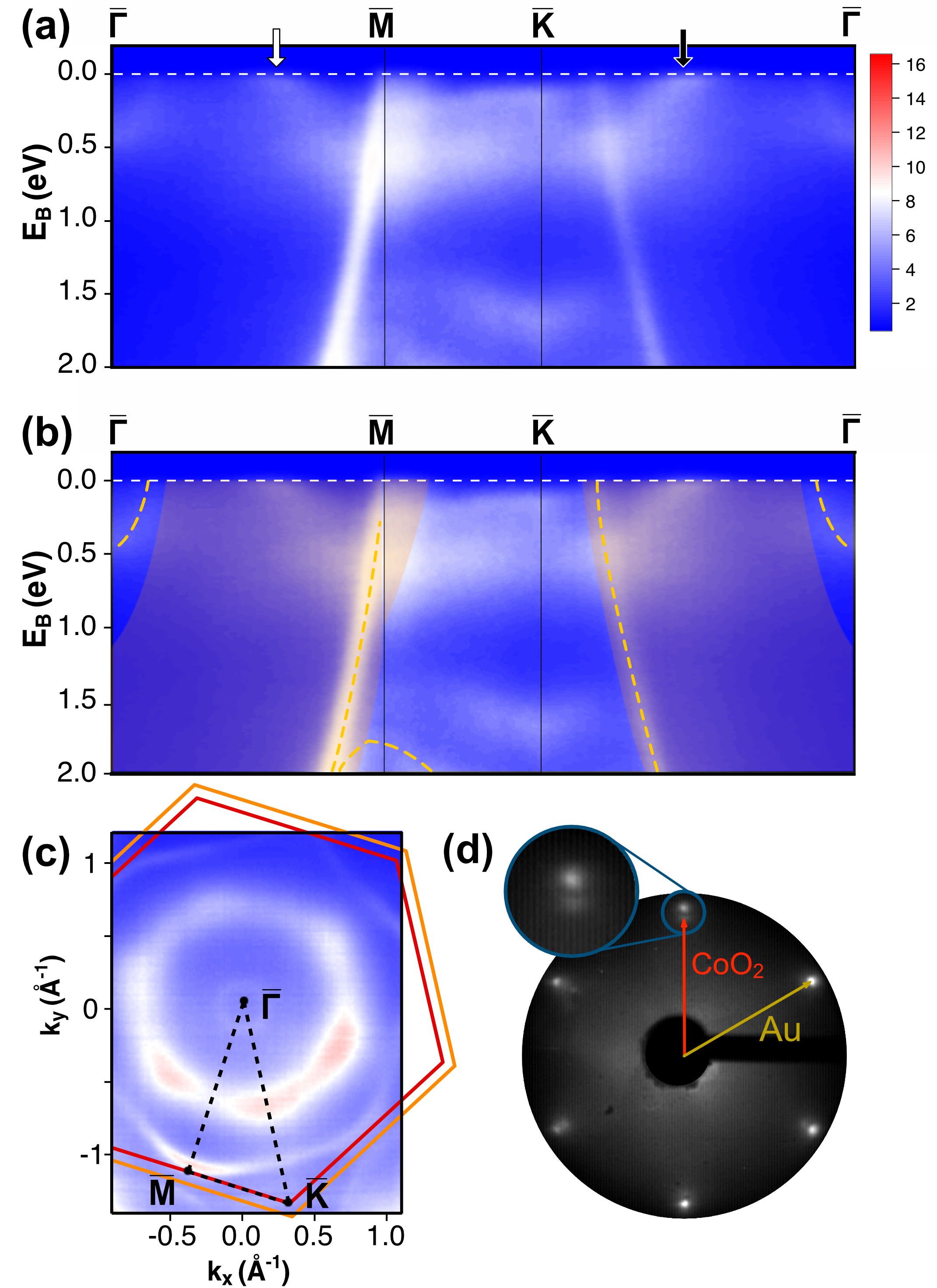
ARPES measurements acquired with photon energy = 89 eV are presented in Fig. 1 for SL CoO2 with a sample coverage of approximately 70%, as determined by in-situ STM (see Sup ). Fig. 1(a) shows the APRES spectrum along cuts through the high-symmetry points , , and of the CoO2 Brillouin zone (BZ). The Au(111) surface state at is faintly visible—presumably deriving from exposed regions of the Au(111) substrate—and so are the sharp, highly dispersive features of the bulk Au(111) sp-band, which cross the Fermi level near and along the cut Takeuchi et al. (1991). In Fig. 1(b), the electron dispersion from Fig. 1(a) is reproduced with the Au features highlighted: the Au surface state and bulk bands are overlaid with orange dashed lines as guides to the eye, and the projected bulk band continuum is shaded orange. Several observed features are not associated with the Au(111) substrate, and we attribute these to the electronic structure of SL CoO2. At first glance, the adlayer band structure shows a metallic character and seems to consist of one band around 1.5 eV binding energy EB (mostly visible between ), and at least two bands close to the Fermi level, at less than 1.0 eV in binding energy. Of these bands close to the Fermi level, the lower one appears to exhibit energy minima at approximately EB = 0.6 eV at and approximately EB = 0.5 eV close to . (As we will see below, however, the actual dispersion is more complicated than this.) A Fermi level crossing at = 0.67(3) Å-1 in the direction is marked with a white arrow in the figure. In the direction, the band crosses the Fermi level at approximately = 0.69(5) Å-1 (black arrow). To identify the Fermi level crossing, the band dispersion near the Fermi level was determined by fitting energy distribution curves (EDCs) to Gaussian-broadened Lorentzians convoluted with the Fermi function, within a range of about 100 meV below the Fermi level. The EDC fits were also found to agree well with Gaussian fits to momentum distribution curves (MDCs) within the same energy range. The bands appear to flatten close to the Fermi level, creating the impression of kinks in the dispersion; this will be further discussed below. The Fermi contour corresponding to the measured dispersion is presented in Fig. 1(c). The overlaid orange hexagon marks the Au(111) surface BZ (SBZ), which is slightly larger than the CoO2 BZ (red hexagon). The high-symmetry directions in Fig. 1(a) are marked with black dashed lines, and the high-symmetry points are labeled. The Au(111) surface state creates a small ring around , and the large, faint hexagonal feature with its vertices at approximately the points of CoO2 arises from Au bulk states. Between these, the SL CoO2 forms a distorted, rounded hexagon centered around ; by referencing the dispersion in panel (a), one can see that this is a large hole pocket. The relative sizes and rotations of the BZs are calculated on the basis of LEED measurements, and a representative measurement is presented in Fig. 1(d). Diffraction spots forming two rotationally aligned hexagons arise from the SL CoO2 (red arrow) and the Au(111) substrate (yellow arrow). By using the known in-plane Au(111) lattice parameter of 2.88 Å Maeland and Flanagan (1964), the in-plane lattice parameter of the SL CoO2 is determined to 3.12(3) Å, which is within the range of values (2.8–3.3 Å) previously reported on the basis of STM measurements Walton et al. (2015); Fester et al. (2017b).

Core level data.
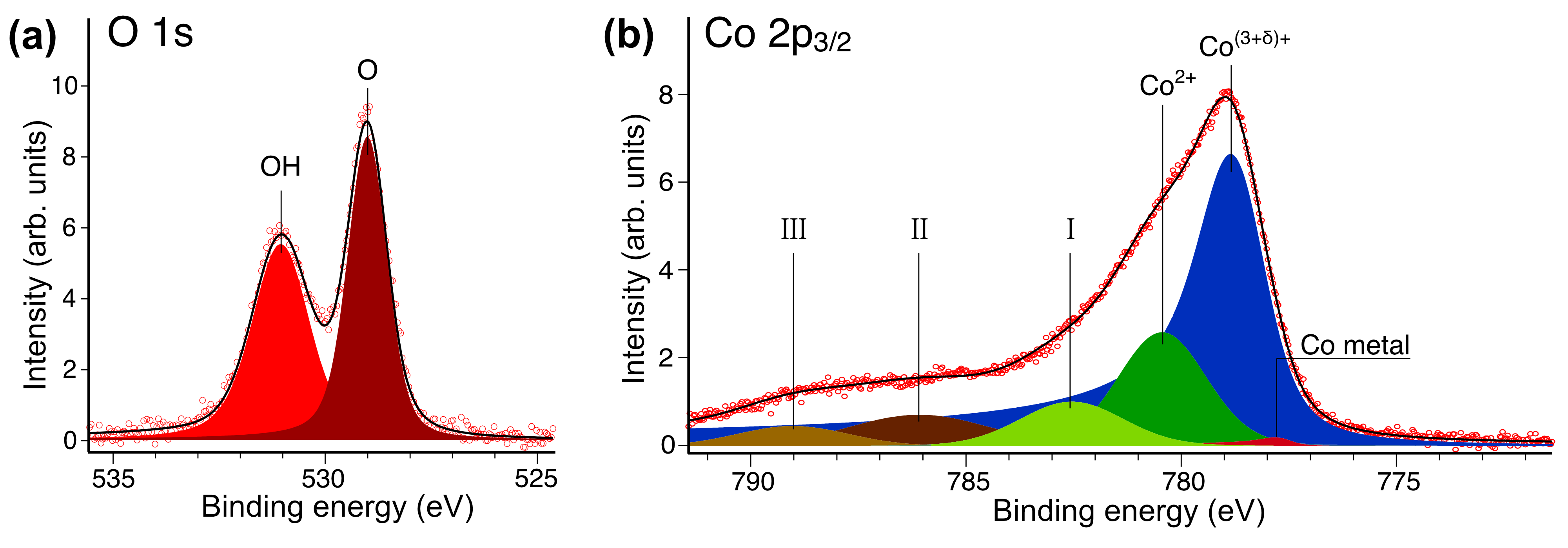
The O 1s spectrum is displayed in Fig. 2(a). The main component (dark red) occurs at a binding energy of EB = 529.0(1) eV (binding energies are referenced to the measured Fermi level). An additional hydroxyl component (bright red) is also present, as expected from previous studies Fester et al. (2017b, 2018b, 2018a). Similarly to what is reported in the earlier-published results, this OH peak is shifted 2.05(5) eV towards higher binding energy relative to the main O peak. Fester, et al., attribute the presence of this component to a partial hydroxyl overlayer formed by H bonding to O at the top of the CoO2 SL (i.e., the side of the SL away from the interface with the Au(111) substrate). They suggest that the hydroxylation results from dissociative adsorption of H2O or H2 rest gas in the vacuum chamber Fester et al. (2017b). The main O peak contains photoemission intensity from unhydroxylated emitters at both the top and the bottom of the layer. These components are known to be very closely separated in energy Fester et al. (2018b) and cannot be resolved.

Fig. 2(b) shows the Co 2p spectrum. It is dominated by a large peak with several closely spaced minor components. The complexity of this spectrum attests to the presence of more than one oxidation state of Co. Freestanding CoO2 might be naïvely expected to exhibit the Co4+ oxidation state; however, we instead observe a Co(3+δ)+ oxidation state—fitted here with an asymmetric main peak (blue) at a binding energy of EB = 778.7 eV—consistent with previously published findings Walton et al. (2015); Fester et al. (2017b). The oxidation state is presumably due to charge transfer from adsorbates (or possibly from the substrate, but see discussion below). We surmise that such charge transfer stabilises the SL; however, the possible mechanism for such stabilisation is beyond the scope of the present work. Some areas of CoO have failed to further oxidize in the second step of the growth procedure, and this is manifest in the persistent presence of a set of peaks arising from the Co2+ oxidation state. Co2+ has a high-spin configuration that generates a complex peak structure with multiplet splitting and shake-up satellites Frost et al. (1974); Chuang et al. (1976); Biesinger et al. (2011); Kim (1975). Here we fit it with just two components, aside from the Co2+ main component at EB = 780.4(1) eV (dark green in the figure): a shake-up satellite, labelled “II” (dark brown), separated from the main peak by 5.7(1) eV, and peak “I” (light green), which arises from multiplet splitting and is fixed to a separation of 2.1 eV from the main peak Biesinger et al. (2011). While the intensities and locations of these two peaks are in relatively good agreement with related peak structures previously identified in the literature Biesinger et al. (2011); Kim (1975); Chuang et al. (1976), our fitting here is phenomenological and does not attempt to capture the details of the complex peak structure of the Co2+ spectrum. Note that this makes it difficult for us to estimate the precise amount of the less-oxidized CoO that remains on this particular sample after the second oxidation step; however, it does not impact the modulation of the Co(3+δ)+ component, which is only associated with photoemission from fully oxidized CoO2.
We identify an additional weak satellite peak (“III,” shown in light brown) at 10.3(1) eV higher binding energy than the Co(3+δ)+ peak. The position and the intensity of this satellite are similar to what has been seen in diverse related compounds such as CoOOH, Co(OH)2, and Co3O4 Chuang et al. (1976); Yang et al. (2010); Biesinger et al. (2011), though its physical interpretation remains uncertain. Finally, a negligible amount of unoxidized Co metal remains, even after the full growth procedure, and leads to the small peak shown in red in the figure11endnote: 1The Co 2p3/2 spectrum for bulk metallic Co is known to display an asymmetric main peak together with surface and bulk plasmon peaks Grosvenor et al. (2005). Here, the plasmon satellites are excluded from the metallic Co fit, due to the very small amount of metallic Co found to be present. The binding energy is set to match the value reported by Walton, et al. Walton et al. (2015), in their measurements of Co metal on Au(111), and the peak shape is asymmetric with both the shape and location similar to what is measured by Walton et al. Walton et al. (2015)..
Crystalline structure determination.
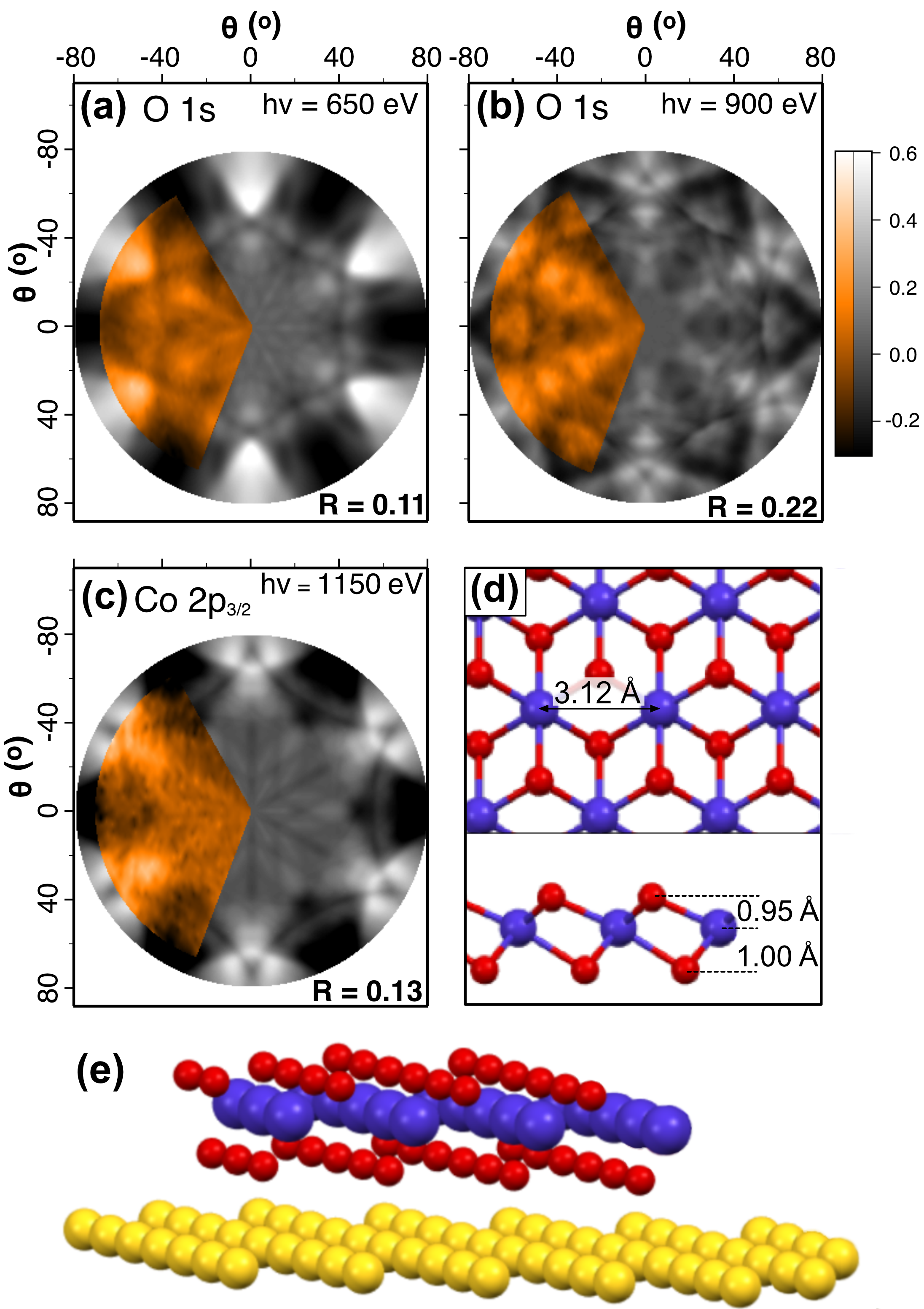
XPD results are presented in Fig. 3. The experimental data are shown in orange, superimposed on the grayscale best-fit multiple-scattering simulations. Fig. 3(a) shows the diffraction pattern of the unhydroxylated O 1s core level peak obtained with a photon energy of = 650 eV. This photon energy was chosen so that the photoelectron kinetic energy would be less than 150 eV, to enhance the cross section for backscattering of photoelectrons from the underlying structure and thus the sensitivity of the measurement to emitters in the top layer of O atoms. By contrast, in Fig. 3(b) a higher photon energy of = 900 eV was used, favouring the forward scattering of photoelectrons and enhancing sensitivity to emitters in the bottom layer of O atoms. The resulting modulation function is, in this latter case, highly dependent on the structure above the bottom layer of O—the arrangement of Co atoms and of O atoms at the top of the layer. The diffraction pattern arising from the Co 2p3/2 core level, acquired from the peak with photon energy h = 1150 eV, is shown in Fig. 3(c). Here, again, forward scattering is favoured. From these three data sets, we obtain the out-of-plane parameters of the crystal lattice from the best-fit structural model to the XPD data, keeping the in-plane parameter in the simulation fixed to the result from LEED. We note that LEED gives sharp diffraction spots, consistent with the presence of well-aligned rotational domains of CoO2. We start by assuming that SL CoO2 has a crystalline structure related to the CdI2 type, in accordance with previous predictions Walton et al. (2015). The CdI2 structure, however, has only three-fold symmetry. We therefore assume that the nearly six-fold symmetry of the XPD patterns derives from the presence of exactly two domains in the sample, and these are related to each other by mirror symmetry (equivalent to 60 in-plane rotation).

The R-factor for the simulations presented in Fig. 3(a–c) is shown at the bottom right of each panel. The low values in each case indicate good agreement between the measurement and the simulation. and were determined by optimizing for all three simulations simultaneously, with the assumption that CoO2 is present on the sample in two equally distributed orientations rotated with respect to one other by 60. The results suggest a slightly smaller distance between Co and top O atoms ( = 0.95(10) Å) than between Co and bottom O atoms ( = 1.00(10) Å). The uncertainties in and are rough estimates of our confidence in identifying the minimum of each of them in terms of all three parameters simultaneously. (We note that these uncertainties span a range of and within which the average value for the three simulations falls mostly within of its minimum, although the average increases more rapidly in the direction of increasing than in the other directions. In any case, the average might not be the most useful way to think about uncertainty in this particular case.) Our values of and are consistent with the asymmetric structure proposed by Walton, et al. Walton et al. (2015). In a qualitative sense, such an asymmetry is not unexpected, considering that one side of the layer is at the interface with the substrate, while the other side is hydroxylated and at the interface with vacuum. However, the range of our uncertainty does also permit the possibility that , which would not affect any of the conclusions we draw in the present study Sup . The atomic structure derived from the XPD simulations is shown from top and side views in Fig. 3(d), and from an oblique view in Fig. 3(e). A hexagonal layer of Au atoms corresponding to the unreconstructed (111) surface is shown in Fig. 3(e) for illustrative purposes, even though the actual XPD simulations do not include the substrate. Because of the lattice mismatch between the SL and the Au(111) substrate, there are many different adsorption sites on the substrate lattice, and thus there is no simple geometric relationship between the emitters in the SL CoO2 and the Au(111); this justifies neglect of the Au(111) in the simulations.
Previously published work has found that adsorbed H at the top of the SL assumes a partially disordered “labyrinth” structure Fester et al. (2017b). In the present study, we observe weak modulation of the hydroxylated O 1s core-level peak: this might arise from small structural changes associated with hydroxylation. However, it is small compared with the modulation of the non-hydroxylated component, and we have not been able to analyse it successfully.
Electronic structure simulations.
Here we analyse the electronic structure of the CoO2 SL. To take into account the electron-correlation effects, we utilize the DFT+GA method Deng et al. (2009); Ho et al. (2008); Lanatà et al. (2015). Specifically, we have utilized the implementation of Lanatà et al. (2015, 2017), using the DFT code Wien2k Schwarz and Blaha (2003), employing the Local Density Approximation (LDA) and the standard “fully localized limit” form for the double-counting functional Anisimov et al. (1997). Our calculations were performed using a -point grid and setting the product of the smallest atomic-sphere radius times the largest plane-wave momentum () to 9. As in Ref. Lanatà et al. (2019), we set the Hund’s coupling constant to eV, while we set the screened Hubbard interaction parameter to eV.
The calculations were performed with the lattice parameters that we measured from LEED and XPD. In the supplementary material Sup we also show that small changes in the out-of-plane parameters—within the precision of XPD measurments—lead to band structures that are also consistent with the ARPES data. In particular, this includes the inversion-symmetric lattice structure with .
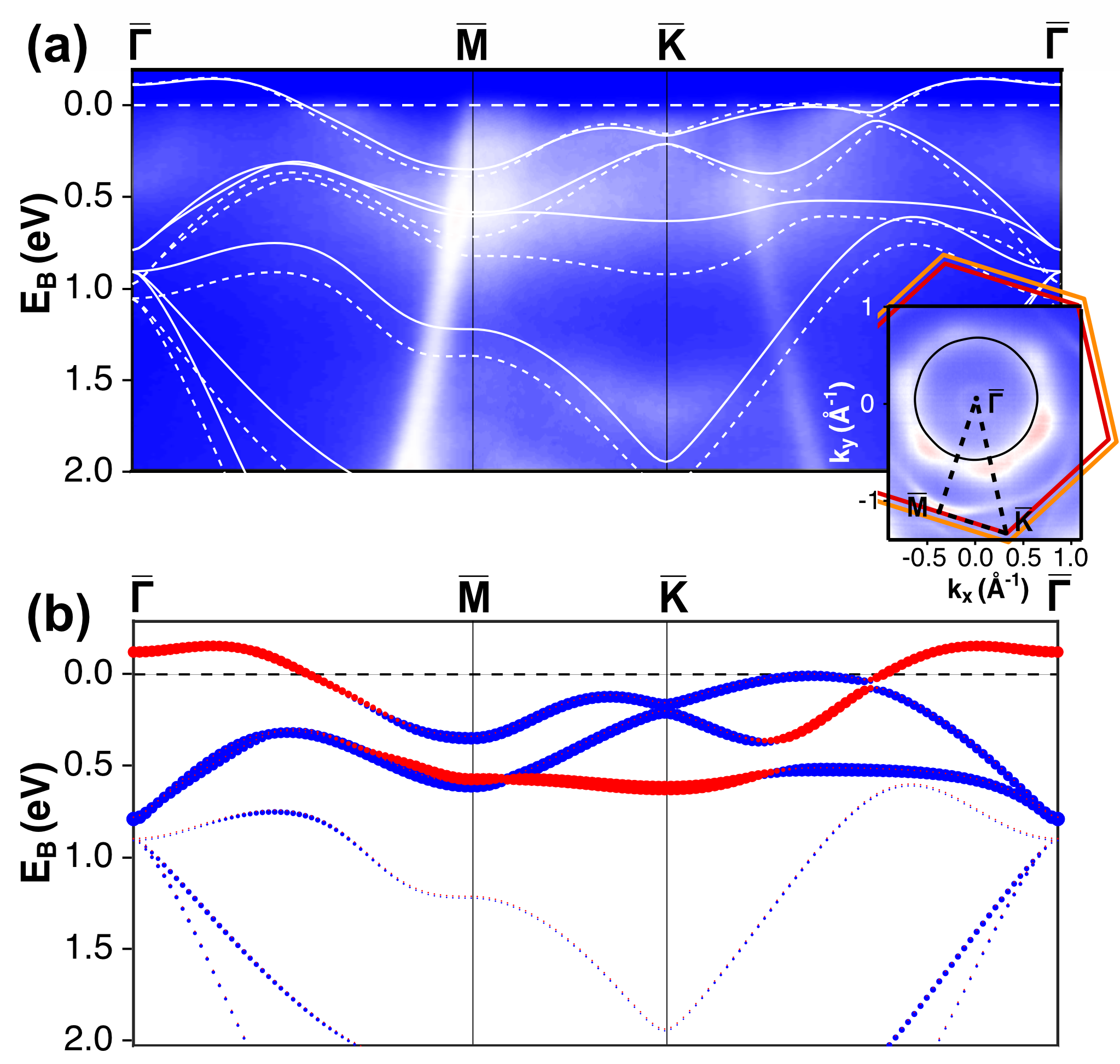
In Fig. 4(a) we compare the DFT and the DFT+GA bands with the dispersion measured by ARPES. Note that in Fig. 4 the theoretical Fermi level of the pure single layer is shifted by approximately 90 meV above the calculated value, as such shift results in a more satisfactory agreement with the experiments. In the supplemental material we argue, based on DFT calculations, that this energy shift may be caused by the adsorption of H atoms on the SL Sup . We observe that the correlation effects captured by DFT+GA improve the agreement with the experiments considerably compared to bare DFT.
Within the multi-orbital GA framework, the correlation effects on the band structure (Fig. 4(b)) are encoded in a linear momentum-independent self energy for the Co 3d electrons, represented as follows Lanatà et al. (2015); Bünemann et al. (2003):
| (3) |
Eq. (3) is characterized by: (i) the so-called “matrix of quasi-particle weights” , whose eigenvalues measure the degree of correlation of the corresponding degrees of freedom, and (ii) the frequency-independent component of the self-energy matrix , inducing interaction-driven d-electron on-site level shifts. The Co 3d manifold is generated by 1 one-dimensional irreducible representation and 2 two-dimensional representations. From standard group-theoretical considerations Lanatà et al. (2017) it follows that has a non-degenerate eigenvalue and two two-fold degenerate eigenvalues .
Based on our calculation, , and , indicating that the SL CoO2 system is a moderately-correlated metal. In Fig. 4(b) we show the Co 3d spectral weight of the bands, resolved with respect to the corresponding point symmetry-group representations. As expected, the correlation effects are particularly important for capturing the experimental behavior of the d-electron bands, such as those at low binding energy close to the -point, which have predominantly character. However, the d-electron correlations considerably influence the whole band structure, including the O 2p band (because of hybridization effects).

IV IV. DISCUSSION
Key questions in 2D-materials research are how the electronic properties of SL systems differ from those of related 3D (bulk) compounds, and how those properties are affected by the environment—for example, by the presence of the substrate, or by adsorbates. Here we consider these questions for the case of SL CoO2.
Influence of Au(111) substrate.
The Au(111) substrate plays a role in catalyzing the growth of CoO2 and in stabilizing the SL Fester et al. (2017a, 2018a). Furthermore, as discussed above, the Au(111) substrate introduces an asymmetry in the out-of-plane direction—not only by interacting directly with the SL, but also by inhibiting hydroxylation at the bottom of the layer. (Full hydroxylation of the top and bottom of the layer—i.e., synthesis of SL Co(OH)2 on Au(111)—has been shown to be possible in the presence of “high” pressures (i.e., 10 mbar) of H2O vapor Fester et al. (2018a); however, to our knowledge, hydroxylation at the bottom of the SL on Au(111) does not occur due to the presence of chamber rest gas alone.)
Nevertheless, despite the impact it has on structure, the Au(111) substrate appears to have a weak influence on the electronic properties of the SL. The most important observation is the fact that calculations neglecting the substrate agree well with the measured ARPES spectra. It is also interesting to observe that the splitting between the two O 1s XPS peaks, which derive from photoemission from the top and the bottom of the layer, is so small that it cannot be resolved by our measurements. If the interaction were strong between the substrate and the oxygen atoms at the bottom of the layer, one might expect to see a significant peak shift, similar to what has been observed for MoS2/Au(111) Bana et al. (2018), and which we do not see in the present case.
Hydroxylation.
To estimate the amount of hydrogen present, we note that in the O 1s core level of Fig. 2(a) the hydroxyl component accounts for 43% of the combined intensity of the two peaks. We assume that there is attenuation of the photoemission intensity from the bottom O layer (due to an inelastic mean free path of = 5.3 Å Qua ; Shinotsuka et al. (2015)) but that the presence of H does not attenuate the photoemission intensity. Furthermore, we assume that hydroxylation happens only at the top of the CoO2 layer Fester et al. (2017b), and that each H is bonded only to a single O. This results in a value of 57% hydroxylated O at the top of the layer. Note, however, that there is a rather high level of uncertainty in this estimate, depending on the validity of our several assumptions. Also, this rough estimate does not take into account the persistance of local regions of CoO after the second oxidation step: these certainly persist on the sample surface, as can be seen in Fig. 2. We have observed—in agreement with previous literature Fester et al. (2018b, a)—that CoO hydroxylates less extensively than CoO2; thus, our estimate represents only a lower bound for the extent of hydroxylation in our sample. Our findings appear roughly consistent with those of previous studies, which have shown that hydroxylation of the CoO2 layer is approximately (i.e., two out of every three O atoms at the top of the layer hydroxylated) after storage in UHV conditions for several hours Fester et al. (2017b). Indeed, the sample that generated the XPS data shown above was stored under UHV conditions for a few days before the measurements in Fig. 2 were acquired.
By contrast, the samples that generated the ARPES data above were stored in UHV conditions for only a few hours before they were measured. They might, therefore, be less hydroxylated than the samples measured with XPS. However, on the basis of the existing literature we would not expect the level of hydroxylation to be less than approximately , as this is the lower bound previously observed in freshly-grown samples Fester et al. (2017b). The measured Fermi contour in Fig. 1 is 70(3) % filled, which indicates a charge transfer to the SL of 0.40(6)e per unit cell (by contrast with undoped CoO2, whose Fermi contour would be half-filled). Thus, the amount of hydroxylation that we estimate using XPS would likely be sufficient to generate the charge transfer that we observe in ARPES, even without charge transfer from the Au(111) substrate.
An obvious question is what influence the hydroxylation has on the electronic dispersion. Previous work Fester et al. (2017b) has shown that the H ions are not well ordered on the surface; therefore, we might expect the OH groups to shift and broaden the band structure rather than to impact the bare-band dispersion. Indeed, the calculations shown in Fig. 4 successfully reproduce most of the main features of the band structure without the inclusion of hydroxylation. Nevertheless, there could, of course, be some level of weak ordering of the H ions, and we consider the implications of this for the band dispersion in Sup ; Kresse and
Furthmüller (1996); Kresse and Joubert (1999); Perdew et al. (1996); Monkhorst and Pack (1976); Demaison et al. (2007).
Comparison of SL CoO2 with related bulk materials.
In light of the apparently weak impact of the substrate on the electronic dispersion, and the likelihood of little if any charge transfer from the substrate, it seems reasonable to consider our CoO2 system as approximately “2D.” Here, then, we discuss the electronic structure of this 2D material in relation to analogous layered bulk systems, such as NaxCoO2 and NaxCoO yH2O. This is a particularly interesting point of consideration, because the superconducting and magnetic properties of the layered bulk materials have been interpreted as arising from the“pseudo-2D” nature of the weakly interacting CoO2 atomic layers: the layers are partially isolated from one another in bulk materials by interleaving layers of Na+, H2O, and H3O+ Takada et al. (2003); Sakurai et al. (2015); Lorenz et al. (2003); Milne et al. (2004); Wang et al. (2005). (We note that superconductivity would not be expected in our samples, because of the proximity effect of the Au(111) substrate.)
The electronic dispersion of the SL is remarkably similar to that of related layered bulk materials Singh (2000); Lee et al. (2004); Hasan et al. (2004); Arakane et al. (2008); Yang et al. (2005); Qian et al. (2006). The large hole pocket around is the most obvious feature of the bulk dispersion, and this is true for the SL, as well. Discussion in the literature has surrounded the question of why six small hole pockets that are predicted to cross the Fermi level along in the bulk dispersion Singh (2000); Lee et al. (2004) are not observed experimentally at any doping level Hasan et al. (2004); Yang et al. (2004, 2005); Arakane et al. (2008). Our results are similar to those of ARPES studies of the bulk, in that no hole pockets rise above the Fermi level along . However, we do note that there are hole pockets just below the Fermi level, and that the measured Fermi contour of the SL exhibits diffuse, elongated intensity in the direction, due to the presence of these shallow states located in close vicinity to, and leaking some intensity across, the Fermi level.
Comparing the properties of the bulk and the SL, it is worth pointing out that there are some structural differences between our samples and the stacked layers of similar bulk systems. The in-plane lattice constant we have identified here, 3.12(3) Å, is larger than that reported for the bulk (2.8222(13) Å Amatucci et al. (1996)). The O height above Co as reported for the bulk (e.g., 0.91 Å for NaCo2O4 Singh (2000)) is within the range of the uncertainty of our measurements of the SL, but the bulk layer would be expected to be symmetrical around the Co plane, whereas our results agree with earlier suggestions that the SL is likely to be asymmetrical around the Co plane Walton et al. (2015).
Kinks and electron-phonon coupling.
A final point of interest relates to electron-phonon coupling, a topic relevant to superconductivity. Several studies of related bulk CoO2-based materials identify kinks in the bands that cross the Fermi level Hasan et al. (2004); Geck et al. (2007); Arakane et al. (2008). In the case of the SL, although at first glance the data in Fig. 1(a) does appear to exhibit kinks at both band crossings, we do not find any decisive indication that such kinks definitely occur. As can be seen from Fig. 4, the sharp downturn in the filled band dispersion just below the Fermi level along the direction seems adequate to explain the shape of the band there, in agreement with Qian et al. Qian et al. (2006). Along the we were not able to convincingly fit any kink in the dispersion, so the situation here remains somewhat unclear.
V V. CONCLUSIONS
Using XPD, we have determined the crystalline structure of SL CoO2 on Au(111), finding good agreement with previous predictions. We have reported the electronic structure on the basis of ARPES measurements and DFT+GA calculations. Our calculations describe the ARPES data well, and indicate that SL CoO2 is characterized by moderate electronic correlations. We have observed significant hydroxylation at the top of the layer, and found that the main effect of the H impurities is a shift of the Fermi level. Our results suggest a weak interaction between the SL and the Au(111) substrate.
VI ACKNOWLEDGMENTS
We gratefully acknowledge financial support from the VILLUM FONDEN via the Centre of Excellence for Dirac Materials (Grant No. 11744) and VILLUM project grant no. 13264 (J.V.L.). Y.-X.Y. was supported by the U.S. Department of Energy, Office of Science, Basic Energy Sciences, as part of the Computational Materials Science Program. C.E.S. received support from the European Community’s Seventh Framework Programme (FP7/2007-2013) CALIPSO under grant agreement no 312284.
References
- Takada et al. (2003) K. Takada, H. Sakurai, E. Takayama-Muromachi, F. Izumi, R. A. Dilanian, and T. Sasaki, Nature 422, 53 (2003).
- Sakurai et al. (2015) H. Sakurai, Y. Ihara, and K. Takada, Physica C 514, 378 (2015).
- Raveau and Seikh (2015) B. Raveau and M. M. Seikh, Zeitschrift fur anorganische und allgemeine Chemie 641, 1385 (2015).
- Lorenz et al. (2003) B. Lorenz, J. Cmaidalka, R. L. Meng, and C. W. Chu, Physical Review B 68, 132504 (2003).
- Milne et al. (2004) C. J. Milne, D. N. Argyriou, A. Chemseddine, N. Aliouane, J. Veira, S. Landsgesell, and D. Alber, Physical Review Letters 93, 247007 (2004).
- Wang et al. (2005) C. H. Wang, X. H. Chen, J. L. Luo, G. T. Liu, X. X. Lu, H. T. Zhang, G. Y. Wang, X. G. Luo, and N. L. Wang, Physical Review B 71, 224515 (2005).
- Nakamura and Uchida (1993) Y. Nakamura and S. Uchida, Physical Review B 47, 8369 (1993).
- Terasaki et al. (1997) I. Terasaki, Y. Sasago, and K. Uchinokura, Physical Review B 56, R12685 (1997).
- Basov et al. (1999) D. N. Basov, S. I. Woods, A. S. Katz, E. J. Singley, R. C. Dynes, M. Xu, D. G. Hinks, C. C. Homes, and M. Strongin, Science 283, 49 (1999).
- Sugiura et al. (2009) K. Sugiura, H. Ohta, S.-i. Nakagawa, R. Huang, Y. Ikuhara, K. Nomura, H. Hosono, and K. Koumoto, Applied Physics Letters 94, 152105 (2009).
- Yang et al. (2005) H.-B. Yang, Z.-H. Pan, A. K. P. Sekharan, T. Sato, S. Souma, T. Takahashi, R. Jin, B. C. Sales, D. Mandrus, A. V. Fedorov, et al., Physical Review Letters 95, 146401 (2005).
- Raghu et al. (2012) S. Raghu, R. Thomale, and T. H. Geballe, Physical Review B 86, 094506 (2012).
- Takahata et al. (2000) K. Takahata, Y. Iguchi, D. Tanaka, T. Itoh, and I. Terasaki, Physical Review B 61, 12551 (2000).
- Schaak et al. (2003) R. E. Schaak, T. Klimczuk, M. L. Foo, and R. J. Cava, Nature 424, 527 (2003).
- Foo et al. (2004) M. L. Foo, Y. Wang, S. Watauchi, H. W. Zandbergen, T. He, R. J. Cava, and N. P. Ong, Physical Review Letters 92, 247001 (2004).
- Rybicki et al. (2016) D. Rybicki, M. Jurkutat, S. Reichardt, C. Kapusta, and J. Haase, Nature Communications 7, 11413 (2016).
- Kuc et al. (2011) A. Kuc, N. Zibouche, and T. Heine, Physical Review B 83, 245213 (2011).
- Zhu et al. (2011) Z. Y. Zhu, Y. C. Cheng, and U. Schwingenschlögl, Physical Review B 84, 153402 (2011).
- Feng et al. (2018) J. Feng, D. Biswas, A. Rajan, M. D. Watson, F. Mazzola, O. J. Clark, K. Underwood, I. Marković, M. McLaren, A. Hunter, et al., Nano Letters 18, 4497 (2018).
- Xi et al. (2015) X. Xi, L. Zhao, Z. Wang, H. Berger, L. Forró, J. Shan, and K. F. Mak, Nature Nanotechnology 10, 765 (2015).
- Walton et al. (2015) A. S. Walton, J. Fester, M. Bajdich, M. A. Arman, J. Osiecki, J. Knudsen, A. Vojvodic, and J. V. Lauritsen, ACS Nano 9, 2445 (2015).
- Fester et al. (2017a) J. Fester, M. Bajdich, A. S. Walton, Z. Sun, P. N. Plessow, A. Vojvodic, and J. V. Lauritsen, Topics in Catalysis 60, 503 (2017a).
- Fester et al. (2018a) J. Fester, A. Makoveev, D. Grumelli, R. Gutzler, Z. Sun, J. Rodríguez-Fernández, K. Kern, and J. V. Lauritsen, Angewandte Chemie 130, 12069 (2018a).
- Nguyen et al. (2019) D.-L. Nguyen, C.-R. Hsing, and C.-M. Wei, Nanoscale 11, 17052 (2019).
- Hoffmann et al. (2004) S. V. Hoffmann, C. Søndergaard, C. Schultz, Z. Li, and P. Hofmann, Nuclear Instruments and Methods in Physics Research, Section A 523, 125430 (2004).
- (26) See supplementary material for information on coverage estimates, discussion of the fitting procedures for XPS and XPD data, and additional discussion about the role of hydroxylation and lattice parameters in the theory calculations.
- Fester et al. (2017b) J. Fester, A. Walton, Z. Li, and J. V. Lauritsen, Physical Chemistry Chemical Physics 19, 2425 (2017b).
- Baraldi et al. (2003) A. Baraldi, G. Comelli, S. Lizzit, M. Kiskinova, and G. Paolucci, Surface Science Reports 49, 169 (2003).
- Bana et al. (2018) H. Bana, E. Travaglia, L. Bignardi, P. Lacovig, C. E. Sanders, M. Dendzik, M. Michiardi, M. Bianchi, D. Lizzit, F. Presel, et al., 2D Materials 5, 035012 (2018).
- Bignardi et al. (2019) L. Bignardi, D. Lizzit, H. Bana, E. Travaglia, P. Lacovig, C. E. Sanders, M. Dendzik, M. Michiardi, M. Bianchi, M. Ewert, et al., Phys. Rev. Materials 3, 014003 (2019).
- Doniach and Sunjic (1970) S. Doniach and M. Sunjic, Journal of Physics C: Solid State Physics 3, 285 (1970).
- García de Abajo et al. (2001) F. J. García de Abajo, M. A. Van Hove, and C. S. Fadley, Physical Review B 63, 075404 (2001).
- Woodruff (2007) D. P. Woodruff, Surface Science Reports 62, 1 (2007).
- Takeuchi et al. (1991) N. Takeuchi, C. T. Chan, and K. M. Ho, Physical Review B 43, 13899 (1991).
- Maeland and Flanagan (1964) A. Maeland and T. B. Flanagan, Canadian Journal of Physics 42, 2364 (1964).
- Fester et al. (2018b) J. Fester, Z. Sun, J. Rodríguez-Fernández, A. Walton, and J. V. Lauritsen, The Journal of Physical Chemistry B 122, 561 (2018b).
- Heimann et al. (1981) P. Heimann, J. van der Veen, and D. Eastman, Solid State Communications 38, 595 (1981).
- Moulder et al. (1992) J. F. Moulder, W. F. Stickle, P. E. Sobol, and K. D. Bomben, Handbook of X-ray Photoelectron Spectroscopy (Physical Electronics Division, Perkin-Elmer Corporation, 1992).
- Frost et al. (1974) D. Frost, C. McDowell, and I. Woolsey, Molecular Physics 27, 1473 (1974).
- Chuang et al. (1976) T. Chuang, C. Brundle, and D. Rice, Surface Science 59, 413 (1976).
- Biesinger et al. (2011) M. C. Biesinger, B. P. Payne, A. P. Grosvenor, L. W. Lau, A. R. Gerson, and R. S. Smart, Applied Surface Science 257, 2717 (2011).
- Kim (1975) K. S. Kim, Physical Review B 11, 2177 (1975).
- Yang et al. (2010) J. Yang, H. Liu, W. N. Martens, and R. L. Frost, The Journal of Physical Chemistry C 114, 111 (2010).
- Deng et al. (2009) X.-Y. Deng, L. Wang, X. Dai, and Z. Fang, Phys. Rev. B 79, 075114 (2009).
- Ho et al. (2008) K. M. Ho, J. Schmalian, and C. Z. Wang, Phys. Rev. B 77, 073101 (2008).
- Lanatà et al. (2015) N. Lanatà, Y. X. Yao, C.-Z. Wang, K.-M. Ho, and G. Kotliar, Phys. Rev. X 5, 011008 (2015).
- Lanatà et al. (2017) N. Lanatà, Y. Yao, X. Deng, V. Dobrosavljević, and G. Kotliar, Phys. Rev. Lett. 118, 126401 (2017).
- Schwarz and Blaha (2003) K. Schwarz and P. Blaha, Computational Materials Science 28, 259 (2003).
- Anisimov et al. (1997) V. I. Anisimov, F. Aryasetiawan, and A. I. Lichtenstein, J. Phys. Condens. Matter 9, 767 (1997).
- Lanatà et al. (2019) N. Lanatà, L. Tsung-Han, V. S. Yong-Xin, Yao, and V. Dobrosavljević, npj Comput. Mater. 5, 30 (2019).
- Bünemann et al. (2003) J. Bünemann, F. Gebhard, and R. Thul, Phys. Rev. B 67, 075103 (2003).
- (52) S Tougaard Quases-Tougaard, Ver. 7.0. Software package for characterizing surface nanostructures by analysis of electron spectra. See http://www.quases.com/ (2019).
- Shinotsuka et al. (2015) H. Shinotsuka, S. Tanuma, C. J. Powell, and D. R. Penn, Surface and Interface Analysis 47, 871 (2015).
- Kresse and Furthmüller (1996) G. Kresse and J. Furthmüller, Comp. Mater. Sci. 6, 15 (1996).
- Kresse and Joubert (1999) G. Kresse and D. Joubert, Physical Review B 59, 1758 (1999).
- Perdew et al. (1996) J. P. Perdew, K. Burke, and M. Ernzerhof, Physical Review Letters 77, 3865 (1996).
- Monkhorst and Pack (1976) H. J. Monkhorst and J. D. Pack, Physical Review B 13, 5188 (1976).
- Demaison et al. (2007) J. Demaison, M. Herman, and J. Lievin, International Reviews in Physical Chemistry 26, 391 (2007).
- Singh (2000) D. J. Singh, Physical Review B 61, 13397 (2000).
- Lee et al. (2004) K.-W. Lee, J. Kuneš, and W. E. Pickett, Physical Review B 70, 045104 (2004).
- Hasan et al. (2004) M. Z. Hasan, Y.-D. Chuang, D. Qian, Y. W. Li, Y. Kong, A. Kuprin, A. V. Fedorov, R. Kimmerling, E. Rotenberg, K. Rossnagel, et al., Physical Review Letters 92, 246402 (2004).
- Arakane et al. (2008) T. Arakane, T. Sato, T. Takahashi, H. Ding, T. Fujii, and A. Asamitsu, Physica B: Condensed Matter 403, 1086 (2008).
- Qian et al. (2006) D. Qian, L. Wray, D. Hsieh, L. Viciu, R. J. Cava, J. L. Luo, D. Wu, N. L. Wang, and M. Z. Hasan, Physical Review Letters 97, 186405 (2006).
- Yang et al. (2004) H.-B. Yang, S.-C. Wang, A. K. P. Sekharan, H. Matsui, S. Souma, T. Sato, T. Takahashi, T. Takeuchi, J. C. Campuzano, R. Jin, et al., Physical Review Letters 92, 246403 (2004).
- Amatucci et al. (1996) G. G. Amatucci, J. M. Tarascon, and L. C. Klein, Journal of The Electrochemical Society 143, 1114 (1996).
- Geck et al. (2007) J. Geck, S. V. Borisenko, H. Berger, H. Eschrig, J. Fink, M. Knupfer, K. Koepernik, A. Koitzsch, A. A. Kordyuk, V. B. Zabolotnyy, et al., Physical Review Letters 99, 046403 (2007).
- Grosvenor et al. (2005) A. P. Grosvenor, S. D. Wik, R. G. Cavell, and A. Mar, Inorganic Chemistry 44, 8988 (2005).