Liver Lesion Segmentation with slice-wise 2D Tiramisu and Tversky loss function
Abstract
At present, lesion segmentation is still performed manually (or semi-automatically) by medical experts. To facilitate this process, we contribute a fully-automatic lesion segmentation pipeline. This work proposes a method as a part of the LiTS (Liver Tumor Segmentation Challenge) competition for ISBI 17 and MICCAI 17 comparing methods for automatic segmentation of liver lesions in CT scans.
By utilizing cascaded, densely connected 2D U-Nets and a Tversky-coefficient based loss function, our framework achieves very good shape extractions with high detection sensitivity, with competitive scores at time of publication. In addition, adjusting hyperparameters in our Tversky-loss allows to tune the network towards higher sensitivity or robustness. The implementation of our method is available online111https://github.com/Confusezius/unet-lits-2d-pipeline.
Index Terms— Deep learning, Convolutional Networks, Liver segmentation, Lesion Segmentation, Tiramisu, U-Net, Tversky
1 Introduction
As neural networks have proven to achieve human-like performance in biomedical image classification [1], and nearly human-like performance for segmentation tasks [2], it stands to reason to construct a segmentation pipeline that applies these methods for automatic lesion detection.
We evaluate what results can be achieved using the deep fully convolutional neural network architectures Tiramisu [3] and the U-Net [4] with standard data preprocessing and data augmentation and without any sophisticated postprocessing.
U-Net-based architectures themselves are widely used for biomedical image segmentation, and have already proven its robustness and strength in many challenges ( segmentation of neuronal structures [5]). The Tiramisu [3] enhances this structure on the basis of densely connected networks created by [6] to achieve better information flow and higher effective depth.
The complete pipeline resembles the cascaded approach proposed in [7] where the authors suggested a U-Net cascade (with an additional subsequent postprocessing step based on conditional random fields). In our case for liver segmentation, a standard U-Net was trained to reduce overall training time, whereas for lesion segmentation, the aforementioned Tiramisu was trained from scratch (without transfer learning) with a Tversky loss function to improve on a simple Dice-based loss function.
2 Dataset and Preprocessing
| Architecture | Dice (A) | Dice (G) | VOE | RVD | ASSD | MSSD | RMSD |
| U-Net LiS (MICCAI 17) | 0.95 | 0.94 | 0.10 | -0.05 | 1.89 | 32.71 | 4.20 |
| U-Net + U-Net LeS (ISBI 17) | 0.42 | - | 0.69 | 103.74 | 32.54 | - | - |
| U-Net + Tiramisu (DL) LeS (MICCAI 17) | 0.45 | 0.44 | 0.33 | -0.16 | 1.28 | 8.85 | 2.07 |
| U-Net + Tiramisu (TL) LeS (MICCAI 17) | 0.57 (14) | 0.66 (16) | 0.34 (1) | 0.02 (4) | 0.95 (1) | 6.81 (6) | 1.60 (3) |
The dataset consists of 200 CT-scans of the abdominal and upper body region such that the liver is fully included as part of the Liver Tumor Segmentation Challenge [8]. The data was provided by different clinics from all over the world and is stored in NIfTI dataformat. For working purposes, the set was divided by the LITS challenge organisers into 130 scans for training (containing liver and lesion masks) and 70 for evaluating the method (without masks). From the 130 training scans, we selected 25 scans for validation purposes during training and 105 for actual training.
Preprocessing on the provided data included value-clipping the volume values (on the Hounsfield scale) to an interval of in order to remove air- and bonelike structures for a more uniform background. The voxel values were then normalized by substracting the training set mean and standardized by dividing through the training set standard deviation. In a final step, the affine matrix was extracted from the NIfTI header to rotate each volume to the same position to make learning easier.
3 Method
As mentioned, the lesion segmentation pipeline includes two different nets: a U-Net to segment the liver from a given volume slice, and the Tiramisu to segment out lesions. By masking the lesion segmentation with the liver mask, we effectively reduce the number of false positives in regions outside the liver. Both architectures will be presented shortly. We implemented our networks by use of the deep learning library Keras [9] on the basis of tensorflow [10].
3.1 U-Net architecture
Our U-Net was implemented with two convolutional layers with padding plus a ReLU-activation layer before max-pooling for the downward movement of the U-branch. These blocks were repeated for a total of four times. We started with 32 filters and a constant 33 filter size. The number of filters was doubled after every convolution-max-pooling-block. Dropout was performed after the ReLU-activation layer with a rate of 0.2. The upsampling branch which performs a transposed convolution to reconstruct the segmentation image was implemented in the same fashion. This totals in roughly network parameters.
3.2 Tiramisu architecture
The Tiramisu was implemented as described in the original paper with 4 denseblocks comprising 4,5,6 and 7 layers respectively in the feature extraction branch and a final bottleneck denseblock with 8 densely connected layers. Each denseblock was created with a growth rate of 12, followed by a maxpooling layer while initializing the network with 32 starting filters. Dropout with probability 0.2 was performed after each denseblock, and the extraction structure was mirrored for the upsampling step. ReLU activation was used and L2-regularization with was added to the convolutional layers. In addition, a constant filter size was implemented. All in all, this results in approx. parameters, i.e. only a fourth of the standard U-Net setup that was used for liver segmentation/ISBI 17. Standard Batch-Normalization was applied before each convolutional layer.
3.3 Liver segmentation training
Training was performed over 50 epochs with a batch size of 5 on a GTX 1070 GPU using an Adam optimizer [11], He uniform initialization [12], standard binary crossentropy loss and a learning rate of which was halved every 15 epochs. The final weights were selected to provide the best validation score. In total, training on 105 randomly selected volumes (including validating on 25) took approximately 80 hours. Note that no data augmentation was performed.
3.4 Lesion segmentation training
The Tiramisu lesion segmentation network was trained for 35 epochs using the Adam optimization method and a tversky loss function [13] based on the Tversky coefficient (through simple negation).
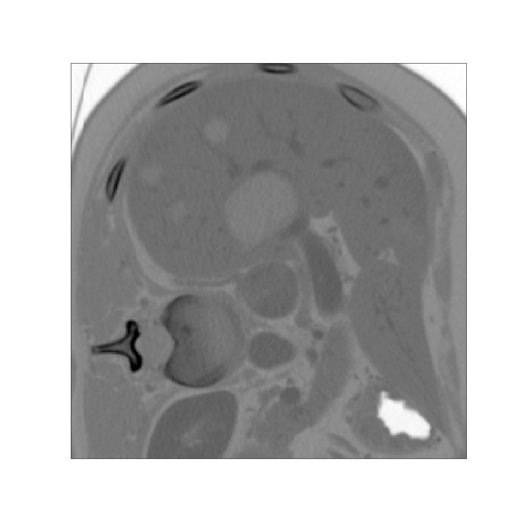
For the loss, the coefficient was negated with false positive penalty and false negative penalty . We penalize false positive predictions higher because training the Tiramisu without transfer learning and simple weighted binary cross entropy or dice loss showed the network being prone to a high false positive rate. An initial learning rate of was chosen, which was halved every 10 epochs. The complete training using a batch-size of 5 required nearly 38 hours, with the usage of very basic data augmentation (minor rotation and translation as well as zooming). During training, the network learned from slices along the coronal axis of which crops were taken in and around the liver. In different runs (see tab.1), a simple Dice loss was also used for training as reference to the Tversky loss under otherwise same conditions.
4 Experiment and results
For the final submission, after each liver segmentation, only the largest connected component was chosen as the final liver mask. In addition to the test results for the introduced pipeline, tab.1 also shows the results for the U-Net-only approach for ISBI 17, and a pipeline with a Tiramisu lesion segmentation network that was trained with a simple dice loss function. Each method was applied on the 70 test volumes and shared the same liver segmentation network. Total inference time including both liver and lesion segmentation took roughly 7s per volume on average, totaling in 8 hour of testing time on a GTX 1070 GPU. For a measurement of tumorburden, the pipeline scores 0.03 on RSME and 0.18 on Max Error.


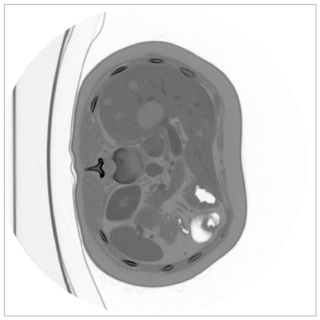
(a) Example slice

(b) Zoomed Liver

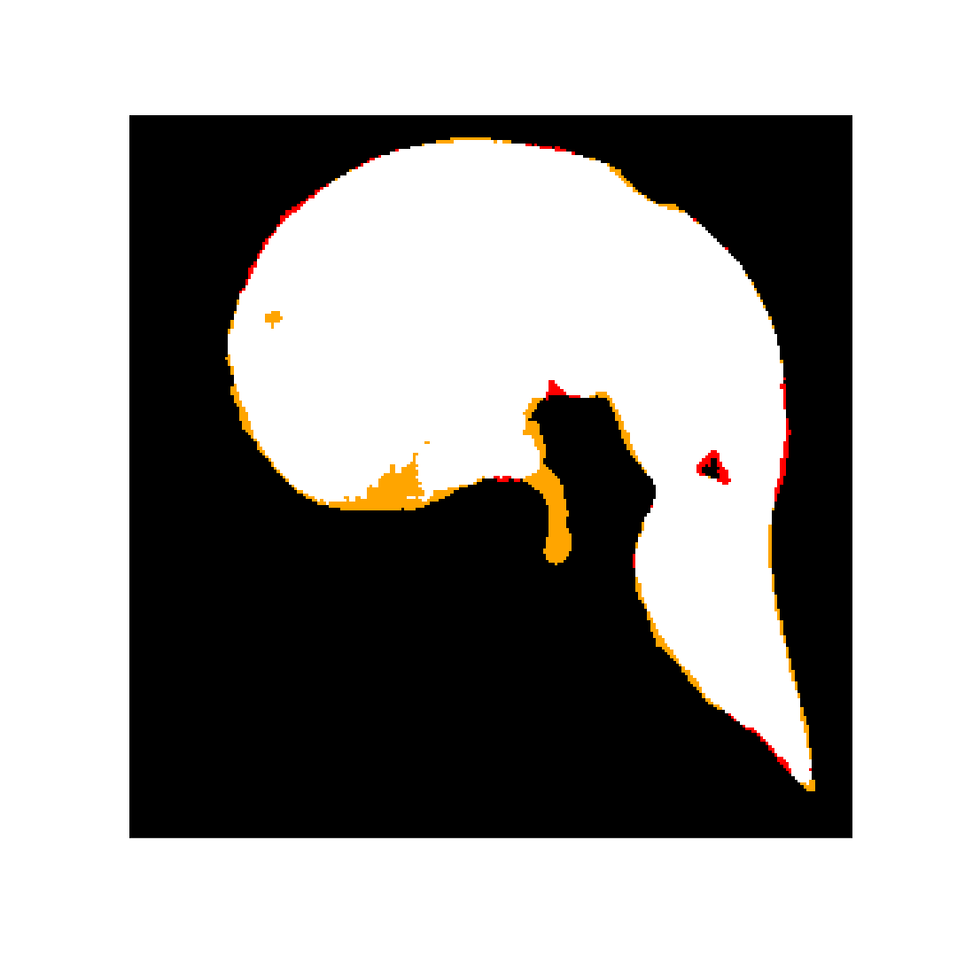
(c) Liver Segmentation

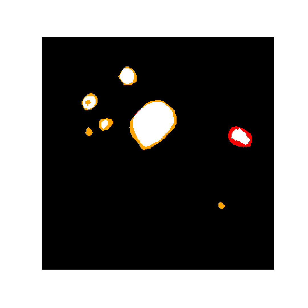
(d) Liver lesion segmentation
5 Discussion
The major strength of this approach is its simplicity, as we use a network-only based segmentation approach to achieve good segmentation results (see tab.1), especially for the Tiramisu-based pipeline when compared to a plain U-Net setup.
Moreover, the proposed pipeline using Tiramisu and Tversky-loss achieves best scores regarding Volume-Overlap Error (VOE) and Average Symmetric Surface Distance (ASSD) as well as scoring high in Relative Volume Difference (RVD) and Maximum Symmetric Surface Distance (MSSD). This insinuates that the Tiramisu performs very well when it comes to actual shape extraction when lesions are detected correctly.
However, there are several issues solving which could improve the segmentation capabilities:
(1) As can be seen from fig.2 and tab.1, our liver segmentation performs reasonably well, but misses a majority of very big and/or lesion at the liver boundary (see fig.1) which then, although maybe detected through the lesion segmentation network, get masked away. Increasing the training effort for our liver segmentation network would therefore allow us to better our current lesion (and liver) segmentation results. We tried to counter this effect by isotropically dilating the liver mask (fig.1), but this only worsened the final results as the number of introduced false positives was higher then the amount of removed false negatives.
(2) Even with our current implementation of the Tversky loss function, the major error source are false positive prediction. Higher penalties, a network ensemble and regularization through, for example, higher dropout or more sophisticated data augmentation would ideally increase the area under the ROC curve and provide better overall scores.
In addition, training a liver segmentation network on the basis of a Tiramisu and using that as weight initialization could potentially help the network to converge faster and to better results.
On a final note, [7] and [14] showed that good postprocessing (for example with 3D conditional random fields and random forests with additional handmade features) could improve the result. We feel this is an avenue worth exploring on this data. Also, segmentation was only performed on a 2D slice-by-slice basis. [15] showed exceptional results through the application of a semi-3D approach by including additional upper and/or lower slices in the segmentation process of the neural network to incorporate more spatial information. Our pipeline could be easily extended to make use of this approach to hopefully improve the results further.
References
- [1] Esteva A, Kuprel B, Novoa RA, Ko J, Swetter SM, Blau HM, and Thrun S., “Dermatologist-level classification of skin cancer with deep neural networks.,” 2017.
- [2] Long J, Shelhamer E, and Darrell T, “Fully convolutional networks for semantic segmentation,” 2015.
- [3] Simon Jégou, Michal Drozdzal, David Vàzquez, Adriana Romero, and Yoshua Bengio, “The one hundred layers tiramisu: Fully convolutional densenets for semantic segmentation,” CoRR, vol. abs/1611.09326, 2016.
- [4] Ronneberger O, Fischer P, and Brox T, “U-net: Convolutional networks for biomedical image segmentation,” 2015.
- [5] Arganda-Carreras I, Turaga SC, Berger DR, Cireşan D, Giusti A, Gambardella LM, Schmidhuber J, Laptev D, Dwivedi S, Buhmann JM, and Liu T. o, “Crowdsourcing the creation of image segmentation algorithms for connectomics,” 2015.
- [6] Gao Huang, Zhuang Liu, and Kilian Q. Weinberger, “Densely connected convolutional networks,” CoRR, vol. abs/1608.06993, 2016.
- [7] Christ PF, Ettlinger F, Grün F, Elshaera ME, Lipkova J, Schlecht S, Ahmaddy F, Tatavarty S, Bickel M, Bilic P, and Rempfler M, “Automatic liver and tumor segmentation of ct and mri volumes using cascaded fully convolutional neural networks,” 2017.
- [8] Patrick Bilic, Patrick Ferdinand Christ, Eugene Vorontsov, Grzegorz Chlebus, Hao Chen, Qi Dou, Chi-Wing Fu, Xiao Han, Pheng-Ann Heng, Jürgen Hesser, Samuel Kadoury, Tomasz K. Konopczynski, Miao Le, Chunming Li, Xiaomeng Li, Jana Lipková, John S. Lowengrub, Hans Meine, Jan Hendrik Moltz, Chris Pal, Marie Piraud, Xiaojuan Qi, Jin Qi, Markus Rempfler, Karsten Roth, Andrea Schenk, Anjany Sekuboyina, Ping Zhou, Christian Hülsemeyer, Marcel Beetz, Florian Ettlinger, Felix Grün, Georgios Kaissis, Fabian Lohöfer, Rickmer Braren, Julian Holch, Felix Hofmann, Wieland H. Sommer, Volker Heinemann, Colin Jacobs, Gabriel Efrain Humpire Mamani, Bram van Ginneken, Gabriel Chartrand, An Tang, Michal Drozdzal, Avi Ben-Cohen, Eyal Klang, Michal Marianne Amitai, Eli Konen, Hayit Greenspan, Johan Moreau, Alexandre Hostettler, Luc Soler, Refael Vivanti, Adi Szeskin, Naama Lev-Cohain, Jacob Sosna, Leo Joskowicz, and Bjoern H. Menze, “The liver tumor segmentation benchmark (lits),” CoRR, vol. abs/1901.04056, 2019.
- [9] Chollet F, “Keras: Deep learning library for theano and tensorflow,” https://github.com/fchollet/keras, 2017.
- [10] Martin Abadi, Ashish Agarwal, Paul Barham, Eugene Brevdo, Zhifeng Chen, Craig Citro, Greg S. Corrado, Andy Davis, Jeffrey Dean, Matthieu Devin, Sanjay Ghemawat, Ian Goodfellow, Andrew Harp, Geoffrey Irving, Michael Isard, Yangqing Jia, Rafal Jozefowicz, Lukasz Kaiser, Manjunath Kudlur, Josh Levenberg, Dan Mané, Rajat Monga, Sherry Moore, Derek Murray, Chris Olah, Mike Schuster, Jonathon Shlens, Benoit Steiner, Ilya Sutskever, Kunal Talwar, Paul Tucker, Vincent Vanhoucke, Vijay Vasudevan, Fernanda Viégas, Oriol Vinyals, Pete Warden, Martin Wattenberg, Martin Wicke, Yuan Yu, and Xiaoqiang Zheng, “Tensorflow: Large-scale machine learning on heterogeneous systems,” 2015, Software available from tensorflow.org.
- [11] Kingma D and Ba J. Adam, “A method for stochastic optimization,” 2014.
- [12] Kaiming He, Xiangyu Zhang, Shaoqing Ren, and Jian Sun, “Delving deep into rectifiers: Surpassing human-level performance on imagenet classification,” CoRR, vol. abs/1502.01852, 2015.
- [13] Seyed Sadegh Mohseni Salehi, Deniz Erdogmus, and Ali Gholipour, “Tversky loss function for image segmentation using 3d fully convolutional deep networks,” CoRR, vol. abs/1706.05721, 2017.
- [14] Grzegorz Chlebus, Hans Meine, Jan Hendrik Moltz, and Andrea Schenk, “Neural network-based automatic liver tumor segmentation with random forest-based candidate filtering,” 2017.
- [15] Xiao Han, “Automatic liver lesion segmentation using A deep convolutional neural network method,” CoRR, vol. abs/1704.07239, 2017.