Particle transport across a channel via an oscillating potential
Abstract
Membrane protein transporters alternate their substrate-binding sites between the extracellular and cytosolic side of the membrane according to the alternating access mechanism. Inspired by this intriguing mechanism devised by nature, we study particle transport through a channel coupled with an energy well that oscillates its position between the two entrances of the channel. We optimize particle transport across the channel by adjusting the oscillation frequency. At the optimal oscillation frequency, the translocation rate through the channel is a hundred times higher with respect to free diffusion across the channel. Our findings reveal the effect of time dependent potentials on particle transport across a channel and will be relevant for membrane transport and microfluidics application.
I Introduction
Transport proteins are ubiquitously expressed in all kingdoms of life and allow for the continuous exchange of ions and nutrients across cell membranes Alberts et al. (1995). A feature common to all transporters is their capability to bind their substrate. The number, position and strength of the substrate-binding sites can be optimized in order to maximize substrate exchange across the cell membrane Kasianowicz et al. (2006). The physical mechanisms underlying transport optimization have been extensively investigated experimentally Benz et al. (1986); Ward and Sze (1992); Meyer and Schulz (1997); Kullman et al. (2002); Pagliara et al. (2014a, 2013); Horner et al. (2015), by molecular dynamics simulations Jensen et al. (2002), and independently rationalized by a continuum diffusion model based on the Smoluchowski equation Berezhkovskii and Bezrukov (2005), a discrete stochastic model Kolomeisky (2007) and a general kinetic model Zilman (2009).
However, these studies do not take into account a fundamental hallmark shared by several transporters that is their capability to alternate their substrate-binding sites between the extracellular and cytosolic side of the membrane according to the alternating access mechanism proposed fifty years ago Jardetzky (1966). A simplified alternated particle transport mechanism can be achieved by modulating the energy landscape in which particles diffuse. Indeed, optical potentials modulated in time have been employed to study particle diffusion Lee and Grier (2006); Juniper et al. (2016), to induce thermal ratchets Faucheux et al. (1995); Lee et al. (2005) to direct Bleil et al. (2007) and sort Brownian particles Gorre-Talini et al. (1997); MacDonald et al. (2003); Jonáš and Zemánek (2008); Xiao and Grier (2010); Ladavac et al. (2004), to study particle escape and synchronization Simon and Libchaber (1992), to investigate stochastic resonance and resonant activation Babič et al. (2004); Schmitt et al. (2006). However, to the best of our knowledge, the effect of oscillating potentials on particle transport across one-dimensional (1D) channels remains to be investigated.
In this letter, inspired by the naturally occurring alternating access mechanism, we use our previously introduced experimental model system Pagliara et al. (2013, 2014a); Misiunas et al. (2015) to couple a modulated potential in a quasi 1D microfluidic channel. Specifically, we use holographic optical tweezers (HOTs) Padgett and Di Leonardo (2011); Bowman et al. (2014) to create an optical potential that oscillates in time between the two entrances of the channel. We find that (i) there is an optimal oscillation frequency that maximizes the particle transport rate through the channel; (ii) at this oscillation frequency, the particle transport rate is two orders of magnitude larger with respect to free diffusion; (iii) channel occupancy increases with oscillation frequency and (iv) the optimal oscillation frequency is the one that resonates with particle diffusion across the region between the centres of the energy well positions.

II Experimental methods
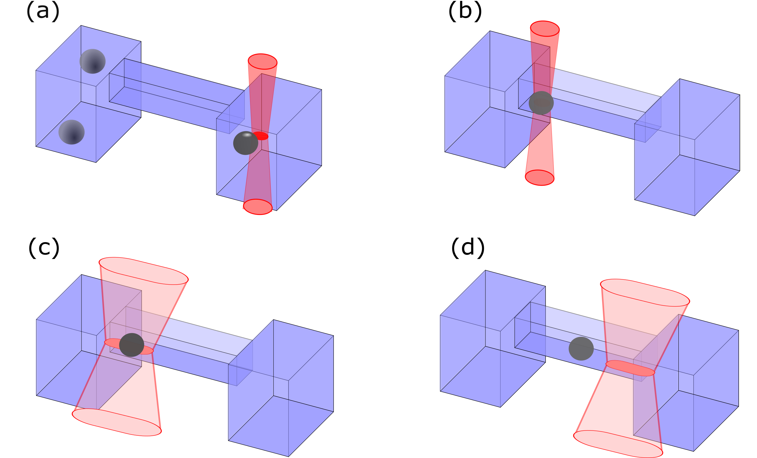
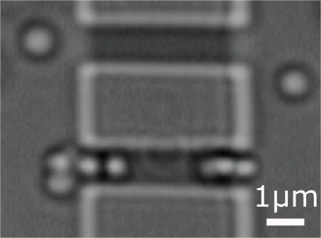
Our microfluidic devices are fabricated as previously reported Pagliara et al. (2011, 2014b). They consist of two 3D reservoirs with a depth of 12 m separated by a polydimethylsiloxane barrier and connected by an array of microfluidic channels. Each channel has a cross section of around 0.90.9 m2 and a length of m. The reservoirs are filled with spherical polystyrene particles of diameter (51010) nm. We use a laser line trap generated by HOTs to create an attractive potential well that extends from the centre of the channel to 1.7 m in the left reservoir (Fig. 1(a), (c) and dotted line in Fig. 1(d)). After a time interval , we switch off this laser line and simultaneously switch on a second laser line trap that extends, for a time interval , from the centre of the channel to 1.7 m in the right reservoir (solid line in Fig. 1(d)). In this way, we produce an attractive potential that oscillates at frequency between the two channel entrances (Fig. 1(a,d) and Video 1). We estimate the extension, depth and shape of the energy wells from the intensity distribution of the line traps generated by HOTs (Fig. 1(d)) Pelton et al. (2004); Hayashi et al. (2008). The deduced potentials are validated by measuring the velocity of a particle dragged through the channels by moving the sample stage at a constant speed Juniper et al. (2012). Experiments are performed over a range of oscillation frequencies and particle concentrations in the reservoirs.
We record videos of particles undergoing Brownian motion in the channels and reservoirs and extract particle trajectories John C. Crocker (1996); Dettmer et al. (2014). We define an attempt as the event for which a particle enters into the channel from either reservoirs and explores it for at least 33 ms, one frame time of the CCD camera that we use Pagliara et al. (2014a). Once a particle has entered the channel, it can either go back to the same reservoir, defined as a return event (Fig. 1(e)), or translocate through the channel and exit to the opposite reservoir, defined as a translocation event (Fig. 1(f)). We determine the attempt rate , the translocation rate and the translocation probability , defined as . Average rate values for each oscillation frequency are obtained from at least five experiments of one hour duration each. In order to collect statistically sufficient samples for the translocation time, we use HOTs for automated drag-and-release experiments in which we trap a single particle in one of the two reservoirs and place it in one of the two channel entrances. At s, we release the particle and simultaneously switch on the oscillating optical potential (sketched in Appendix A and Video 2).

III Results and discussion
III.1 Dependence of translocation rate and probability on the frequency of the oscillating potential
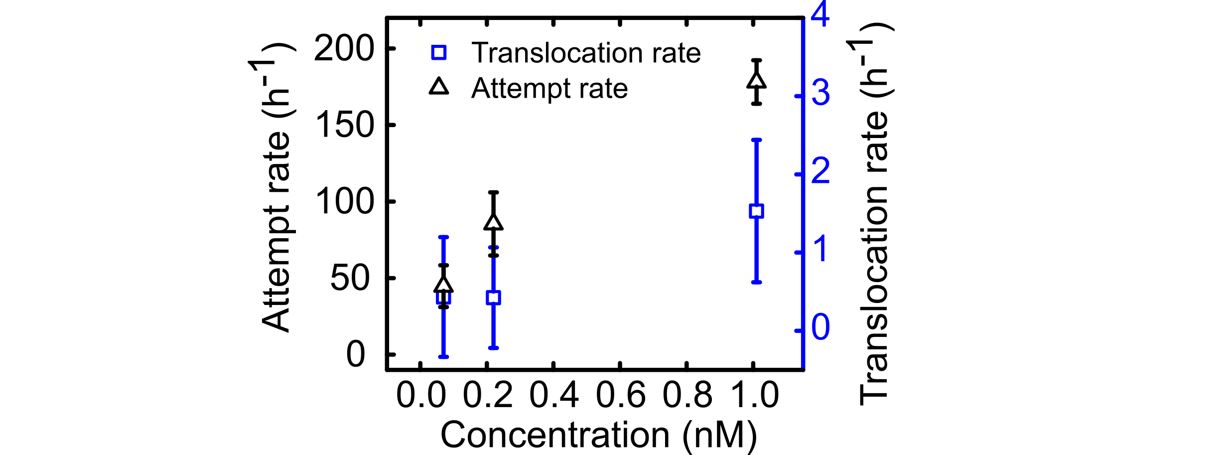
For nM, the attempt rate increases with the oscillation frequency up to particles (h-1) at a frequency of 0.5 Hz (circles in Fig. 2(a)). The translocation probability instead sharply decreases down to 0.02 for Hz (circles in Fig. 2(b)). These two effects cancel each other and as a consequence the translocation rate has a weak dependence on the oscillation frequency (circles in Fig. 2(c)).
At higher particle concentrations, the frequency of the oscillating potential strongly affects particle transport across the channel. For nM, the attempt rate increases with frequency up to a maximum of particles (h-1) for Hz (triangles in Fig. 2(a) and Fig. 3(a)). The translocation probability and the translocation rate instead first increase with frequency and peak at oscillation frequencies of 0.1 Hz and 0.16 Hz, respectively (triangles in Fig. 2(b,c) and Fig. 3(b,c)).

At even higher particle concentrations nM, the attempt rate is not significantly affected by the frequency of the oscillating potential (squares in Fig. 2(a)). The translocation probability and rate first increase with frequency peaking at an optimal oscillation frequency of 0.15 Hz and 0.19 Hz, respectively, and then decrease at higher frequencies (squares in Fig. 2(b) and (c)).
We find that the attempt and translocation rate increase with the concentration of the particles in the reservoirs for all tested oscillation frequencies (Fig. 2(a,c)), but interestingly, this is not the case for the translocation probability (Fig. 2(b)). Moreover, for nM the translocation rate at the optimal oscillation frequency = 0.1 Hz is 102 times higher than the one measured in free diffusion (dashed line in Fig. 3(c) and Appendix B) and twice the one measured for a static potential constantly switched on (dotted lines in Fig. 3(c)). For = 1.01 nM at Hz, the translocation rate (squares in Fig. 2(c)) is 65 times higher than in free diffusion and 4 times higher than in the presence of the static double well potential.
III.2 Dependence of channel occupancy on the frequency of the oscillating potential
In order to gain more insight on the presence of an optimal oscillation frequency, we measure the channel occupation probability , which is the probability to simultaneously find particles in the channel. At high particle concentrations, we measure that the probability to find one particle in the channel is at a maximum for frequencies close to the optimal oscillation frequency (triangles and squares for and 1.07 nM, respectively, in Fig. 4(b)). Notably, the channel is predominantly empty at low frequencies (e.g. p(0)=0.58 for nM and Hz, triangles in Fig. 4(a)) whereas at high frequency and concentration the channel is crowded (Fig. 4(c,d)), e.g. for nM and Hz. Therefore, the optimal oscillation frequency is the one that allows for populating the channel without overcrowding it Pagliara et al. (2014a).


III.3 Dependence of the channel translocation time on the frequency of the oscillating potential
We perform drag-and-release experiments to measure the translocation time of a particle across the channel (Video 2). This experiment is repeated at least 300 times for each and performed at nM. In free diffusion the translocation time can be calculated as previously reported Redner (2001):
| (1) |
where is half the length of the channel and is the particle diffusion coefficient. In the presence of an external potential in the channel, the translocation time is given by Berezhkovskii et al. (2017):
| (2) |
where is the transition length, with and denoting the Boltzmann constant and absolute temperature.
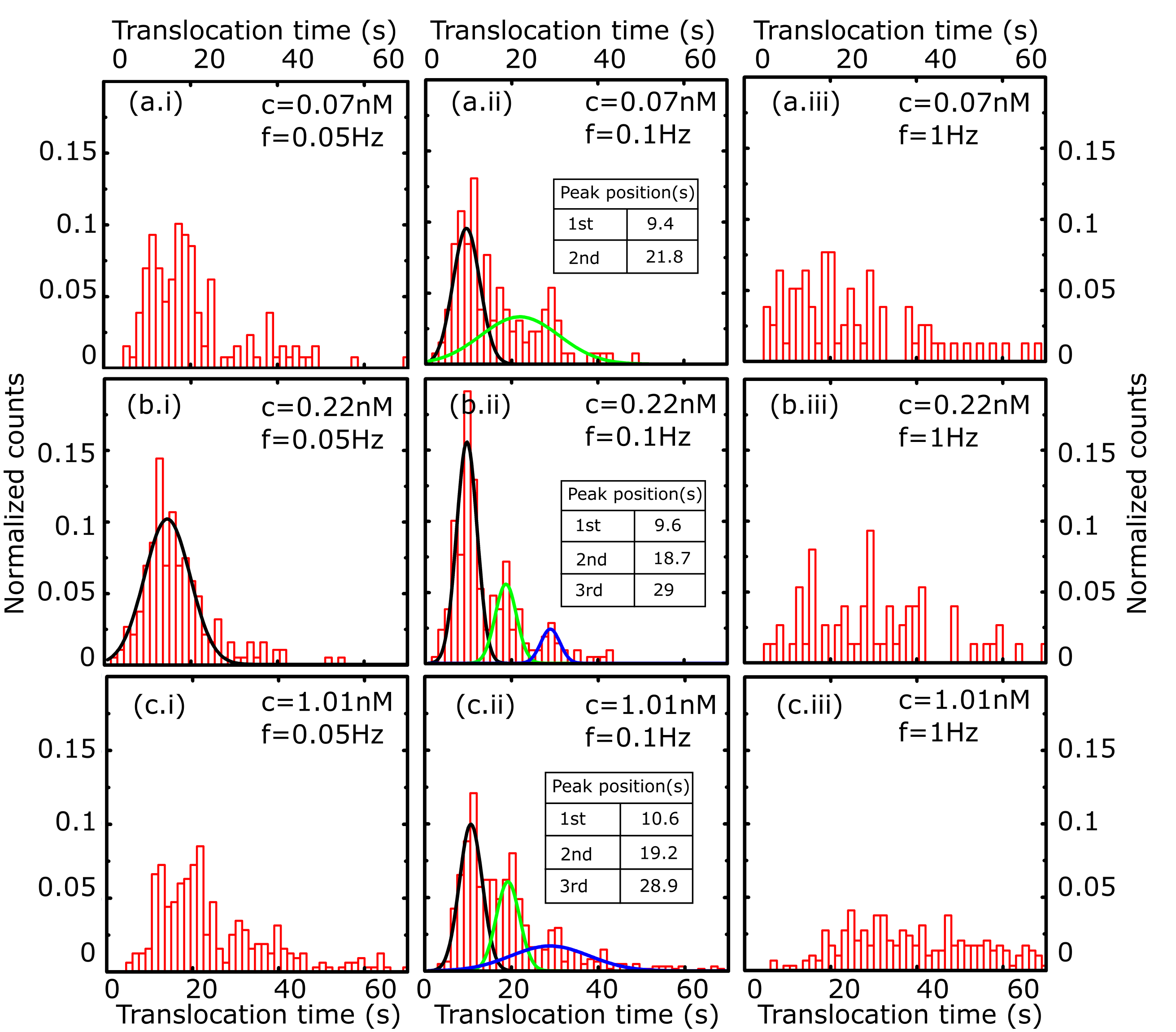
We measure the distribution of translocation times for each oscillation frequency. We find resonance-like peaks with the first maximum located at 40.5, 20.5, 10.5 and 4.5 s for and 0.33 Hz, respectively (red bins in Fig. 5(a-e) and Appendix E). Due to the oscillating nature of our potential, Eq. (1) and Eq. (2) can not fully describe our experimental data. However, we performed 1D Brownian dynamics simulations by using the experimentally measured oscillating potential (details in Appendix F) and find translocation time values that favourably compare with the experimental values (Fig. 5 (a-e)). Moreover, our Brownian dynamics simulations confirm the frequency dependence of the translocation probability with an optimal oscillation frequency of Hz close to the experimentally measured one (black dots and red triangles, respectively, in Fig. 5(f)).

III.4 Optimal oscillation frequency is defined by diffusion between potential wells
By measuring the transition times across portions of the channel, we provide an intuitive explanation of the optimal oscillation frequency. Firstly, let us consider a representative translocation from the left to the right reservoir. Upon entering the channel, the particle may diffuse to the minimum of the left attractive potential well, while the left laser line is switched on (Region I in Fig. 6(a)). The particle is trapped close to this position until, at , the left laser line is switched off and the right line is turned on. At this time the particle is free to diffuse either towards the left or right entrance of the channel. In the most efficient scenario in terms of particle transport, the particle travels in free diffusion across region II (Fig. 6(b)) and region III where reaches the right-hand side potential minimum when the right laser line is still on (Fig. 6(c)). Finally, when this line is switched off the particle is free to diffuse through region IV out of the channel (Fig. 6(d)). We perform drag-and-release experiments to evaluate the transition time across each of the four regions above. The particle’s first transition time by free diffusion in channel portions of different length is plotted in Fig. 7(a). The first transition time through region II and III is s at the oscillation frequency Hz (Fig. 7(b), a particle is released from the HOTs at the left entrance of the channel with the right-hand side potential constantly on). s at Hz is smaller than the corresponding s calculated according to Eq. 1 due to the presence of the external potential. s at Hz is in agreement with the value calculated according to Eq. 2 ( s) by using the experimentally measured potential. is close to s indicating that the optimal oscillation frequency is the one that matches the transition time through regions II and III. Notably, for higher than the optimal oscillation frequency, particle’s transition through regions II and III is interrupted by the potential oscillation. For lower than the optimal oscillation frequency, a particle has a higher chance to exit the channel through region I resulting in a return event, although the chance for a particle to be transported through regions II and II is increased. Overall for frequencies different from the optimal frequency, particle diffusion through regions II and III does not synchronize with the time scale defined by the oscillation frequency. This explains the observed decrease in translocation rate and probability at lower and higher than the optimal frequency (Fig. 2).
Optical potentials modulated in time have been extensively employed to direct particle motion Lee and Grier (2006); Juniper et al. (2016); Faucheux et al. (1995); Lee et al. (2005); Bleil et al. (2007); Gorre-Talini et al. (1997); MacDonald et al. (2003); Jonáš and Zemánek (2008); Xiao and Grier (2010); Ladavac et al. (2004); Simon and Libchaber (1992); Babič et al. (2004); Schmitt et al. (2006), including in microfluidic applications MacDonald et al. (2003). However, these have yet to be implemented for enhancing particle transport across a quasi 1D microfluidic channel connecting two reservoirs. In this paper we create a modulated optical potential consisting of a laser line that alternates its position between the two entrances of a microfluidic channel. We optimize the oscillation frequency (Fig. 2) and explain the physical mechanism underlying the optimal oscillation frequency (Fig. 4-7). Particle transport in the presence of the modulated optical potential is two orders of magnitudes higher than in free diffusion (Fig. 3).
Our experiment indicates that oscillating potentials may be an additional avenue for enhancing transport across synthetic channels or pores. In order to mimic membrane transport in living cells, we are planning to scale our synthetic platform down to the nanoscale Bell et al. (2011) where the characteristic diffusion time is closer to the one observed in protein transporter. Furthermore, it is possible to explore asymmetric systems with charged particles only in one of the two reservoirs. Thus our system will be mimicking electrochemical gradients and even exhibit effects like charge polarisation under applied external driving forces. Finally, an avenue with exciting phenomena will involve mixing several types of particles with different sizes and surface charges, exploring competitive effects and thus mimicking the mechanisms of secondary active transporters, where the transport of a substrate is coupled to the transport of a second substrate.

IV Summary
We studied the effect of a time dependent potential on particle transport through a microfluidic channel. Inspired by the alternating access mechanism, we coupled an energy well that oscillates between the two entrances of a microfluidic channel. We found that particle transport through the channel can be maximized by optimizing the oscillation potential frequency. Importantly, the optimal oscillation frequency makes the alternating access channel more efficient in terms of transport compared to static channels where particles are either in free diffusion or can simultaneously bind to the ends of the channel. We found that the optimal frequency is the one that allows synchronizing alternating access with particle diffusion across the region of the channel between the two oscillating energy well positions. We anticipate that our findings will stimulate further investigation on mimicking the functioning of membrane protein transporters Caspi et al. (2008), on synchronized oscillations Doering and Gadoua (1992); Schmitt et al. (2006); Hayashi et al. (2012); Juniper et al. (2015) and on the use of modulated potentials for particle control in microfluidics applications MacDonald et al. (2003).
Acknowledgements.
We thank Shayan Lameh for proofreading. This work was supported by a Royal Society Research Grant, a Wellcome Trust Strategic Seed Corn Fund and a Start up Grant from the University of Exeter awarded to S.P. U.F.K was funded by an ERC Consolidator Grant (Designerpores 67144). Y.T. was supported by scholarship from Cavendish-NUDT, Lundgren and Pannett Fund, Churchill College. J.G. acknowledges the support of the Winton Programme for the Physics of Sustainability and the European Union’s Horizon 2020 research and innovation programme under ETN grant 674979-NANOTRANS.Appendix A Drag and release experiment

Appendix B Transport of particles through the channel in free diffusion

Appendix C Fitting attempt and translocation rate
Attempt rate, translocation rate and translocation probability in Fig. 2 and 3 are fitted by a two-term exponential model via the nonlinear least-squares method, where are the fitting parameters. The values for these parameters estimated by the fitting are reported in Tables I-III.
| (nM) | a | b | c | d |
|---|---|---|---|---|
| 0.07 | 9853436.5 | -2.1 | -9853424.6 | -2.1 |
| 0.22 | 83037687.0 | -0.3 | -83037511.7 | -0.3 |
| 1.01 | 3013.6 | 0.65 | 10014.3 | -73.7 |
| (nM) | a | b | c | d |
|---|---|---|---|---|
| 0.07 | 26.7 | -1.9 | -19.0 | -12.7 |
| 0.22 | 77.4 | -2.0 | -86.7 | -14.5 |
| 1.01 | 137.6 | -1.0 | -187.9 | -17.0 |
| (nM) | a | b | c | d |
|---|---|---|---|---|
| 0.07 | 0.2 | -3.9 | -0.1 | -45.0 |
| 0.22 | 0.2 | -3.9 | -0.2 | -23.9 |
| 1.01 | 0.05 | -1.8 | -0.1 | -20.7 |
Appendix D Channel jamming with static double well potential

Appendix E Transition and translocation times
For a particle in free diffusion in the channel the translocation time calculated according to Eq. 1 using m and = 0.25 Dettmer et al. (2014) , is 15.36 s. This is also in agreement with the value obtained via Brownian simulation (14.99 s).

Appendix F Brownian dynamics simulation
We carried out Brownian dynamics simulations in a 1D channel with length .
Particle trajectories start at and are terminated at their first contact with each of the perfect absorbing boundaries set at and . In simulations we track the fraction of particles that end at as well as their transit time defined as the time that a particle takes to reach for the first time.
The actual particle’s position, , is given by , where is the previous position, is a pseudo random number generated with a Gaussian distribution with average position displacement and standard deviation , and Force was derived from the potential depicted in Fig. 1(d). When running simulations we set s and we average over 100 millions random walkers.
References
- Alberts et al. (1995) B. Alberts, D. Bray, J. Lewis, M. Raff, K. Roberts, J. D. Watson, and A. Grimstone, Molecular Biology of the Cell (3rd edn) (Garland Science, 1995).
- Kasianowicz et al. (2006) J. J. Kasianowicz, T. L. Nguyen, and V. M. Stanford, Proceedings of the National Academy of Sciences of the United States of America 103, 11431 (2006).
- Benz et al. (1986) R. Benz, A. Schmid, T. Nakae, and G. H. Vos-Scheperkeuter, Journal of bacteriology 165, 978 (1986).
- Ward and Sze (1992) J. M. Ward and H. Sze, Plant Physiology 99, 925 (1992).
- Meyer and Schulz (1997) J. E. W. Meyer and G. E. Schulz, Protein Science 6, 1084 (1997).
- Kullman et al. (2002) L. Kullman, M. Winterhalter, and S. M. Bezrukov, Biophysical Journal 82, 803 (2002).
- Pagliara et al. (2014a) S. Pagliara, S. L. Dettmer, and U. F. Keyser, Physical Review Letters 113, 048102 (2014a).
- Pagliara et al. (2013) S. Pagliara, C. Schwall, and U. F. Keyser, Advanced materials 25, 844 (2013).
- Horner et al. (2015) A. Horner, F. Zocher, J. Preiner, N. Ollinger, C. Siligan, S. A. Akimov, and P. Pohl, Science Advances 1, 1 (2015).
- Jensen et al. (2002) M. Ø. Jensen, S. Park, E. Tajkhorshid, and K. Schulten, Proceedings of the National Academy of Sciences of the United States of America 99, 6731 (2002).
- Berezhkovskii and Bezrukov (2005) A. M. Berezhkovskii and S. M. Bezrukov, Biophysical Journal 88, L17 (2005).
- Kolomeisky (2007) A. Kolomeisky, Physical Review Letters 98, 048105 (2007).
- Zilman (2009) A. Zilman, Biophysical Journal 96, 1235 (2009).
- Jardetzky (1966) O. Jardetzky, Nature 211, 969 (1966).
- Lee and Grier (2006) S.-H. Lee and D. G. Grier, Physical Review Letters 96, 190601 (2006).
- Juniper et al. (2016) M. P. N. Juniper, A. V. Straube, D. G. A. L. Aarts, and R. P. A. Dullens, Physical Review E 93, 012608 (2016).
- Faucheux et al. (1995) L. P. Faucheux, L. S. Bourdieu, P. D. Kaplan, and A. J. Libchaber, Physical Review Letters 74, 1504 (1995).
- Lee et al. (2005) S.-H. Lee, K. Ladavac, M. Polin, and D. G. Grier, Physical Review Letters 94, 110601 (2005).
- Bleil et al. (2007) S. Bleil, P. Reimann, and C. Bechinger, Physical Review E 75, 1 (2007).
- Gorre-Talini et al. (1997) L. Gorre-Talini, S. Jeanjean, and P. Silberzan, Physical Review E 56, 2025 (1997).
- MacDonald et al. (2003) M. P. MacDonald, G. C. Spalding, and K. Dholakia, Nature 426, 421 (2003).
- Jonáš and Zemánek (2008) A. Jonáš and P. Zemánek, Electrophoresis 29, 4813 (2008).
- Xiao and Grier (2010) K. Xiao and D. G. Grier, Physical Review E 82, 051407 (2010).
- Ladavac et al. (2004) K. Ladavac, K. Kasza, and D. G. Grier, Physical Review E 70, 010901 (2004).
- Simon and Libchaber (1992) A. Simon and A. Libchaber, Physical Review Letters 68, 3375 (1992).
- Babič et al. (2004) D. Babič, C. Schmitt, I. Poberaj, and C. Bechinger, Europhysics Letters 67, 158 (2004).
- Schmitt et al. (2006) C. Schmitt, B. Dybiec, P. Hänggi, and C. Bechinger, Europhysics Letters 74, 937 (2006).
- Misiunas et al. (2015) K. Misiunas, S. Pagliara, E. Lauga, J. R. Lister, and U. F. Keyser, Physical Review Letters 115, 038301 (2015).
- Padgett and Di Leonardo (2011) M. Padgett and R. Di Leonardo, Lab on a Chip 11, 1196 (2011).
- Bowman et al. (2014) R. W. Bowman, G. M. Gibson, A. Linnenberger, D. B. Phillips, J. a. Grieve, D. M. Carberry, S. Serati, M. J. Miles, and M. J. Padgett, Computer Physics Communications 185, 268 (2014).
- Pagliara et al. (2011) S. Pagliara, C. Chimerel, R. Langford, D. G. a. L. Aarts, and U. F. Keyser, Lab on a chip 11, 3365 (2011).
- Pagliara et al. (2014b) S. Pagliara, S. L. Dettmer, K. Misiunas, L. Lea, Y. Tan, and U. F. Keyser, The European Physical Journal Special Topics 223, 3145 (2014b).
- Pelton et al. (2004) M. Pelton, K. Ladavac, and D. G. Grier, Physical Review E 70, 031108 (2004).
- Hayashi et al. (2008) Y. Hayashi, S. Ashihara, T. Shimura, and K. Kuroda, Optics Communications 281, 3792 (2008).
- Juniper et al. (2012) M. P. N. Juniper, R. Besseling, D. G. a. L. Aarts, and R. P. a. Dullens, Optics express 20, 28707 (2012).
- John C. Crocker (1996) D. G. G. John C. Crocker, Journal of Colloid and Interface Science 310, 298 (1996).
- Dettmer et al. (2014) S. L. Dettmer, S. Pagliara, K. Misiunas, and U. F. Keyser, Physical Review E 89, 062305 (2014).
- Redner (2001) S. Redner, A guide to first-passage processes (Cambridge University Press, 2001).
- Berezhkovskii et al. (2017) A. M. Berezhkovskii, L. Dagdug, and S. M. Bezrukov, The Journal of Physical Chemistry B 121, 5455 (2017).
- Bell et al. (2011) N. A. Bell, C. R. Engst, M. Ablay, G. Divitini, C. Ducati, T. Liedl, and U. F. Keyser, Nano letters 12, 512 (2011).
- Caspi et al. (2008) Y. Caspi, D. Zbaida, H. Cohen, and M. Elbaum, Nano Letters 8, 3728 (2008).
- Doering and Gadoua (1992) C. R. Doering and J. C. Gadoua, Physical Review Letters 69, 2318 (1992).
- Hayashi et al. (2012) K. Hayashi, S. de Lorenzo, M. Manosas, J. M. Huguet, and F. Ritort, Physical Review X 2, 1 (2012).
- Juniper et al. (2015) M. P. N. Juniper, A. V. Straube, R. Besseling, D. G. A. L. Aarts, and R. P. A. Dullens, Nature communications 6, 7187 (2015).