The interaction of intense femtosecond laser pulses with argon microdroplets studied near the soft x-ray emission threshold
Abstract
The extreme ultraviolet plasma emission from liquid microsized argon droplets exposed to intense near-infrared laser pulses has been investigated. Emission from the warm dense matter targets is recorded in a spectral range in between 16 and 30 nm at laser intensities of W/ cm2. Above the emission threshold, soft x-ray radiation exponentially increases with the pulse energy whereby a strong dependence of the yields on the pulse duration is observed, which points at an effective electron collisional heating of the microplasma by inverse bremsstrahlung. Accompanying hydrodynamic simulations reveal the temporal and spatial development of the microplasma conditions. The good agreement in between the measured and calculated emission spectra as well as the extracted electron temperatures confirm that hydrodynamic simulations can be applied in the analysis of strongly excited droplets.
1 Introduction
The interaction of intense near-infrared (NIR) laser light with small particles has been of particular interest in the last decades, since it allows to study matter of finite size under extreme conditions [1, 2, 3, 4, 5, 6, 7]. Due to the strong absorption capabilities, nanoparticles are effectively transformed into nanoplasmas [8], which results in the emission of highly charged and energetic ions [9, 10, 11] and fast electrons [12, 13]. The generation of short wavelength radiation [14, 15] and high harmonics [16, 17] allows to resolve further details of the size-limited plasma conditions [4, 18, 19, 20, 21].
With microdroplets, the dimension of the target becomes comparable to NIR laser wavelength and propagation phenomena show up. The laser light wave penetrates into the droplet up to a region where the critical density in the plasma is achieved. In addition, the spatial density scale length is much lower compared to, e.g., the Ti:sapphire laser wavelength of 800 nm. Hence, absorption of radiation occurs only near the surface of the droplet. The resulting hot plasma is far from equilibrium. As a consequence, the dynamics of the laser-produced plasma layer has to be simulated in order to resolve details of the processes involved. The microplasma may retroact on the light propagation, further influencing the absorption properties including extreme ultraviolet (EUV) re-absorption. As a result of strong electron density gradients, wave propagation phenomena show up, e.g., shock waves [22], wave breaking and turbulence [23]. The limited size of the target space charge confinement of hot electrons will lead to an efficient coupling of the laser radiation into the particle [24]. Particle-in-cell simulations show that an inhomogeneous plasma layer develops which extends deeper into the droplets than expected from classical predictions [25].
Beside particle plasma diagnostics, radiation has proven to give meaningful information to characterize the complex dynamics in microplasmas [26, 27]. For example, intense short-wavelength radiation from extended-ultraviolet free-electron lasers can isochorically heat particles, whereby self-Thomson scattering can be exploited to characterize the resulting warm dense matter (WDM) state [28]. EUV-pump–EUV-probe studies were successful in mapping the solid-to-plasma transition in real time [29]. Combining time-resolved soft x-ray diagnostics with microplasma generation by intense NIR laser pulses remains a challenge due to the initially incomplete spatial heating. WDM targets also represent laboratory systems to simulate conditions found in planets and are thus relevant for astrophysical research [30]. Finally, the short wavelength emission from particles in a molecular beam have potential for applications like high repetition rate table-top x-ray sources [31], because the technique provides a regenerative target and nearly debris-free conditions.
In this contribution we address in a joint experimental and theoretical project the generation of the transient plasma state by analyzing the resulting EUV radiation from an NIR laser-induced argon microplasma. In a simplified view of the interaction, ionization of the liquid microdroplets is triggered by tunnel ionization. As the electron quiver amplitude at W/ cm2 [32] exceeds the interatomic distances in the liquid [33], efficient collisional ionization into high charge states will take place producing a plasma at near solid-state density. The thin plasma sheet provides in the following for an efficient heating by inverse bremsstrahlung leading to a strong nonlinear dynamics. After the impact of the laser pulse, the outward expansion of the plasma layer leads to a decrease in the plasma density and an adiabatic cooling [34]. Soft x-ray emission as a characteristic fluorescence signature of the plasma conditions occurs on a pico- to nanosecond time scale.
In contrast to other experiments on droplets at near relativistic intensities, e.g. [5, 20], moderate laser intensities are applied in order to study the dynamics close to the argon tunnel ionization threshold of W/ cm2 [35]. To solidify the investigation the experiments are accompanied by theoretical work, i.e the experimental average electron temperatures extracted from the EUV spectra will be compared to simulations. The good agreement of the theoretical results to the experiments point out that the microplasma dynamics can be described well by 1D hydrodynamical codes.
2 Experiment
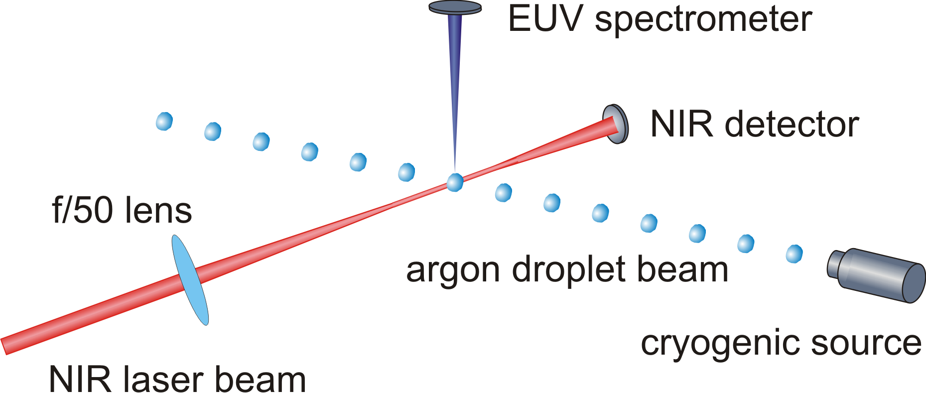
To effectively conduct the experiment, a regenerative high-density target is utilized, i.e., a liquid jet source similar to the one used in [36]. In short, the microdroplet beam is generated by expanding liquefied argon gas (100 K at 5 bar) through a 5 m nozzle into vacuum forming a continuous filament. Due to surface vibrations, the filament undergoes a Rayleigh breakup and splits into monodisperse droplets [37] with diameters of approximately 9 m. The droplets are irradiated by pulses from a Ti:sapphire laser system with a center wavelength at 810 nm and a pulse repetition rate of 1 kHz. The laser beam is focused with a f/50 lens, see Fig. 1. Experiments are performed with pulse durations between 100 fs and 800 fs and pulse energies up to 1.5 mJ, which corresponds to a maximal pulse intensity of W/cm2 in the interaction region. The EUV emission produced in the strong-field interaction with the droplets is recorded by a home-build EUV spectrometer aligned to both laser and droplet beam. The spectrometer consists of an aberration corrected flat-field grating as single optical element [38]. A back-illuminated CCD camera (Andor, model Newton 940 DO) serves for EUV photon detection. A 200 nm aluminum foil is used to suppress scattered laser light. With the instrument, spectra in a range between 16 and 30 nm can be recorded.
As the Rayleigh breakup takes place spontaneously, a temporal synchronization between droplets and laser pulses cannot be achieved. In order to estimate the efficiency at which droplets are hit by laser radiation, the transmitted energy of single pulses after passing the interaction region is determined (NIR detector in Fig. 1). From the analysis we found that about 70% of the pulses interact with a droplet. To suppress signal fluctuations spectra are integrated over laser pulses.
3 Simulation
The interaction of intense femtosecond laser pulses with microdroplets is studied using the radiation-hydrodynamic HELIOS code [39]. Briefly, HELIOS provides a Lagrangian reference frame where electrons and ions are assumed to be co-moving. Pressure contributions to the equation of motion stem from electrons, ions and radiation. Separate ion and electron temperatures and flux-limited Spitzer thermal conductivity are assumed. Deviations from local thermodynamic equilibrium conditions are accounted for by solving multi-level atomic rate equations at each time step in the simulation. The laser energy is deposited via inverse bremsstrahlung as well as bound-bound and bound-free transitions using an SESAME-like equation of state. The EUV emission of the microdroplets is calculated using SPECT3D based on the temperature and density of the plasma plume expected via radiation-hydrodynamic simulations. SPECT3D is a collisional-radiative code [40] whereas the radiation incident at a detector is estimated by solving the radiative transfer equation along a series of lines-of-sight through the plasma grid. Atomic cross section data are generated using a collection of publicly available codes [41, 42, 43, 44, 45], where different processes are considered, such as excitation, electron-impact ionization, photoionization, radiative recombination, autoionization, and dielectronic recombination. In addition, continuum lowering effects are implemented using an occupation probability model.
4 Experimental results
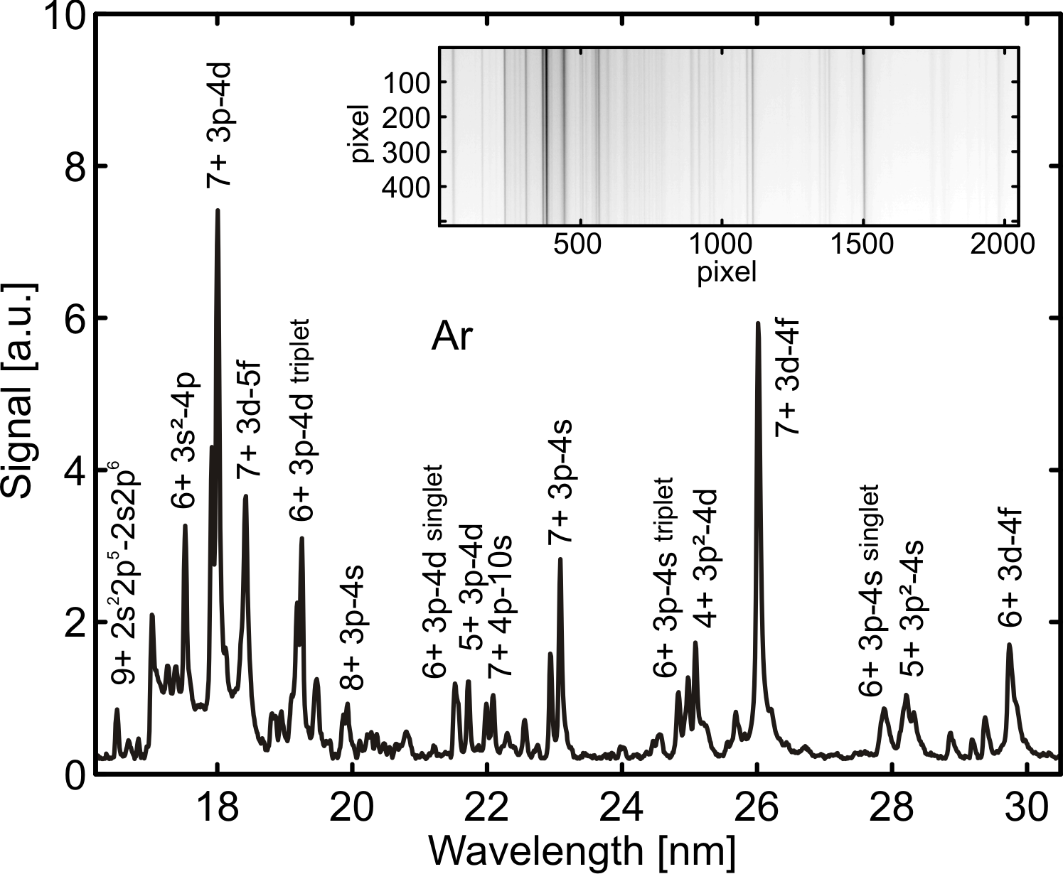
Inset: Raw data of the flat-field spectrometer.
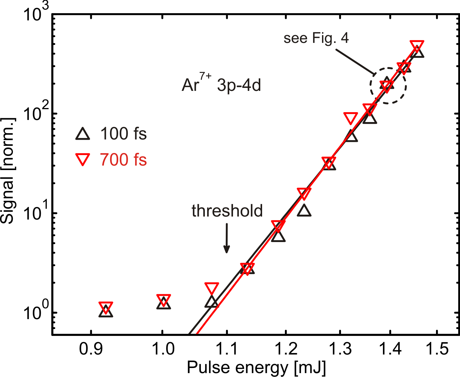
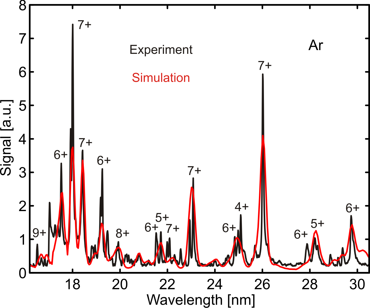
Fig. 2 shows an EUV spectrum recorded after exposure of microdroplets to 100 fs laser pulses at intensities of W/cm2. The characteristic line pattern can be assigned to atomic transitions from Ar4+ to Ar9+ [19, 46]. Note that under the given intensity conditions, the laser field ionization of single atoms only leads to charge states up to Ar3+ [35] indicating that anticipated processes like collisional ionization must contribute. In the following we concentrate on the ion species Ar7+ as it represents a characteristic and dominant fingerprint of the microplasma. Fig. 3 shows the normalized yields of the Ar7+ 3p-4d transition line depending on the pulse energy for pulse durations of 100 fs and 700 fs. A strong increase of the EUV signal is observed beyond a threshold of 1.1 mJ. At 700 fs and 1.1 mJ, the laser intensity matches the argon tunnel ionization threshold [35], but is a factor of 7 higher for 100 fs pulses. One obtains no significant change in the emission threshold behavior if shorter pulses are used. Charging of the droplet by only tunnel ionization is thus not sufficient to cause plasma EUV emission. The strong nonlinear dependence around the EUV threshold points to an avalanche-like process whereby collisional ionization and inverse bremsstrahlung efficiently heat the target. For both pulse durations, the yields enhance by more than two orders of magnitude, although the pulse energy only increases from 1.1 to 1.5 mJ.
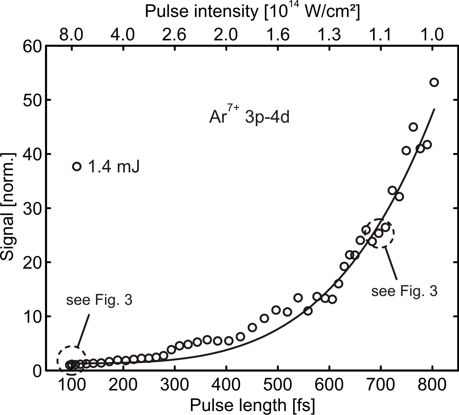
Whereas the plot in Fig. 3 appears to suggest that the pulse duration is of little importance, a closer look reveals a differentiated perspective. Fig. 4 shows the dependence of the Ar7+ 3p-4d emission on the pulse duration for a given pulse energy of 1.4 mJ. Apparently, an extended heating period with stretched pulses at energies above the emission threshold is more effective even though the laser intensity decreases. We emphasize that similar findings have been obtained in experiments on small clusters [47].
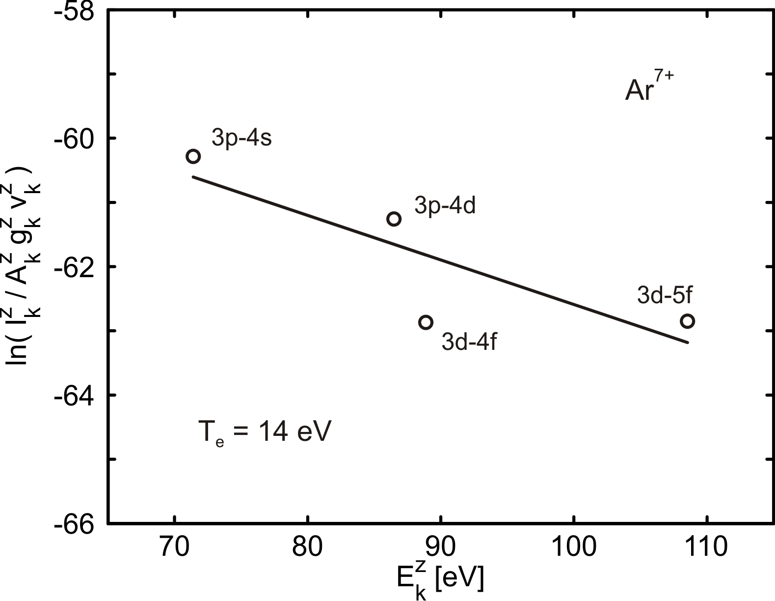
From the emission spectra obtained in the experiment, relevant plasma parameters as for example the electron temperature can be extracted. We apply the Boltzmann plot method to determine from line intensities [48] assuming the microplasma to be at local thermodynamic equilibrium (LTE) at the time of the EUV emission. Briefly, the density of a certain ion species as function of can be described by a Boltzmann distribution , where is the total density, the statistical weight, the partition function, the upper energy level and the Boltzmann constant. The relative line intensity of a certain transition in the charge state scales by , with and the frequency and Einstein coefficients, respectively. One can derive a linear equation in the form of . A linear regression taking into account the emission lines of a given charge state allows to extract . Fig. 5 shows the Boltzmann plot analysis for Ar7+ giving a value of of about 14 eV () in this case. The accuracy in the determination of is comparable to other work in the warm dense matter regime, e.g. [28].
5 Computational results
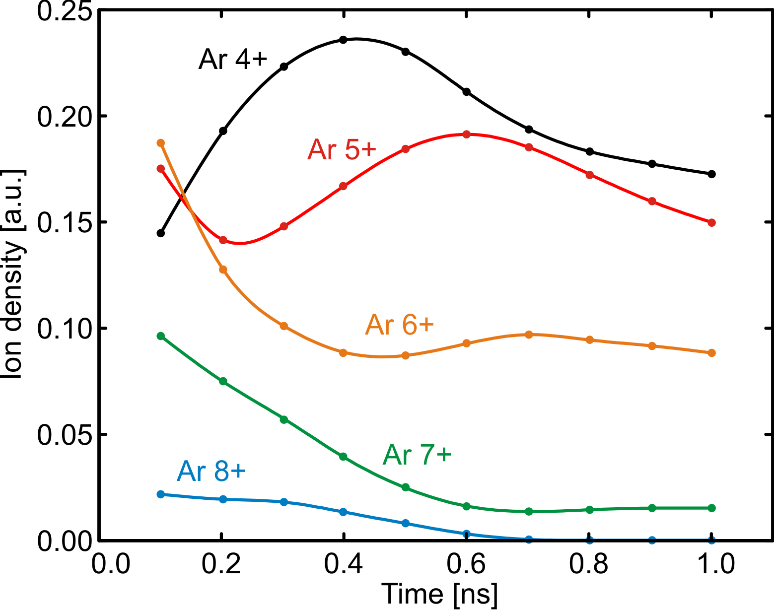
In the HELIOS calculations, argon droplets are considered with an initial diameter of 10 m and a mass density of g/cm3 irradiated by intense laser pulses at 800 nm center wavelength. Fig. 6 shows the temporal ion density distributions of various charge states for laser pulses with 100 fs pulse length and 2.1 mJ pulse energy. The charge state abundances are captured up to nanoseconds after the laser pulse exposure at . The ionization kinetics are caused by collisional excitation and recombination as well as collective processes in the plasma layer. In the expansion, electron-ion recombination leads to a general decrease of . The short-term enhancement in the ion densities of certain charge states after some hundred picoseconds can be traced back to electron localization reducing the value of in higher charged Arq+.
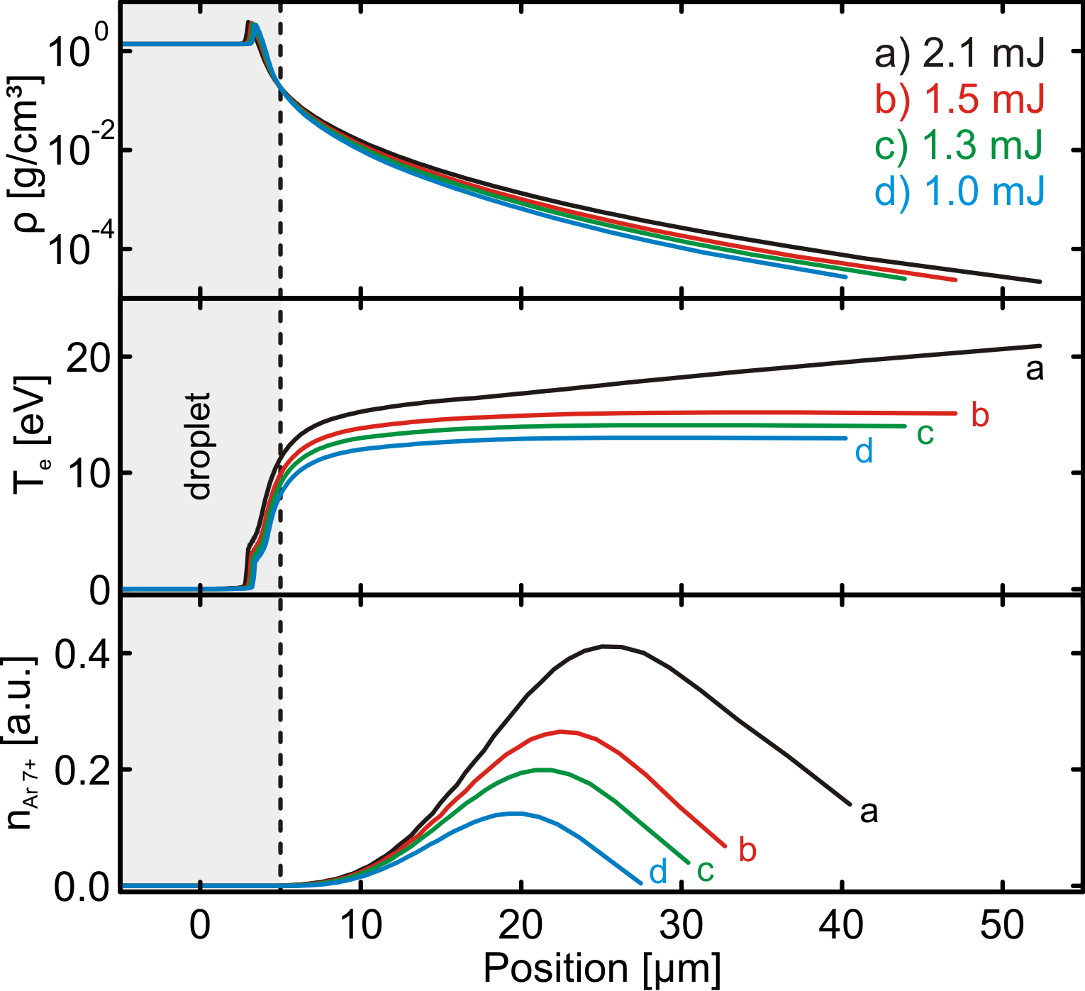
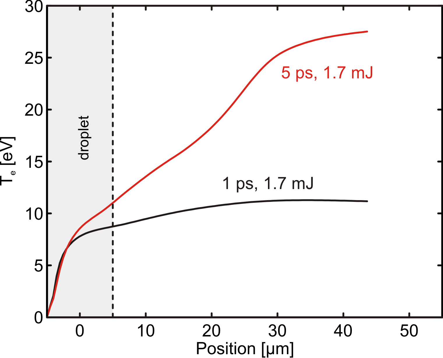
Fig. 7 displays the spatial evolution of the mass density and electron temperature as well as the ionization fraction of Ar7+ at ps. Most of the inner part of the droplet (left to dashed line) has a low temperature as the laser radiation is shielded by the plasma layer. As a consequence, the mass density inside the droplet remains at its initial value . A plasma compression is observed in the surface region. A hot, highly ionized plasma layer extends over tens of micrometers outwards. The mass density drops - depending on the laser pulse energy - to values as low as g/cm3 beyond 40 m. The composition of the plasma varies with distance. An enhanced abundance of Ar7+ (Fig. 7 bottom) is observed in a layer between 20 and 40 m approaching values of 10 to 40 % of the total density in the plasma plume. In general, only a minor dependence of and on the pulse energy is obtained for the considered range of parameters. The inner region of the microdroplet is still almost unaffected. However, in particular hot electrons penetrate and slowly heat-up the droplet core leading to an increasing ionization. After ns, the droplet is almost fully ionized as illustrated in Fig. 8. Interestingly and in accordance with the experimental findings, see Fig. 4, longer pulses extend the heating period and lead to an increase in the temperature of the microplasma and, thereby, enhanced population of higher ionization states. Whereas the inner region of the droplets is nearly unaffected by a variation of the pulse duration, the temperature near the surface and, especially, in the outer plasma region increases applying longer pulses.
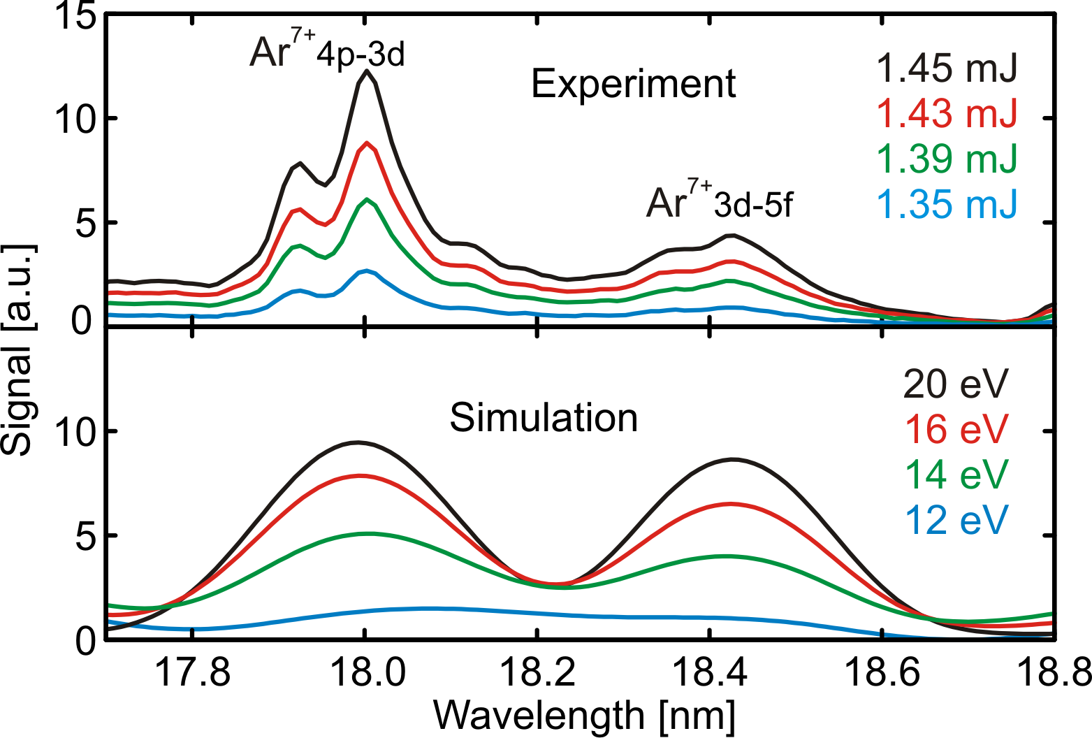
Finally we like to compare the experimental findings with the results obtained in the simulations. For this purpose the resulting soft x-ray spectra using the SPECT3D software package are computed [40]. For given the relative line intensities are calculated and compared to the measurement, see Fig. 9. For a complete description, one should consider that the EUV plasma emission will originate from the full period of the microplasma expansion. Hence, only averaged values of, e.g., can be extracted from the measurements. Taking this into account, a good agreement is found between experiment and theory as details of the spectra match almost consistently, see Fig. 10. The corresponding plasma electron temperatures also agree although the calculations have been performed using a one-dimensional hydrodynamic code which cannot reveal the full spatial evolution of the plasma.
6 Conclusion
Soft x-ray radiation from argon microdroplets irradiated by intense femtosecond pulses has been analyzed near the EUV emission threshold in a joint experimental and theoretical study. At given pulse energy, charging of the droplets can be controlled by the pulse duration. The longer impact time supports plasma heating leading to an enhancement of the EUV emission. Hydrodynamic simulations reveal that plasma formation initially takes place on the surface of the droplet. In the expansion of the plasma layer spatial and temporal charge state distributions change up to nanoseconds. Whereas the outer plasma cools down in the adiabatic expansion, the inner region of the droplet heats up on a comparable time scale. The experimental spectrum contains contributions from the full development of the microplasma expansion. Nevertheless average electron temperatures extracted from the EUV emission signal are found to be in good agreement with hydrodynamical (HELIOS) and collisional-radiative (SPECT3D) simulations. In further work the analysis of the plasma parameters may be improved by applying the Saha-Boltzmann distribution instead of the Boltzmann distribution.
7 Acknowledgment
We gratefully acknowledge financial support by the DFG within the SFB 652 and by the BMBF within the FSP 302.
References
- [1] Krainov V P and Smirnov M B 2002 Phys. Rep. 370 237–331
- [2] Saalmann U, Siedschlag C and Rost J M 2006 J. Phys. B 39 R39
- [3] Fennel T, Meiwes-Broer K H, Tiggesbäumker J, Reinhard P G, Dinh P M and Suraud E 2010 Rev. Mod. Phys. 82 1793
- [4] McNaught S J, Fan J, Parra E and Milchberg H M 2001 Appl. Phys. Lett. 79 4100–4102
- [5] Wieland M, Wilhein T, Faubel M, Ellert C, Schmidt M and Sublemontier O 2001 Appl. Phys. B 72 591–597
- [6] Parra E, McNaught S J, Fan J and Milchberg H M 2003 Appl. Phys. A 77 317–323
- [7] Garnov S V, Bukin V V, Malyutin A A and Strelkov V V 2009 Ultrafast space-time and spectrum-time resolved diagnostics of multicharged femtosecond laser microplasma AIP Conf. Proc. vol 1153 (AIP) pp 37–48
- [8] Ditmire T, Smith R A, Tisch J W G and Hutchinson M H R 1997 Phys. Rev. Lett. 78 3121
- [9] Snyder E M, Buzza S A and Castleman Jr A W 1996 Phys. Rev. Lett. 77 3347
- [10] Lezius M, Dobosz S, Normand D and Schmidt M 1998 Phys. Rev. Lett. 80 261–264
- [11] Köller L, Schumacher M, Köhn J, Teuber S, Tiggesbäumker J and Meiwes-Broer K H 1999 Phys. Rev. Lett. 82 3783–3786
- [12] Springate E, Aseyev S A, Zamith S and Vrakking M J J 2003 Phys. Rev. A 68 053201
- [13] Passig J, Irsig R, Truong N X, Fennel T, Tiggesbäumker J and Meiwes-Broer K H 2012 New J. Phys. 14 085020
- [14] McPherson A, Thompson B D, Borlsov A B, Boyer K and Rhodes C K 1994 Nature 370 631–634
- [15] Issac R C, Vieux G, Ersfeld B, Brunetti E, Jamison S P, Gallacher J, Clark D and Jaroszynski D A 2004 Phys. Plas. 11 3491–3496
- [16] Tisch J W G, Ditmire T, Fraser D J, Hay N, Mason M B, Springate E, Marangos J P and Hutchinson M H R 1997 J. Phys. B 30 L709
- [17] Bulanov S V, Naumova N M and Pegoraro F 1994 Phys. Plas. 1 745–757
- [18] Ditmire T, Donnelly T, Rubenchik A M, Falcone R W and Perry M D 1996 Phys. Rev. A 53 3379–3402
- [19] Zweiback J, Ditmire T and Perry M D 1999 Phys. Rev. A 59 R3166–R3169
- [20] Parra E, Alexeev I, Fan J, Kim K Y, McNaught S J and Milchberg H M 2000 Phys. Rev. E 62 R5931–R5934
- [21] Prigent C, Deiss C, Lamour E, Rozet J P, Vernhet D and Burgdörfer J 2008 Phys. Rev. A 78 053201
- [22] Zeld’ovich Y B and Raizer Y P 1966 Physics of shock waves and high-temperature hydrodynamic phenomena (Academic Press)
- [23] Varin C, Peltz C, Brabec T and Fennel T 2012 Phys. Rev. Lett. 108 175007
- [24] Sperling P, Liseykina T, Bauer D and Redmer R 2013 New J. Phys. 15 025041
- [25] Liseykina T V and Bauer D 2013 Phys. Rev. Lett. 110 145003
- [26] Glenzer S H and Redmer R 2009 Rev. Mod. Phys. 81 1625–1663
- [27] Ostrikov K, Neyts E C and Meyyappan M 2013 Adv. Phys. 62 113–224
- [28] Fäustlin R R, Bornath T, Döppner T, Düsterer S, Förster E, Fortmann C, Glenzer S H, Göde S, Gregori G, Irsig R, Laarmann T, Lee H J, Li B, Meiwes-Broer K H, Mithen J, Nagler B, Przystawik A, Redlin H, Redmer R, Reinholz H, Röpke G, Tavella F, Thiele R, Tiggesbäumker J, Toleikis S, Uschmann I, Vinko S M, Whitcher T, Zastrau U, Ziaja B and Tschentscher T 2010 Phys. Rev. Lett. 104 125002
- [29] Zastrau U, Sperling P, Harmand M, Becker A, Bornath T, Bredow R, Dziarzhytski S, Fennel T, Fletcher L B, Förster E, Göde S, Gregori G, Hilbert V, Hochhaus D, Holst B, Laarmann T, Lee H J, Ma T, Mithen J P, Mitzner R, Murphy C D, Nakatsutsumi M, Neumayer P, Przystawik A, Roling S, Schulz M, Siemer B, Skruszewicz S, Tiggesbäumker J, Toleikis S, Tschentscher T, White T, Wöstmann M, Zacharias H, Döppner T, Glenzer S H and Redmer R 2014 Phys. Rev. Lett. 112 105002
- [30] Guillot T 1999 Science 286 72–77
- [31] Stiel H, Vogt U, Ter-Avetisyan S, Schnurer M, Will I and Nickles P V 2002 Proc. SPIE 4781 26–34
- [32] Krainov V P, Smirnov B M and Smirnov M B 2007 Physics-Uspekhi 50 907
- [33] Henshaw D G, Hurst D G and Pope N K 1953 Phys. Rev. 92 1229
- [34] Shihab M, Abou-Koura G H and El-Siragy N M 2016 Appl. Phys. B 122 146
- [35] Augst S, Strickland D, Meyerhofer D D, Chin S L and Eberly J H 1989 Phys. Rev. Lett. 63 2212
- [36] Toleikis S, Bornath T, Döppner T, Düsterer S, Fäustlin R R, Förster E, Fortmann C, Glenzer S H, Göde S, Gregori G, Irsig R, Laarmann T, Lee H J, Li B, Meiwes-Broer K H, Mithen J, Nagler B, Przystawik A, Radcliffe P, Redlin H, Redmer R, Reinholz H, Röpke G, Tavella F, Thiele R, Tiggesbäumker J, Uschmann I, Vinko S M, Whitcher T, Zastrau U, Ziaja B and Tschentscher T 2010 J. Phys. B 43 194017
- [37] Toennies J P 2013 Mol. Phys. 111 1879–1891
- [38] Kita T, Harada T, Nakano N and Kuroda H 1983 Appl. Opt. 22 512–513
- [39] MacFarlane J J, Golovkin I E and Woodruff P R 2006 J. Quant. Spectr. Rad. Transf. 99 381–397
- [40] MacFarlane J J, Golovkin I E, Wang P, Woodruff P R and Pereyra N A 2007 High Energ. Dens. Phys. 3 181–190
- [41] Wang P 1991 Computation and application of atomic data for inertial confinement fusion plasmas Ph.D. thesis University of Wisconsin
- [42] Fischer C F 1978 Comp. Phys. Comm. 14 145–153
- [43] Abdallah Jr J, Clark R E H and Cowan R D 1988 Theoretical atomic physics code development I. cats: Cowan atomic structure code (la–11436-m-vol1)
- [44] Wang P, MacFarlane J J and Moses G A 1992 Rev. Sci. Instr. 63 5059–5061
- [45] Wang P, MacFarlane J J and Moses G A 1993 Phys. Rev. E 48 3934–3942
- [46] Kramida A, Ralchenko Y, Reader J and and NIST ASD Team 2017 NIST Atomic Spectra Database (ver. 5.3), [Online]. Available: http://physics.nist.gov/asd. National Institute of Standards and Technology, Gaithersburg, MD.
- [47] Köller L, Schumacher M, Köhn J, Teuber S, Tiggesbäumker J and Meiwes-Broer K H 1999 Phys. Rev. Lett. 82 3783–3786
- [48] Griem H R 1997 Principles of Plasma Spectroscopy (Cambridge University Press)