A review of geometric, topological and graph theory apparatuses for the modeling and analysis of biomolecular data
Abstract
Geometric, topological and graph theory modeling and analysis of biomolecules are of essential importance in the conceptualization of molecular structure, function, dynamics, and transport. On the one hand, geometric modeling provides molecular surface and structural representation, and offers the basis for molecular visualization, which is crucial for the understanding of molecular structure and interactions. On the other hand, it bridges the gap between molecular structural data and theoretical/mathematical models. Topological analysis and modeling give rise to atomic critical points and connectivity, and shed light on the intrinsic topological invariants such as independent components (atoms), rings (pockets) and cavities. Graph theory analyzes biomolecular interactions and reveals biomolecular structure-function relationship. In this paper, we review certain geometric, topological and graph theory apparatuses for biomolecular data modeling and analysis. These apparatuses are categorized into discrete and continuous ones. For discrete approaches, graph theory, Gaussian network model, anisotropic network model, normal mode analysis, quasi-harmonic analysis, flexibility and rigidity index, molecular nonlinear dynamics, spectral graph theory, and persistent homology are discussed. For continuous mathematical tools, we present discrete to continuum mapping, high dimensional persistent homology, biomolecular geometric modeling, differential geometry theory of surfaces, curvature evaluation, variational derivation of minimal molecular surfaces, atoms in molecule theory and quantum chemical topology. Four new approaches, including analytical minimal molecular surface, Hessian matrix eigenvalue map, curvature map and virtual particle model, are introduced for the first time to bridge the gaps in biomolecular modeling and analysis. Emphasis is given to the connections of existing biophysical models/methods to mathematical theories, such as graph theory, Morse theory, Poicaré-Hopf theorem, differential geometry, differential topology, algebraic topology and geometric topology. Potential new directions and standing open problems are briefly discussed.
Key words: Molecular bioscience, Molecular biophysics, Graph theory, Graph spectral theory, Graph Laplacian, Differential geometry, Differential topology Persistent homology, Morse theory, Conley index, Poincaré-Hopf index, Laplace-Beltrami operator, Dynamical system.
1 Introduction
Life science is regarded as the last forefront in natural science and the 21st century will be the century of biological sciences. Molecular biology is the foundation of biological sciences and molecular mechanism, which is governed by all the valid mechanics, including quantum mechanics when it is relevant, and is the ultimate truth of life science. One trend of biological sciences in the 21st century is that many traditional disciplines, such as epidemiology, neuroscience, zoology, physiology and population biology, are transforming from macroscopic and phenomenological to molecular-based sciences. Another trend is that biological sciences in the 21st century are transforming from qualitative and descriptive to quantitative and predictive, as many other disciplines in natural science have done in the past. Such a transformation creates unprecedented opportunities for mathematically driven advances in life science [287].
Biomolecules, such as proteins and the nucleic acids, including DNA and RNA, are essential for all known forms of life, such as animals, fungi, protists, archaea, bacteria and plants. Indeed, proteins perform a vast variety of biological functions, including membrane channel transport, signal transduction, organism structure supporting, enzymatic catalysis for transcription and the cell cycle, and immune agents. In contrast, nucleic acids function in association with proteins and are essential players in encoding, transmitting and expressing genetic information, which is stored through nucleic acid sequences, i.e., DNA or RNA molecules and transmitted via transcription and translation processes. The understanding of biomolecular structure, function, dynamics and transport is a fundamental issue in molecular biology and biophysics. A traditional dogma is that sequence determines structure, while structure determines function [6]. This, however, has been undermined by the fact that many intrinsically disordered proteins can also be functional [212, 289, 246, 67]. Disordered proteins are associated with sporadic neurodegenerative diseases, including Alzheimer’s disease, Parkinson’s disease and mad cow disease [67, 268]. Randomness in disordered proteins is a consequence of protein flexibility, which is an intrinsic protein function. In general, the understanding of protein structure-function relationship is also crucial for shedding light on protein specification, protein-protein interactions, protein-drug binding that are essential to drug design and discovery, and improving human health and wellbeing.
Much of the present understanding of biomolecular structures and functions and their relationship come from experimental data that are collected from a number of means, such as macromolecular X-ray crystallography, nuclear magnetic resonance (NMR), cryo-electron microscopy (cryo-EM), electron paramagnetic resonance (EPR), multiangle light scattering, confocal laser-scanning microscopy, scanning capacitance microscopy, small angle scattering, ultra fast laser spectroscopy, etc. The major players for single macromolecules are X-ray crystallography and NMR. For example, advanced X-ray crystallography technology is able to offer decisive structural information at Armstrong and sub-Armstrong resolutions, while an important advantage of NMR experiments is that they are able to provide biomolecular structural information under physiological conditions. The continuously effort in the past few decades has made X-ray crystallography and NMR technologically relatively well developed, except for their use in special circumstances, such as the study of membrane proteins. However, theses approaches are not directly suitable for proteasomes, subcellular structures, organelles, cells and tissues, whose study has become increasingly popular in structural biology. Currently, a unique experimental tool for imaging subcellular structures, organelles, multiprotein complexes and even cells and tissues is cryo-EM [271].
The rapid advances of experimental technology in the past few decades have led to the accumulation of vast amount of three-dimensional (3D) biomolecular structural data. The Protein Data Bank (PDB) has collected more than one hundred twenty thousands of 3D biomolecular structures. Biomolecular geometric information holds the key to our understanding of biomolecular structure, function, and dynamics. It has wide spread applications in virtual screening, computer-aid drug design, binding pocket descriptor, quantitative structure activity relationship, protein design, RNA design, molecular machine design, etc. In general, the availability of biomolecular structural data has paved the way for the transition from the traditional qualitative description to quantitative analysis and prediction in biological sciences. An essential ingredient of quantitative biology is geometric, topological and graph theory modeling, analysis and computation. Aided by increasingly powerful high performance computers, geometric, topological and graph theory modeling, analysis and computation have become indispensable apparatuses not only for the visualization of biological data, but also filling the gap between biological data and mathematical models of biological systems [308, 307, 104, 103, 291, 60, 239, 191, 248, 180, 88, 181].
One of the simplest molecular geometric models, or molecular structural models, is the space-filling Corey-Pauling-Koltun (CPK) theory, which represents an atom by a solid sphere with a van der Waals (VDW) radius [162]. The outer boundary of CPK model gives rise to the van der Waals (vdW) surface, which is composed of piece-wise unburied sphere surfaces. Solvent accessible surface (SAS) and solvent-excluded surface (SES) have also been introduced to create smooth molecular surfaces by rolling a probe molecule over the vdW surface [236, 74]. SESs have widely been applied to protein folding [256], protein surface topography [166], protein-protein interactions [78], DNA binding and bending [96], macromolecular docking [147], enzyme catalysis [182], drug classification [29], and solvation energies [235]. The SES model also plays a crucial role in implicit solvent models [16, 54], molecular dynamics simulations [116] and ion channel transports [54, 314, 315]. Computationally, efficient algorithms for computing SES are developed or introduced, such as alpha-shapes [61] and marching tetrahedra [50]. A popular software for the Lagrangian representation of SESs, called MSMS, has been developed [243]. Recently, a software package, called Eulerian solvent excluded surface (ESES), for the Eulerian representation of SESs [184], has also been developed. However SAS and SES are still not differentiable and have geometric singularities, i.e., cusps and tips. To construct smooth surface representation of macromolecules, Gaussian surface (GS) has been proposed to represent each atom by a C∞ Gaussian function, while accounting their overlapping properties [124]. Differential geometry theory of surfaces provides a natural approach to describe biomolecular surfaces and boundaries. Utilizing the Euler-Lagrange variation, a differential geometry based surface model, the minimal molecular surface (MMS), has been introduced for biomolecular geometric modeling [20, 21]. Differential geometry based variational approach has been widely applied to biophysical modeling of solvation [284, 62, 63, 274], ion channel [288, 55, 56] and multiscale analysis [284], in conjugation with other physical models, such as electrostatics, elasticity and molecular mechanics [286]. Geometry modeling and annotation of biomolecular surfaces together with physical features, such as electrostatics and lipophilicity, provide some of the best predictions of biomolecular solvation free energies [276, 277], protein-drug binding affinities [278], protein mutation energy changes [41] and protein-protein interaction hot spots [85, 89].
Theoretical modeling of the structure-function relationship of biomolecules is usually based on fundamental laws of physics, i.e., quantum mechanics (QM), molecular mechanism (MM), continuum mechanics, statistical mechanics, thermodynamics, etc. QM methods are indispensable for chemical reactions and protein degradations [282, 79, 310]. Molecular dynamics (MD) [193] is a powerful tool for the understanding of the biomolecular conformational landscapes and elucidating collective motion and fluctuation. Currently, MD is a main workhorse in molecular biophysics for biomolecular modeling and simulation. However, all-electron or all-atom representations and long-time integrations lead to such an excessively large number of degrees of freedom that their application to real-time large-scale dynamics of large proteins or multiprotein complexes becomes prohibitively expensive. For instance, current computer simulations of protein folding take many months to come up with a very poor copy of what Nature administers perfectly within a tiny fraction of a second. Therefore, in the past few decades, many graph theory based biomolecular models, including normal mode analysis (NMA) [123, 264, 36, 177], elastic network model (ENM) [15, 14, 9, 138, 261, 179] become very popular for understanding protein flexibility and long time dynamics. In these models, the diagonalization of the interaction matrix or Hamiltonian of a protein is a required procedure to obtain protein eigenmodes and associated eigenvalues. The low order eigenmodes can be interpreted as the slow motions of the protein around the equilibrium state and the Moore-Penrose pseudo-inverse matrix can be used to predict the protein thermal factors, or B-factors. However, NMA interaction potentials are quite complicated. Tirion simplified its complexity by retaining only the harmonic potential for elasticity, which is the dominant term in the MD Hamiltonian [266]. Network theory [106] has had a considerable impact in protein flexibility analysis. The combination of elasticity and coarse-grained network gives rise to elastic network model (ENM) [138]. In this spirit, Gaussian network model (GNM) [15, 14, 80] and anisotropic network model (ANM) [9] have been proposed. Yang et al. [303] have shown that the GNM is about one order more efficient than most other flexibility approaches. The above graph theory based methods have been improved in a number of aspects, including crystal periodicity and cofactor corrections [168, 163, 139, 255], and density - cluster rotational - translational blocking [90]. These approaches have many applications in biophysics, including stability analysis [186], molecular docking [118], and viral capsid analysis [233, 260]. In particular, based on spectral graph theory that the behavior of the second eigenmode can be used for clustering, these methods have been utilized to unveil the molecular mechanism of the protein domain motions of hemoglobin [301], F1 ATPase [317, 81], chaperonin GroEL [157, 316] and the ribosome [262, 281]. The reader is referred to reviews for more details [190, 303, 253, 80].
Note that ENM type of methods is still too expensive for analyzing subcellular organelles and multiportein complexes, such as HIV and Zika virus, and molecular motors, due to their matrix decomposition procedure which is of the order of in computational complexity, where is the number of network nodes, or protein atoms. An interesting and important mathematical issue is how to reduce the computational complexity of ENM, GNM and ANM for handling excessively large biomolecules. Flexibility-rigidity index (FRI) has been developed as a more accurate and efficient method for biomolecular graph analysis [293, 213]. In particular, aided with a cell lists algorithm [4], the fast FRI (fFRI) is about ten percent more accurate than GNM on a test set of 364 proteins and is orders of magnitude faster than GNM on a set of 44 proteins, due to its computational complexity. It has been demonstrated that fFRI is able to predict the B-factors of an entire HIV virus capsid with 313,236 residues in less than 30 seconds on a single-core processor, which would require GNM more than 120 years to accomplish if its computer memory were not a problem [213].
Topological analysis of molecules has become very popular since the introduction of the theory of atoms in molecules (AIM) for molecular electron density data by Bader and coworkers [11, 12]. AIM was proposed to quantitatively define the atomic bonds and interatomic surfaces (IASs) by employing a topology based partition of electron density. It has two main strands: the scalar field topology of molecular electron density maps and the scalar field topology of the local Laplacian of electron density [230, 229]. The former characterizes chemical bonds and atoms, and the latter provides a new procedure to analyze electron pair localization. Electron localization function (ELF) [250] was proposed for the study of electron pairing. ELF utilizes the gradient vector field topology to partition the electron density map into topological basins. In fact, a general theory called quantum chemical topology (QCT) [229] has been developed for the topological analysis of electron density functions. Apart form the above mentioned AIM and ELF, QCT also includes the electrostatic potential [172], electron localizability indicator (ELI) [161], localized orbital locator (LOL) [245], the virial field [156], the magnetically induced current distribution [155], the total energy (catchment regions) [196] and the intracule density [69]. QCT has proved to be very effective in analyzing interactions between atoms in molecular systems, particularly the covalent interactions and chemical structure of small molecular systems. Many software packages have been developed for QCT analysis [31, 137].
The essential idea behind QCT is the scalar field topology analysis [23, 128], which includes several major components such as critical points (CPs) and their classification, zero-flux interface (interatomic interface), gradient vector field, etc. In fact, when vector field topology is applied to the gradient of a scalar function, it coincides with scalar field topology. Mathematically, this topological analysis is also known as the Morse theory, which describes the topological structure of a closed manifold by means of a nondegenerate gradient vector field. Morse theory is a powerful tool for studying the topology of molecular structural data through critical points of a Morse function. A well-defined Morse function needs to be differentiable and its CPs are isolated and non-degenerated. It can be noticed that all the above-mentioned scalar fields in QCT are Morse functions and more can be proposed as long as they satisfy the Morse function constraints. In the Morse theory, critical points are classified into minima, maxima, and saddle points based on their indices. In AIM, the three types of CPs are associated with chemical meanings. A maximal CP is called a nucleic critical point (NCP). A minimal CP is related with cage critical point (CCP). Finally, saddle points can be further classified into two types, i.e., bond critical points (BCPs) and ring critical points (RCPs). Current research issues in QCT include how to reduce the computational complexity and extend this approach for biomolecules [120, 128]. Additionally, its connection to scalar field topology and vector field topology needs to be further clarified so that related mathematical theories, including Poincaré - Hopf theorem [227], Conley index theory [73], Floer homology, etc., and algorithms developed in computer science can be better applied to molecular sciences.
In additional to differential topology, algebraic topology, specifically, persistent homology, has drawn much attention in recent years. Persistent homology has been developed as a new multiscale representation of topological features. The 0th dimensional version was originally introduced for computer vision applications under the name “size function” [108, 109] and the idea was also studied by Robins [238]. Persistent homology theory was formulated, together with an algorithm given, by Edelsbrunner et al. [99], and a more general theory was developed by Zomorodian and Carlsson [319]. There has since been significant theoretical development [27, 70, 71, 72, 52, 53, 46, 49, 87, 48], as well as various computational algorithms [217, 92, 199, 265, 204, 22]. Often, persistent homology can be visualized through barcodes [47, 119], in which various horizontal line segments or bars are the homology generators that survive over filtration scales. Persistence diagrams are another equivalent representation [101]. Computational homology and persistent homology have been applied to a variety of domains, including image analysis [45, 219, 252, 26, 110], chaotic dynamics verification [198, 153], sensor network [249], complex network [174, 142], data analysis [44, 211, 279, 237, 185], shape recognition [94, 1] and computational biology [154, 113, 82, 224, 223]. Compared with traditional computational topology [165, 305, 51] and/or computational homology, persistent homology inherently has an additional dimension, the filtration parameter, which can be utilized to embed some crucial geometric or quantitative information into topological invariants. The importance of retaining geometric information in topological analysis has been recognized [30], and topology has been advocated as a new approach for tackling big datasets [28, 34, 111, 44, 119]. Most recently, persistent homology has been developed as a powerful tool for analyzing biomolecular topological fingerprints [296, 300, 299], quantitative fullerene stability analysis [292], topological transition in protein folding [297], cryo-EM structure determination [298], and in conjugation with machine learning for protein classification [43] and protein-ligand/drug binding affinity prediction [42]. Differential geometry based topological persistence [275] and multidimensional persistence [297] have also been developed for biomolecules analysis and modeling. It is worthy to mention that persistent topology along is able to outperform all the eminent methods in computational biophysics for the blind binding affinity prediction of protein-ligand complexes from massive data sets [42].
The objective of this paper is threefold. First, the main objective is to provide a review of some widely used geometric, topological and graph theory apparatuses for the modeling and analysis of biomolecular data. We keep our description concise, elementary and accessible to upper level undergraduate students in mathematics and most researchers in computational biophysics. We point out some open problems and potential topics in our discussions. Our goal is to provide a reference for mathematicians who are interested in mathematical molecular bioscience and biophysics (MMBB), an emergent field in mathematics [287], and for biophysicists and theoreticians who are interested in mathematical foundations of many theoretical approaches in molecular biology and biophysics. Obviously, our topic selection is limited by our knowledge, experience and understanding, and for the same reason, we might have missed many important results and references on the selected topics as well. Additionally, inspired by the success of QCT and persistent homology, the density filtration for Hessian matrix eigenvalue maps and molecular curvature maps has been introduced. Both maps are constructed from molecular rigidity density obtained via a discrete to continuum mapping (DCM) technique, which transfers atomic information in a molecule to atomic density distribution, a continuous scalar function. In this approach, a series of isosurfaces are generated and systematically studied for eigenvalue and curvature maps. Geometric and topological (Geo-Topo) fingerprints are identified to characterize unique patterns within eigenvalue and curvature maps, specifically, the maps of three eigenvalues derived from local Hessian matrix at each location of the rigidity density and the maps of Gaussian, mean, maximal and minimal curvatures computed everywhere of the rigidity density. Topological properties of eigenvalue and curvature maps are classified by their critical points. The evolution of isosurfaces during the filtration process is found to be well characterized by CPs. Different behaviors are found in different types of maps. Persistent homology analysis is also employed for eigenvalue map analysis to reveal intrinsic topological invariants of three Hessian matrix eigenvalues. Finally, a new minimal molecular surface, called analytical minimal molecular surface (AMMS) via the zero-value isosurface of the mean curvature map, has been introduced. It is found that this new surface definition can capture the topological property, such as the inner bond information. It also offers an efficient geometric modeling of biomolecules.
The rest of this paper is organized as following: Section 2 is devoted to a review of some discrete mathematical apparatuses, namely, graph theory and persistent homology, for the analysis and modeling of biomolecular data. More specifically, we illustrate the applications of graph theory, Gaussian network model, anisotropic network model, normal mode analysis, flexibility and rigidity index, spectral graph theory, and persistent homology to biomolecular data analysis, such as protein B-factor prediction, domain separation, anisotropic motion, topological fingerprints, etc. The review of some continuous geometric and topological apparatuses for scalar field topology and geometry are given in Section 3. We examine the basic concepts in differential geometry, biomolecular surfaces, curvature analysis, and theory of atoms in molecules. Discrete to continuum mapping and two algorithms for curvature evaluation are discussed. Further, we introduce two new approaches, i.e., Hessian matrix eigenvalue maps and curvature maps, for geometric-topological fingerprint analysis of biomolecular data. The relation between scalar field geometry and topological CPs are discussed in detail. A new analytical minimal molecular surface is also introduced. Virtual particle model is proposed to analyze the anisotropic motions of continuous scalar fields, such as cryo-EM maps. Finally, persistent homology analysis for eigenvalue scalar field is discussed. This paper ends with some concluding remarks.
2 Discrete apparatuses for biomolecules
One of the major challenges in the biological sciences is the prediction of protein functions from protein structures. One function prediction is about protein flexibility, which strongly correlates with biomolecular enzymatic activities, such as allosteric transition, ligand binding and catalysis, as well as protein stiffness and rigidity for structural supporting. For instance, in enzymatic processes, protein flexibility enhances protein-protein interactions, which in turn reduce the activation energy barrier. Additionally, protein flexibility and motion amplify the probability of barrier crossing in enzymatic chemical reactions. Therefore, the investigation of protein flexibility at a variety of energy spectra and time scales is vital to the understanding and prediction of other protein functions. Currently, the most important technique for protein flexibility analysis is X-ray crystallography. Among more than one hundred twenty thousand structures in the protein data bank (PDB), more than eighty percent structures are collected by X-ray crystallography. The Debye-Waller factor, or B-factor, can be directly computed from X-ray diffraction or other diffraction data. In the PDB, biomolecular structures are recorded in terms of (discrete) atomic types, atomic positions, occupation numbers, and B-factors. Although atomic B-factors are directly associated with atomic flexibility, they can be influenced by variations in atomic diffraction cross sections and chemical stability during the diffraction data collection. Therefore, only the B-factors for specific types of atoms, say Cα atoms, can be directly interpreted as their relative flexibility without corrections. Another important method for accessing protein flexibility is nuclear magnetic resonance (NMR) which often provides structural flexibility information under physiological conditions. NMR spectroscopy allows the characterization of protein flexibility in diverse spatial dimensions and a large range of time scales. About six percent of structures in the PDB are determined by electron microscopy (EM) which does not directly offer the flexibility information at present. Therefore, it is important to have mathematical or biophysical methods to predict their flexibility.
2.1 Graph theory related methodologies
With the development of experimental tools, vast amount of data for biomolecular structures and interaction networks are available and provide us with unprecedented opportunities in mathematical modeling. The graph theory and network models have been widely used in the study of biomolecular structures and interactions and found many applications in drug design, protein function analysis, gene identification, RNA structure representation, etc. [86, 114, 105] Generally speaking, biomolecular graph and network models can be divided into two major categories, namely, abstract graph/network models, which include biomolecular interaction-network models, and geometric graph/network models, where the distance geometry plays an important role.
The geometric graph models or biomolecular structure graph models construct unique graphs based on biomolecular 3D structural data. The graph theory is then employed to analyze biomolecular properties in four major aspects: flexibility and rigidity analysis, protein mode analysis, protein domain decomposition and biomolecular nonlinear dynamics. Many other network based approaches, including GNM [15, 14] and ANM [9], have been developed for protein flexibility analysis. More recently, FRI has been proposed as a matrix-decomposition-free method for flexibility analysis, [293, 213]. The fundamental assumptions of the FRI method are as follows. Protein functions, such as flexibility, rigidity, and energy, are fully determined by the structure of the protein and its environment, and the protein structure is in turn determined by the relevant interactions. Therefore, whenever a native protein structure is available, there is no need to analyze protein flexibility and rigidity by tracing back to the protein interaction Hamiltonian. Consequently, the FRI bypasses the matrix diagonalization. In fact, FRI does not even require the 3D geometric information of the protein structure. It assesses graphic connectivity of the protein distance geometry and analyzes the geometric compactness of the protein structure. It can be regarded as a kernel generalization of the local density model [131, 178, 183].
Another very important application of biomolecular structure graph model is the protein mode analysis. As stated above, the low order eigenmodes provide information of the protein dynamics at equilibrium state. Normal mode analysis (NMA) plays important roles in mode analysis. However, its potential function involves too many interactions and it is very inefficient for large biomolecular systems. Anisotropic network model (ANM) has dramatically reduced the complexity of the potential function by representing the biological macromolecule as an elastic mass-and-spring network. In the network each node is a Cα atom of the associated residue and springs represent the interactions between the nodes. The overall potential is the sum of harmonic potentials between interacting nodes. To describe the internal motions of the spring connecting two atoms, there is only one degree of freedom. Qualitatively, this corresponds to the compression and expansion of the spring in a direction given by the locations of the two atoms. In other words, ANM is an extension of the GNM to three coordinates per atom, thus accounting for directionality.
The biomolecular structure graph models can also be used in protein domain decomposition. The biomolecular structural domains are stable and compact units of the structure that can fold independent of the rest of the protein and perform a specific function. A domain usually contains a hydrophobic core and a protein is usually formed by the combination of two or several domains. There are many methods that decompose a protein structure into domains [3, 129, 140, 169, 203, 216, 270]. Some of them are done manually through structure visualization. One of them is the structural classification of proteins (SCOP) database, where data are largely manually classified into protein structural domains based on similarities of their structures and amino acid sequences. With the surge of protein structure data, efficient and robust computational algorithms are developed. They have demonstrated a high level of consistency and robustness in the process of partitioning a structure into domains. Graph theory is also used in RNA structure analysis, particularly in RNA motif representation and RNA classification[114, 105, 158]. Spectral graph theory is widely used for clustering. The essential idea is to study and explore graphs through the eigenvalues and eigenvectors of matrices naturally associated with these graphs.
Molecular nonlinear dynamics (MND) models can be naturally derived from biomolecular graph models [295]. Essentially, each node in the graph is an atom and represented by a nonlinear oscillator. These nonlinear oscillators are connected through the graph connectivity. In this manner, one can study protein structure and function through the nonlinear dynamics theory widely used in chaos, synchronization, stability, pattern formation, etc.
 |
2.1.1 Elementary graph theory
Graph theory deals with a set of discrete vertices (or atoms) and their connectivity (or bonds). Normally, an undirected graph can be denoted as a pair , where denotes its set of vertices (or protein atoms), . Here denotes its set of edges, which can be understood as certain covalent or noncovalent bonds among atoms in a molecule. Each edge in is an unordered pair of vertices, with the edge connecting distinct vertices and written as . Then the adjacency matrix of is given by [68, 202, 201, 273]
| (1) |
The degree of a vertex is defined as , which is the total number of edges that are connected to node . The degree matrix can be defined as
| (2) |
With these two matrices, one can defined Laplacian matrix as . The Laplacian matrix is also known as admittance matrix, Kirchhoff matrix or discrete Laplacian. It is widely used to represent a graph. More specifically, it can be expressed as,
| (3) |
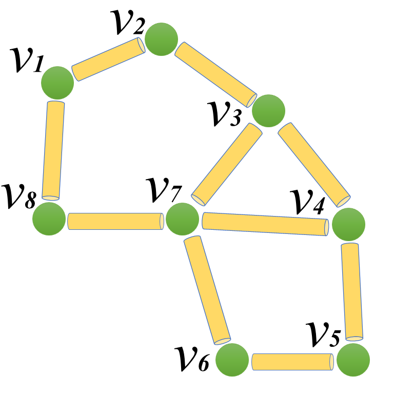
For example, the adjacency, degree and Kirchhoff matrices for the graph in Fig. 1 are, respectively
| (28) |
The Laplacian matrix has several basic properties. It is a symmetric and semi-positive definite. The rank of the Laplacian matrix is with the number of connected components. Its second smallest eigenvalue is known as the algebraic connectivity (or Fiedler value)[68, 273].
More generally, one can assign weights to edges to construct a weighted graph . Here is the associated unweighted graph, and is the weighted adjacent matrix. The weight is also known as pairwise distance or pairwise affinity. The new degree of vertex is . The weighted degree matrix and weighted Laplacian matrix can be defined accordingly.
Normally, a graph structure is not given. Instead, one may have the information of nodes and general weight functions. In this case there are several general ways to construct a graph[273]:
-
1.
-neighborhood graph: connect all points whose pairwise distances are smaller than ;
-
2.
-nearest neighbor graph: connect vertex with vertex , if is among the -nearest neighbors of ; and
-
3.
fully connected graph: connect all points with positive similarity with each other.
In biomolecular structure graph models, coordinates for atoms in molecules are available. Therefore, distances and distance-based functions can be used to construct structure graphs. The simplest way is to use a cutoff distance and build up edges between atoms or residues within the cutoff distance only. This approach has been used in GNM, which is an important tool for the study of protein flexibility and rigidity.
To unify the notation, in the following discussion, one can consider an -particle representation of a biomolecule. Here a particle can be an ordinary atom in a full atomic representation or a Cα atom in a coarse-grained representation. One can denote the coordinates of these particles and the Euclidean space distance between th and th particles. More specifically, the coordinate is a position vector .
2.1.2 Gaussian network model (GNM)
 |
Gaussian network model (GNM) [15, 14, 80, 179, 304] can be viewed as a special graph model using the Kirchhoff matrix. It was proposed for biomolecular flexibility and long-time scale dynamics analysis, particularly, the prediction of the Debye-Waller factor or B-factor. Experimentally, B-factor is an indication of the relative thermal fluctuations of different parts of a structure. Atoms with small B-factors belong to a part of the structure that is very rigid. Atoms with large B-factors generally belong to part of the structure that is very flexible. The B-factor information can be found in the structural data downloaded from the Protein Data Bank (PDB).
As stated above, the graph or network in GNM is constructed by using a cutoff distance . If the distance between two atoms are less than the cutoff distance, an edge is formed between them. Otherwise, no edge is built. The corresponding discrete Laplacian matrix describes the relative connectivity within a protein structure, and thus, it is also called a connectivity matrix.
| (29) |
In a nutshell, the GNM prediction of the th B-factor of the biomolecule can be expressed as [15, 14, 220]
| (30) |
where is a fitting parameter that can be related to the thermal energy and is the th diagonal element of the Moore-Penrose pseudo-inverse of graph Laplacian matrix . More specifically, , where denotes the transpose and and are the th eigenvalue and eigenvector of , respectively. The summation omits the first eignmode whose eigenvalue is zero.
2.1.3 Anisotropic network model (ANM)
In Gaussian network model [15, 14, 80, 179, 304], only the distance information is used with no consideration about the anisotropic properties in different directions. It should be noticed that in GNM, the Kirchhoff matrix is of the dimension with being the total number of atoms. In order to introduce the anisotropic information, one has to discriminate the distance between atoms in three different directions. To this end, at each label of , a local Hessian matrix is constructed [9]
| (34) |
As the same in the GNM, the diagonal part is the negative summation of the off diagonal elements:
| (35) |
This approach, called anisotropic network model (ANM), can be used to generate the anisotropic motion of biomolecules. It is noticed that the dimension of the Hessian matrix is no longer , instead it is . The dimension of an eigenvector is . Therefore, for each atom, one now has a vector associated with it, which gives an direction in the . The norm of this vector gives a relative amplitude. This eigenvector is also called eigenmode. It describes the relative motion of the protein near its equilibrium state.
Generalized GNM and generalized ANM
In both Gaussian network model and anisotropic network model, a cutoff distance is used to construct their connectivity matrices, i.e., Laplacian matrix and Hessian matrix, respectively. However, physically, the correlation between any two particles normally decays with respect to distance. To account for this effect, a correlation function is introduced. In general, it is a real-valued monotonically decreasing radial basis function satisfying [293, 213],
| (36) | |||||
| (37) |
In this function, the parameter is a characteristic distance between particles and . It can also be simplified to atomic parameter , which depends only on the atomic type. In coarse-grained models, only atom is considered. Therefore, one can further simplify it to a constant . This parameter can also be viewed as a resolution parameter.
 |
Delta sequences of the positive type discussed in an earlier work [283] are all good choices. For example, one can use generalized exponential functions
| (38) |
or generalized Lorentz functions
| (39) |
Using these correlation functions, one can obtain a weighted graph representation or weighted graph Laplacian as [294],
| (40) |
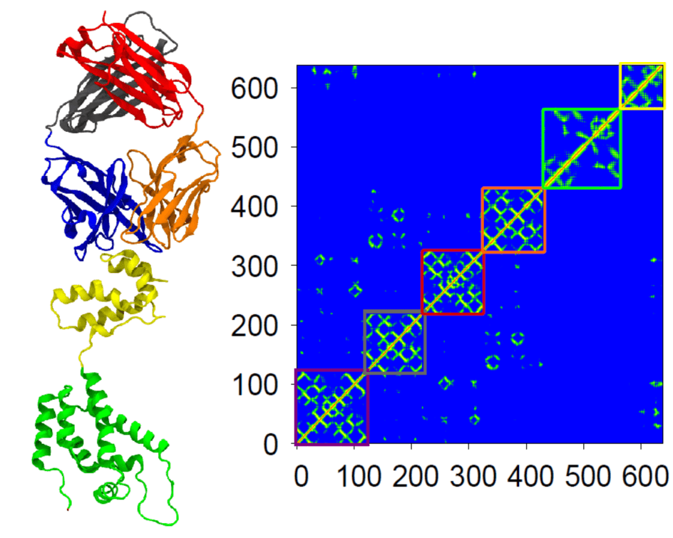
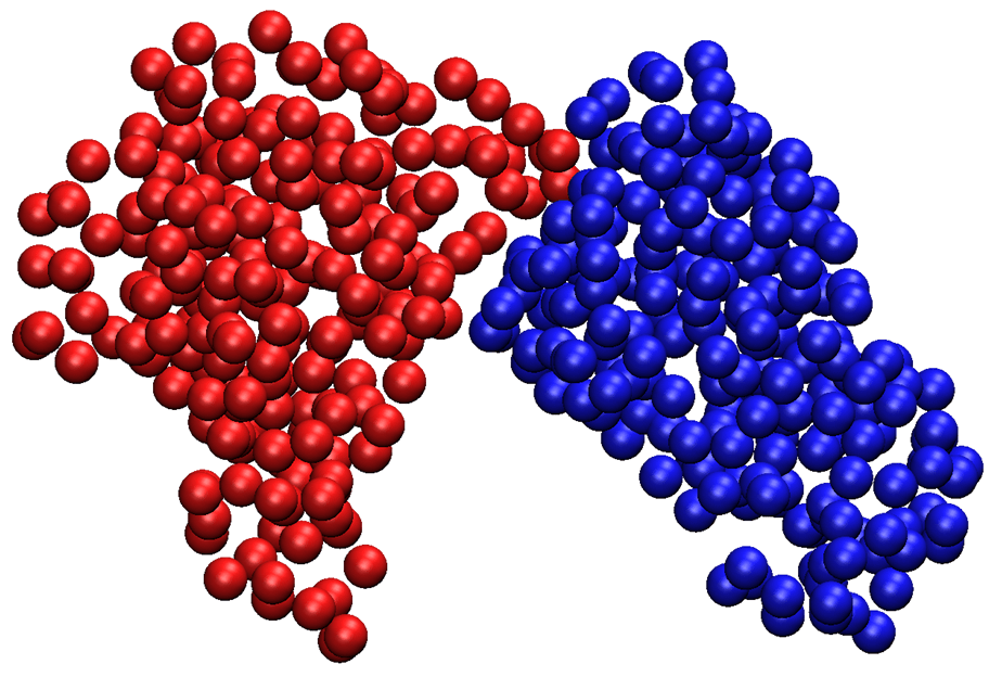
It is found that the weighted graph can deliver a better prediction of B-factors. This weighted graph approach is called the generalized GNM (gGNM). Figure 3 shows the protein domain separation obtained with gGNM.
The local Hessian matrix in Eq. (34) can also be generated to consider the distance effect to obtain a generalized form [294],
| (44) |
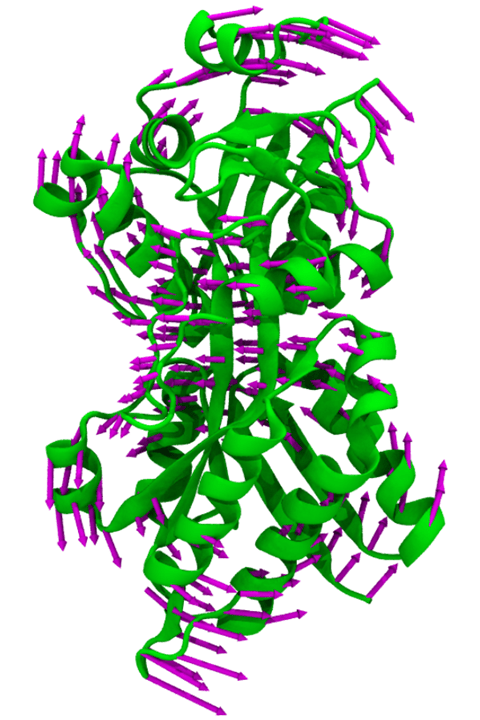
Again the diagonal part is the negative summation of the off-diagonal elements the same as Eq. (35). Note that Hinsen [138] has proposed a special case: , where is a constant. It was shown that gGNM and generalized anisotropic network model (gANM) outperform the original GNM and ANM respectively in B-factor predictions [294]. Figure 4 illustrates an eigenmode for protein 2ABH obtained with gANM. There will be a continuous interest in design new and optimal graph theory approaches for biomolecular analysis.
 |
2.1.4 Normal mode analysis (NMA) and quasi-harmonic analysis
Normal mode analysis (NMA) is one of the major tools for the study of biomolecular motions [123, 176, 35, 187, 134]. It is found that protein normal modes with the largest fluctuation or the lowest frequency are functionally relevant. Mathematically, NMA has its root in harmonic analysis. It assumes that conformational energy surface at an energy minimum can be approximated by some harmonic functions.
In normal mode analysis, one usually needs the atomic coordinates and a force field describing the interactions between constituent atoms. Typically, there are three major steps in applying NMA [134]. Firstly, one needs to carry out molecular dynamics simulations to minimize the conformational potential energy to obtain an equilibrium state. Secondly, one needs to calculate the second derivatives of the potential energy to construct the Hessian matrix. Finally, one needs to perform the eigenvalue decomposition of the Hessian matrix.
Originally, NMA uses exactly the same force fields as used in molecular dynamics simulations. Due to the computational inefficiency, elastic network models (ENMs) was proposed. Generally speaking, ENM is just the NMA with a simplified force field and associated coarse-grained representation. It has two major advantages. Firstly, there is no need for energy minimization as the distances of all of the elastic connections are taken to be at their minimal energy lengths. Secondly, due to the coarse-grained representation, the eigenvalue decomposition is much efficient. Even through ENM is a much simplified model, it is found that ENM is able to reproduce the NMA results with a respectable degree of similarity.
Standard NMA
For a mechanical system consisting of atoms , its Hamiltonian is given by the sum of kinetic energy and potential energy :
| (45) |
If the structure has an equilibrium conformation , one can have the Taylor expansion of the potential energy
| (46) |
Since the biomolecular system achieves a minimum of the energy at the equilibrium conformation , the related derivative functions vanishes. If one uses the mass-weighted coordinates , the potential function becomes
| (47) | |||
The related kinetic energy is
| (48) |
The Hamiltonian is
| (49) |
where indicates the derivative of with respect to time.
It can be seen that the oscillatory motions in this system are coupled and thus the movement of one atom depends on that of others. However, one can decompose the motion into independent harmonic oscillators with an appropriate normal mode coordinates. This is done by the eigenvalue decomposition of the Hessian matrix . Here is obtained from the second order derivative of the potential function. The matrix contains the eigenvectors and . The diagonal matrix contains the corresponding eigenvalues. The mass-weighted Cartesian and normal mode coordinates are linearly related by . Finally, the Hamiltonian is expressed in the form,
| (50) |
Essential dynamics and quasi-harmonic analysis
Due to the complexity of biomolecular systems, it is notoriously difficult to carry out molecular dynamics simulations over the relevant biological time scales. However, it has been found that the vast majority of protein dynamics can be described by a surprisingly low number of collective degrees of freedom. In this manner, a principal components analysis (PCA) is often employed to analyze the simulation results[115, 160]. Mathematically, similar to NMA, PCA also employs the eigenvalue decomposition as it assume that that the major collective modes of fluctuation dominate the functional dynamics. In contrast to NMA, PCA of a molecular dynamics simulation trajectory does not rest on the assumption of a harmonic potential. Modes in PCA are usually sorted according to variance rather than frequency. As the collective motion is highly related to biomolecular functions, the dynamics in the low-dimensional subspace spanned by these modes was termed “essential dynamics” [5]. A major advantage of PCA is that individual modes can be visualized and studied separately.
PCA on the mass weighted MD trajectory is also called quasi-harmonic analysis [37], which typically consists of three steps [134]. Firstly, one can superimpose all biomolecular configurations from the simulation trajectory to remove the internal rotation and translation. Secondly, one can perform an average over the regularized trajectory to construct a covariance matrix. Thirdly, an eigenvalue decomposition is employed on the covariance matrix. The original trajectory can then be analyzed in terms of principal components.
For a -dimensional vector trajectory , the correlation between atomic motions can be expressed in the covariance matrix :
| (51) |
where is the statistical covariance and denote the average over time. The correlation matrix is symmetric and can be diagonalized by an orthogonal transformation,
| (52) |
with being eigenvectors. The original configurations can be projected into principal components , i.e., . For visualization, one can transform principal components into the Cartesian coordinates: .
2.1.5 Flexibility rigidity index (FRI)
Due to the involved matrix diagonalization, the computational complexity of GNM is of the order of , which is intractable for large biomolecules, such as viruses and subcellular organelles. Therefore, it is both important and desirable to have a method whose computational complexity scales as or better, as . This order reduction is a standard mathematical issue and is mathematically challenging. However, by examining Eq. (30), one notices that what is used in the GNM theoretical prediction is the diagonal elements of the inverse of the graph Laplacian matrix. Mathematically, a good approximation is given by the inverse of the diagonal elements of the graph Laplacian matrix, providing that the matrix is diagonally dominant. Flexibility rigidity index (FRI) [293, 213] is such a method and has several major characteristics. FRI provides a more straightforward and computationally-efficient way to predict B-factors. A major advantage of the FRI method is that it does not resort to mode decomposition and its computational complexity can be reduced to by means of the cell lists algorithm used in fast FRI (fFRI) [213].
The fundamental assumptions of the FRI method are as follows. Protein functions, such as flexibility, rigidity, and energy, are fully determined by the structure of the protein and its environment, and the protein structure is in turn determined by the relevant interactions. Therefore, whenever the protein structural data is available, there is no need to analyze protein flexibility and rigidity by tracing back to the protein interaction Hamiltonian. Consequently, the FRI bypasses the matrix diagonalization.
In a nutshell, the FRI prediction of the th B-factor of the biomolecule can be given by [293, 213]
| (53) |
where and are fitting parameters, is the th flexibility index and
| (54) |
is the th rigidity index. Here, is an atomic number depended weight function that can be set to and for a Cα network. The correlation function can be chosen from any monotonically decreasing function satisfying Eqs. (38) and (75). FRI was shown to outperform GNM and ANM in B-factor predictions based on hundreds of biomolecules [293, 213].
Multiscale FRI
Biomolecules are inherently multiscale in nature due to their multiscale interactions. For example, proteins involve covalent bonds, hydrogen bonds, van der Walls bonds, electrostatic interactions, dipolar and quadrupole interactions, hydrophobic interactions, domain interactions, and protein-protein interactions. Therefore, their thermal motions are influenced by the multiscale interactions among their particles. Multiscale FRI (mFRI) was proposed to capture biomolecular multiscale behavior [215]. Essential idea is to build multiscale kernels, i.e., kernels parametrized at multiple scales. Multiscale flexibility index can be expressed as
| (55) |
where , and are the corresponding quantities associated with the th kernel. Then, one organizes these kernels in a multi-parameters minimization procedure
| (56) |
where are the experimental B-factors. In principle, all parameters can be optimized. For simplicity and computational efficiency, one only needs to determine and in the above minimization process. For each kernel , and will be selected according to the type of particles.
Specifically, for a simple Cα network (graph), one can set and choose a single kernel function parametrized at different scales. The predicted B-factors can be expressed as
| (57) |
The difference between Eqs. (55) and (57) is that, in Eqs. (55), both the kernel and the scale can be changed for different . In contrast, in Eq. (57), only the scale is changed. One can use a given kernel, such as
| (58) |
to achieve good multiscale predictions. It was demonstrated that mFRI is about 20% more accurate than GNM in the B-factor predictions [215]. Parameters learned from mFRI were incorporated in GNM and ANM to create multiscale GNM (mGNM) and multiscale ANM (mANM) [215].
Consistency between GNM and FRI
 |
To further explore the relation between GNM and FRI, let us examine the parameter limits of generalized exponential functions (38) and generalized Lorentz functions (75)
| (59) | |||||
| (60) |
where and is the ideal low-pass filter (ILF) used in the GNM Kirchhoff matrix
| (61) |
Relations (59) and (60) unequivocally connect FRI correlation functions to the GNM Kirchhoff matrix.
It has been observed that GNM-ILF and FRI-ILF provide essentially identical predictions when the cutoff distance is equal to or larger than 20Å[294]. This phenomenon indicates that when the cutoff is sufficiently large, the diagonal elements of the gGNM inverse matrix and the direct inverse of the diagonal elements of the FRI correlation matrix become linearly strongly dependent. To understand this dependence at a large cutoff distance, an extreme case is considered when the cutoff distance is equal to or even larger than the protein size so that all the particles within the network are fully connected. In this situation, one can analytically calculate th diagonal element of the GNM inverse matrix
| (62) |
and the FRI inverse of the th diagonal element
| (63) |
These two expressions have the same asymptotic behavior as , which explains numerical results. However, mathematically, it is still an open problem to estimate the error bound between gGNM and FRI methods, i.e., the difference between the direct inverse of a diagonal element of a given weighted graph Laplacian matrix and the diagonal element of its matrix inverse.
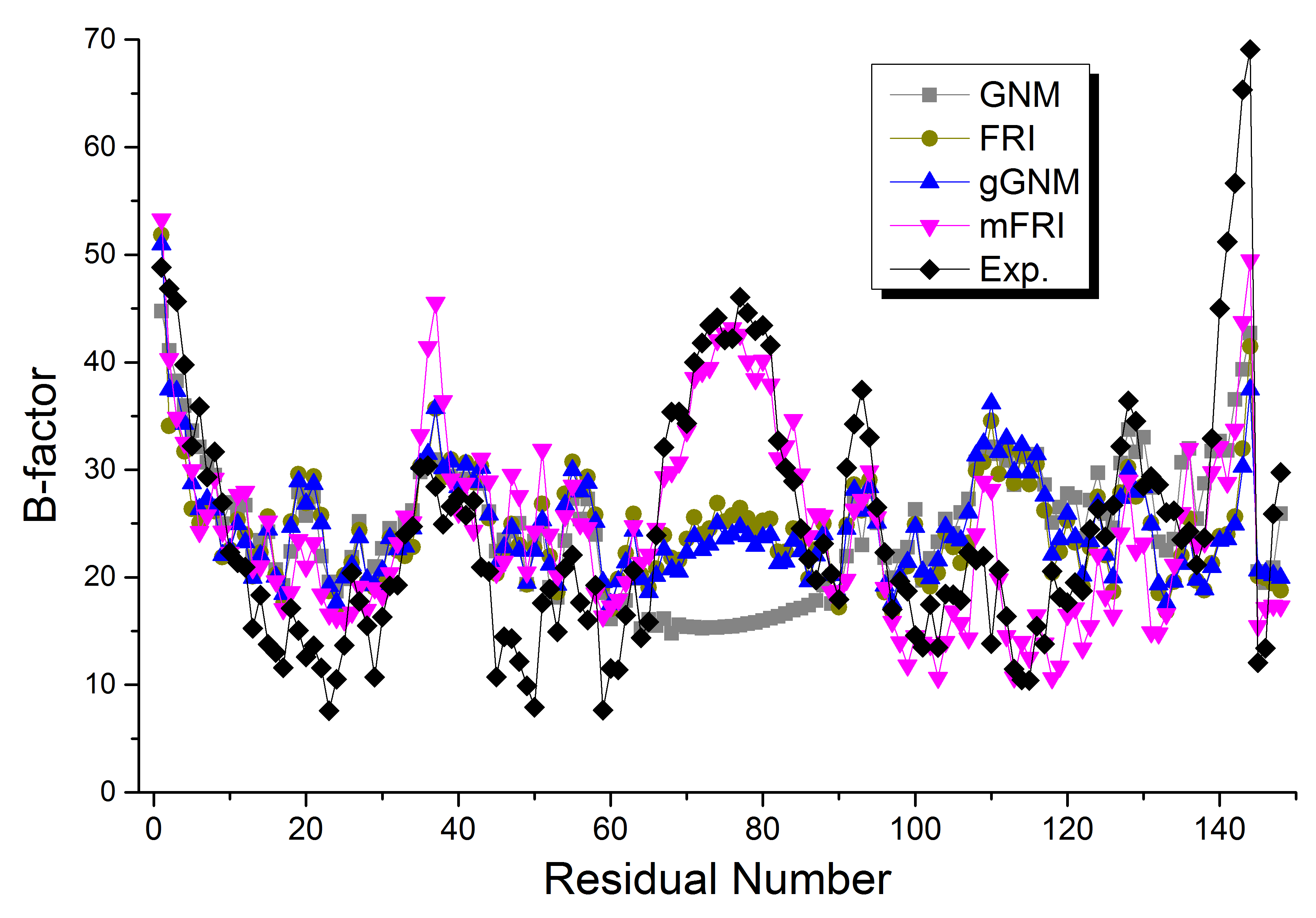
A comparison of the performances of GNM, FRI, gGNM and mFRI is illustrated in Fig. 5. The hinge around 75th residue is well captured by mFRI. Indeed, mFRI has the best accuracy in B-factor prediction based on the test of 364 proteins [215]. The possible application to FRI in other systems, such as social networks, genetic networks, cellular networks and tissue networks, is still an open problem.
Anisotropic FRI
The anisotropic FRI (aFRI) has been proposed for mode analysis [213]. In this model, depending on one’s interest, the size of the Hessian matrix can vary from for a completely local aFRI to for a completely global aFRI. To construct such a Hessian matrix, one can partition all atoms in a molecule into a total of clusters . Each cluster with has atoms so that . For convenience, one can denote
| (64) |
Note that for each given , one can define as a local anisotropic matrix
| (65) |
In the anisotropic flexibility and rigidity (aFRI) approach, a flexibility Hessian matrix for cluster is defined by
| (66) | |||||
| (67) | |||||
| (68) |
where denotes the adjoint of matrix such that , here is identity matrix.
Another representation for the flexibility Hessian matrix can be defined as follows
| (69) | |||||
| (70) | |||||
| (71) |
where is a matrix with every element being one.
One can achieve eigenvectors for atoms in cluster by diagonalizing , . Note that, the diagonal part , , has inherent information of all atoms in the system. As a result, the B-factors can be predicted by the following form:
| (72) | |||||
| (73) |
It was found that aFRI is much more accurate than ANM in protein B-factor prediction [213]. The anisotropic cluster analysis was found to play a significant role in study the local motion of RNA polymerase II translocation [214].
2.1.6 Spectral graph theory
Spectral graph theory [68, 202, 201, 273] concerns the study and exploration of graphs through the eigenvalues and eigenvectors of matrices naturally associated with those graphs [247, 194, 208, 10, 309]. Therefore, widely used GNM and ANM methods make use of spectral graph theory.
For a given graph with nodes, one is interested in its matrix representation. Matrices and correspond to weighted adjacent matrix and weighted degree matrix, respectively. With this notation, one has the unnormalized graph Laplacian
It is often called admittance matrix, Kirchhoff matrix or discrete Laplacian. The graph matrix has several interesting properties. Firstly, it is symmetric and positive semi-definite. Secondly, it has non-negative, real-valued eigenvalues. Thirdly, the smallest eigenvalue is 0 and the corresponding eigenvector is constant vector . Fourthly, for every vector , one has , which can be derived from
| (74) | |||
where is the degree of the th vertex.
In spectral graph theory, two other kinds of Laplacian matrices [68] are also widely used. They are the normalized Laplacian matrix
and random-walk normalized Laplacian matrix [194, 189, 2]
where is an identity matrix. Three different Laplacian matrices are tightly related to different ways of graph decompositions.
Graph decomposition and graph cut
A protein may have different domains. Identifying protein domains and analyzing their relative motions are important for studying protein functions. A protein complex involves different proteins. The study of protein complex can often be formulated as a graph decomposition problem as well.
In many situations, for a given graph , one wants to partition it into subgraphs, so that nodes within a subgroup are of similar properties and nodes in different subgroups are of different properties. Mathematically, if the weight is a measurement of similarity, i.e., large weight means a great similarity, an optimized partition means that edges within the same subgroup should have large weights and edges across subgroups should have small weights. State differently, one wants to find a way to cut the graph so that it will minimize the weights of edges connecting vertices in different subgroups.
Let us first consider a simple situation, divide into two subgroup and . The notation denotes the complementary of . One can define a partition as,
| (75) |
Therefore, if two nodes and are in the same subgroup, one will have , otherwise . In this way, a , which is the total weights of edges connecting two subgroups, can be defined as following,
| (76) | |||
It can be also noticed that if one defines , then one has .
To obtain an optimized partition means to minimize the value of . However, the way of cut stated above does not consider the size of the subgraphs. In this way, the cut or graph composition can be very uneven in terms of the number of nodes in subgroup. For example, one extreme situation is that one of the subgraph may only have a few nodes (i.e., one or two nodes), while the other subgroup may have all the rest of nodes. To avoid this problem, three commonly defined cuts, namely, ratio cut, normalized cut and min-max cut, are proposed in the literature [130, 247, 95]. To facilitate the description, one can define as the total number of nodes in graph and is the summation of all weights in , i.e., .
-
•
Ratio cut is defined as [130]
(77) -
•
Normalized cut is defined as [247]
(78) -
•
Min-Max cut is given by [95]
(79)
These decompositions have found many applications in image segmentation [247]. However, the impact of these cuts to protein domain partition is yet to be examined. An important issue is how to cut a given biomolecule to elucidate its biological function and predict its chemical and biological behavior.
Ratio cut and Laplaician matrix
To solve the optimization problem
| (80) |
one can define the vector as
| (81) |
One can have
One also has
| (83) |
Therefore, the vector is orthogonal to the vector with common components (leading eigenvector of the Laplacian matrix). It is noted that
| (84) |
In this way, the minimization is equivalent to
| (85) |
As the entries of the solution vector are only allowed to take two particular values, Eq. (85) is a discrete optimization problem. One can discharge the discreteness condition and allow the vector to take any arbitrary values. This results in a relaxed problem
| (86) |
From the Rayleigh-Ritz theorem, it can be seen that the solution of this problem is the second smallest eigenvector of the Laplacian matrix. In order to obtain a partition of the graph, one can choose,
| (87) |
Mathematically, the second smallest eigenvalue is known as the algebraic connectivity (or Fiedler value) of a graph. The corresponding eigenvector of the second eigenvalue offers a near optimized partition. It becomes an interesting issue to design certain weighted Laplacian matrix so that a protein domain partition is optimal with respect to protein functions. Transferring the discrete Laplacian matrix to a continuous Laplacian operator and casting the domain separation problem into an optimization one are promising approaches. Certainly, these issues are also biologically significant in the exploration of protein structure-function relationship.
Normalized cut and normalized Laplacian matrix
One can define the vector as
| (88) |
One can have
Additionally, it is easy to see that
| (90) |
This means that vector is orthogonal to constant one vector. Moreover, one can evaluate
| (91) |
In this way, the minimization process is equivalent to
| (92) |
This is also a discrete optimization problem, because the entries of the solution vector are only allowed to take two particular values. By discarding the discreteness condition and allowing the vector to be any arbitrary values, one results in a relaxed problem
| (93) |
Now one can substitute . After substitution, the problem is
| (94) |
It can been seen that and is the first eigenvector of . From the Rayleigh-Ritz theorem, it can be seen that the solution of this problem is the second smallest eigenvalue of .
Graph Laplacian and continuous Laplace operator
It has been found that there is a connection between graph Laplacian and the continuous Laplace operator [24, 171, 25, 135, 121]. Roughly speaking, if one chooses with as the distance between node and node , one can have
| (95) |
The term can be roughly viewed as a discretization of . In this way, there is a connection between graph Laplacian and the continuous Laplace operator through this functional formulation,
| (96) |
More specifically, one can set as defined in Eqs. (36) and (37)
| (97) |
where and is the coordinate of th node. This operator can be naturally extended to an integral operator
| (98) |
If data points are sampled from a uniform distribution on a -dimensional manifold , let set , with , where , and assume . Belkin [25] found that there is a constant , such that in probability [24, 25]
| (99) |
The continuous Laplace operator has been widely utilized in many biophysical models, such as Poisson-Boltzmann theory for electrostatics [141], Laplace-Beltrami equation for molecular surface modeling [21, 284], Poisson-Nernst-Planck equation for ion channel modeling [146, 288], and elasticity equation for macromolecular conformational change induced by electrostatic forces [318]. Obviously, these issues are associated with a graph problem. The modeling of biomolecular structure, function, dynamics and transport by combining graph theory and partial differential equation (PDE) is an open problem.
Modularity
Modularity is total summation of the weights within the group minus the expected one in an equivalent network with weight randomly placed.
 |
Mathematically, the modularity is defined as [207, 206, 107, 148]
| (100) |
where is a resolution parameter, which is designed to change the scale at which a network is clustered [107]. Here if and are in the same subgroup (), otherwise it equals to 0 (). The term is the weighted adjacent matrix component.
The modularity matrix is defined as:
| (101) |
then the above equation can be further simplified as
| (102) |
Again one assume that graph can be divided into two parts and
| (103) | |||
One can define the total variation (TV): , weighted -norm , and mean .
One can also define to be a function . This is the indicator function of a subsect . In this manner, one has
| (104) | |||
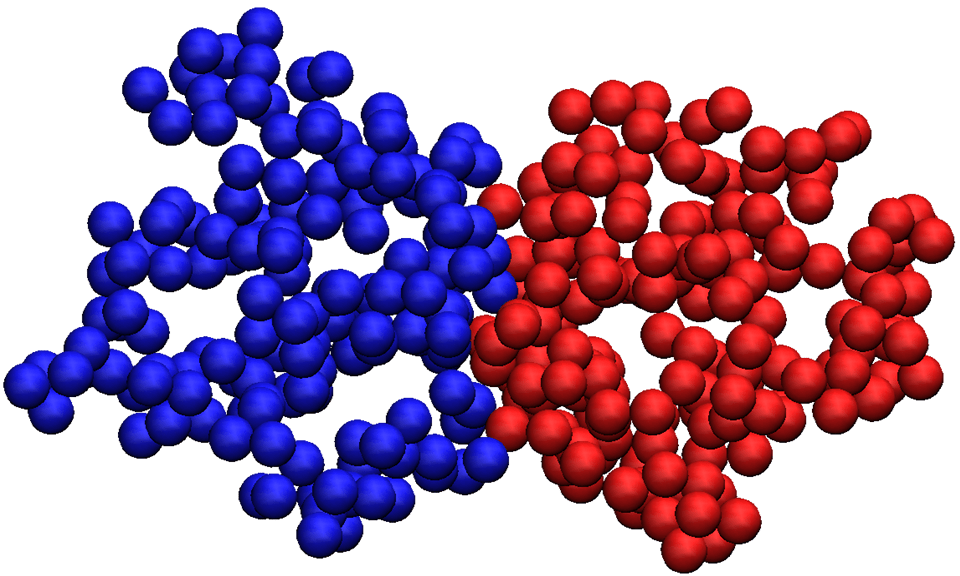
Figure 6 illustrates the modularity matrix, second-eigenvector formed matrix and domain indication matrix for protein 2ABH. Figure 7 shows the protein domain decomposition using a modularity matrix.
 |
One can have the theorem: maximizing the modularity functional over all the partitions is equivalent to minimize [144] . Essentially, this is equal to a balance cut problem
| (105) |
One can let by with [144] . In this way, for each , it has only a single entry equals 1. The minimizing problem can be solved through the following equation [195, 144]
| (106) |
Here is the composition of function and . Normally, the function is a multi-well potential [195].
The field of spectral graph, modularity and related variation formation for biomolecular systems is completely open. There is much to be done on this interesting field. For example, one can formulate molecular design, such as drug design, protein design, and the design of protein-DNA and/or protein-RNA complexes, as graph cut problems. In drug design, one would like to optimize the protein-drug binding affinity for a given drug candidate and its target protein. If the selection of graph notes is also a part of optimization, one then would like to optimize protein-drug binding affinity, drug target selectivity, drug pharmacokinetics, drug toxicity, etc. To be more specific, one needs to consider a minimization process,
| (107) |
Here is the total number of atoms in the complex, the parameter is a scale parameter and is the free energy change. The function is denoted as the graph representation of protein-protein or protein-ligand complexes. The functions and are graph models for the corresponding protein or ligand. They are all position-dependent and the minimization process is to find the best fitting configuration so that one can achieve a cut that minimizes the free energy change . In fact, biomolecular free energy minimization often leads to PDE based models for solvation, ion channel, membrane protein interaction, molecular machine assembly, to name only a few [284]. It should be noticed that in Eq. (107), all the three types of graph cuts might be used. This approach can be combined with techniques in other mathematical disciplines, such as those in dynamical systems, stochastic analysis, and differential equation, to address complex design problems, namely, drug design, protein design, RNA design, molecular machine design etc, in biomolecular systems.
2.1.7 Molecular nonlinear dynamics
To introduce molecular nonlinear dynamics, one can consider a folding protein that constitutes particles and has the spatiotemporal complexity of . Assume that the molecular mechanics of the protein is described by molecular nonlinear dynamics having a set of nonlinear oscillators of dimension , where is the dimensionality of a single nonlinear oscillator. Let us consider an -dimensional nonlinear system for interacting chaotic oscillators [295]
| (108) |
where is an array of state functions for nonlinear oscillators, is an -dimensional nonlinear function for the th oscillator, is an array of nonlinear functions of oscillators, and . Here, is the overall interaction strength, is an weighted Laplacian matrix and is an linking matrix. Essentially, for each node in the biomolecular graph, one has an -dimensional nonlinear oscillator. These oscillators are connected by the Laplacian matrix and a fixed linking matrix.
For example, one can choose a set of Lorenz attractors [188] and a simple link matrix as following: ,
| (112) |
If the Laplacian matrix shown in Fig. 1 is used as the connectivity matrix, the nonlinear dynamic system for the first node is
Stability analysis
One can use the FRI kernel weighted Laplacian matrix to define the driving and response relation of nonlinear chaotic oscillators. Due to the synchronization of chaotic oscillators, an -time reduction in the spatiotemporal complexity can be achieved, leading to an intrinsically low dimensional manifold (ILDM) of dimension . Formally, the -dimensional ILDM is defined as
| (113) |
where is a synchronous state or reference state.
To understand the stability of the ILDM of protein chaotic dynamics, one can define a transverse state function as , where is a vector of identical components . Obviously, the invariant ILDM is given by . Therefore, the stability of the ILDM can be analyzed by , which can be studied by the following linearized equation [221, 143]
| (114) |
where is the Jacobian of .
To further analyze the stability of Eq. (114), one can diagonalize connectivity matrix
| (115) |
where are eigenvectors and are the associated eigenvalues. These eigenvectors span a vector space in which a transverse state vector has the expansion [221, 143]
| (116) |
Therefore, the stability problem of the ILDM is equivalent to the following stability problem
| (117) |
where is the diagonal component of . The stability of Eq. (117) is determined by the largest Lyapunov exponent , namely, , which can be decomposed into two contributions
where is the largest Lyapunov exponent of the original dimensional chaotic system , which can be easily computed for most chaotic systems. Here, depends on and . The largest eigenvalue equals 0, and its corresponding eigenvector represents the homogeneous motion of the ILDM, and all of other eigenvalues govern the transverse stability of the ILDM. Let us consider a simple case in which the linking matrix is the unit matrix (). Then stability of the ILDM is determined by the second largest eigenvalue (algebraic connectivity, or Fiedler value), which enables us to estimate the critical interaction strength in terms of and [295],
| (118) |
The dynamical system reaches the ILDM when and is unstable when . The eigenvalues of protein connectivity matrices are obtained with a standard matrix diagonalization algorithm. Molecular nonlinear dynamics has been recently developed as an efficient means for protein B-factor prediction [295]. It can be potentially used for protein domain separation without resorting to the matrix diagonalization.
The availability of more than a hundred thousands of interacting protein networks in the PDB provides living example problems for analyzing dynamical systems. Indeed, the connection between dynamical systems and spectral graph theory in mathematics, and the structure and function of macromolecules gives rise to exciting opportunities to further study dynamical systems and graph theory, and better analyze biomolecules.
2.2 Persistent homology
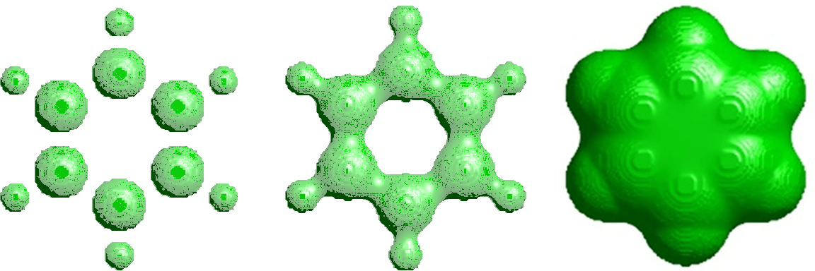 |
As a branch of algebraic topology, persistent homology is a topological approach that utilizes algebraic algorithms to compute topological invariants in data[44, 211, 279, 237, 185]. It is a working horse in the popular topological data analysis and has found its success in qualitative characterization or classification. Specifically, persistent homology describes geometric features of biomolecular date with topological invariants that persist over the systematic change of a scale parameter relevant to topological events. Such a change is called filtration. Figure 8 illustrates the change of the topology of benzene molecule over different density thresholds. The idea is to capture topological structures continuously over a range of spatial scales during the filtration process. Unlike computational homology that is based on truly metric free or coordinate free representations, persistent homology can embed geometric information from protein data into topological invariants so that “birth” and “death” of geometric features, such as circles, rings, loops, pockets, voids and cavities can be monitored by topological measurements during the filtration process[99, 319]
2.2.1 Simplicial homology and persistent homology
An essential ingredient of persistent homology is simplicial homology which is built on simplicial complex. Simplicial complex is a finite set that consists of discrete vertices (nodes or atoms in a protein), edges (line segments or bonds in a biomolecule), triangles, and their high dimensional counterparts. Simplicial homology can be defined on simplicial complex to analyze and extract topological invariants. Then a filtration process is used to establish topological persistence from simplicial homology analysis[99, 319].
Simplicial complex
A key component of simplicial complex is a -simplex, , defined as the convex hall of affine independent nodes in (). Let be affine independent points (or atoms in a biomolecule) and express a -simplex as
| (119) |
Moreover, let us define an -dimensional face of as the convex hall formed by the nonempty subset of vertices from (). Clearly, a 0-simplex is a vertex, a 1-simplex is an edge, a 2-simplex is a triangle, and a 3-simplex represents a tetrahedron. One can also define the empty set as a (-1)-simplex.
A simplicial complex is constructed to combine these geometric components, including vertices, edges, triangles, and tetrahedrons together under certain rules. More specifically, a simplicial complex is a finite set of simplicies that satisfy two conditions. One is that any face of a simplex from is also in and the other is that the intersection of any two simplices in is either empty or shared faces. The dimension of a simplicial complex is defined as the maximal dimension of its simplicies. The underlying topological space is a union of all the simplices of , i.e., . Further, the concept of chain is introduced to associate this topological space with algebra groups.
Homology
One can denote a linear combination of -simplex as a -chain . The coefficients can be chosen from different fields, such as rational field , real number field and complex number field, and from integers and prime integers with prime number . For simplicity, one can consider the coefficients are chosen from , for which the addition operation between two chains is the modulo 2 addition for the coefficients of their corresponding simplices. The set of all -chains of simplicial complex and the addition operation form an Abelian group . Therefore, the homology of a topological space is represented by a series of Abelian groups.
A boundary operation is defined as . Without orientation, the boundary of a -simplex is
| (120) |
where the notation means that the -simplex is generated by eliminating vertex from the sequence. When a boundary operator is applied twice, any -chain will be mapped to a zero element, i.e., . As a special case, one has . The th cycle group and the th boundary group are the subgroups of and can be defined by means of the boundary operator,
| (121) | |||
| (122) |
An element in the th cycle group or the th boundary group is called the th cycle or the th boundary. One has since the boundary of a boundary is always empty . Geometrically, the th cycle is a dimensional loop or hole.
With all the above definitions, one can define the homology group. Specifically, the th homology group is defined as the quotient group of the th cycle group and th boundary group : . Two th cycle elements are called homologous if they are different by a th boundary element. The th Betti number represents the rank of the th homology group,
| (123) |
From the fundamental theorem of finitely generated Abelian groups, the th homology group can be given as a direct sum,
| (124) |
where is the rank of the subgroup and is th Betti number. Here is torsion subgroup with torsion coefficients , the power of prime number.
Topologically, cycle element in forms a -dimensional loop or ring that is not from the boundary of a higher dimensional chain element. The geometric meanings of Betti numbers in are the follows: represents the number of isolated components (i.e., protein atoms), is the number of one-dimensional loop or ring, and describes the number of two-dimensional voids or cavities. Together, the Betti number sequence gives the intrinsic topological property of biomolecular data.
ech complex, Rips complex and alpha complex
A key concept for the construction of simplicial complex from a point set of a given topological space is nerve. Basically, given an index set and open set that is a cover of a point set , i.e., , the nerve N of U should satisfy two basic conditions. One is that . Additionally, if for , then . Usually, for a given biomolecular dataset, the simplest way to construct a cover is to assign a ball of certain radius around each atom. If the biomolecular dataset is dense enough and the radius is large enough, then the union of all the balls has the capability to recover the underlying space for the biomolecule.
The nerve of a cover of the biomolecule constructed from the union of atomic balls is a ech complex for the biomolecule. More specifically, for a biomolecular dataset , one defines a cover of closed atomic balls with radius and centered at . The ech complex of with radius is denoted as , which is the nerve of the closed ball set B,
| (125) |
One can relax ech complex conditions to generate a Vietoris-Rips complex, in which, a simplex is constructed if the largest distance between any two atoms is at most . One can denote the Vietoris-Rips complex, or Rips complex [100]. There is a sandwich relation for these abstract complexes,
| (126) |
In practical applications, Rips complex is preferred due to its computational convenience.
Another important geometric concept in computational geometry is alpha complex. Let be a biomolecular dataset in Euclidean space and define the Voronoi cell of a point as
| (127) |
Then the collection of all Voronoi cells for the biomolecule forms a Voronoi diagram. Further, the nerve of the biomolcular Voronoi diagram generates a Delaunay complex.
One can define as the intersection of Voronoi cell with ball , i.e., . The alpha complex of the dataset is defined as the nerve of cover ,
| (128) |
Therefore, an alpha complex is a subset of the Delaunay complex.
 |
General filtration processes
In order to construct a simplicial homology from a dataset, a special parameter (like the radius mentioned above) is commonly used. However, to find a “suitable” value for this parameter, so that it can reveal the underlying manifold, is not straightforward. An elegant alternative approach is to carry out a filtration process [39, 101, 91, 93, 199]. A suitable filtration is vital to the resulting persistent homology, which will be described in the following section. In practice, two commonly used filtration algorithms are the Euclidean-distance or correlation matrix based and density based ones. These basic filtration algorithms can be modified to achieve different goals in data analysis [296, 297, 300].
In the Euclidean-distance based filtration, one associates each atom with an ever-increasing radius to form an ever-growing ball for each atom. Various aforementioned complex construction algorithms can be utilized to identify the corresponding complexes. This filtration process can be formalized by the use of a distance matrix . Here the matrix element represents the distance between atom and atom . For diagonal terms, one assumes . With a filtration threshold , a 1-simplex is generated between atoms and if . Higher dimensional complexes can also be created similarly.
Another important filtration process is the density based filtration process. In this process, the filtration goes along the increase or decrease of the density value. In this way, a series of isosurfaces are generated. Morse complex is used for the characterization of their topological invariants.
Persistent homology
Persistent homology is an elegant mathematical theory to describe topological invariants from a series of topological spaces in various scales, that are generated by the filtration process. Persistent homology concerns a family of homologies, in which the connectivity of the given dataset is systematically reset according to a (scale) parameter. For a simplicial complex , the filtration is defined as a nested sub-sequence of subcomplexes,
| (129) |
The introduction of filtration leads to the creation of persistent homology. When the filtration parameter is a scale parameter, simplicial complexes generated from a filtration give a multiscale representation of the corresponding topological space, from which related homology groups can be evaluated to reveal topological features of the given dataset. Furthermore, the concept of persistence is introduced to measure the persistent length of topological features. The -persistent th homology group is
| (130) |
Through the study of the persistent pattern of these topological features, the so called persistent homology is capable of capturing the intrinsic properties of the underlying protein topological space solely from the protein atomic coordinates.
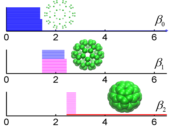
To visualize the persistent homology results, many elegant representation methods have been proposed, including persistent diagram[98], persistent barcode[119], persistent landscape [38], etc. In this paper, a barcode representation is used. The persistent barcode of an Euclidean-distance based filtration process of a fullerene molecular is shown in Fig. 9.
The combination of optimization and persistent homology was discussed in a recent work for biomolecular data analysis [275]. The essentially idea to create an object functional for extracting certain geometric features in data. Then the use of variational principle to result in a differential equation, which is subsequently utilized to filtrate the biomolecular data. In this work, the minimization of the surface energy of biomolecules was the objective, which leads to the Laplace-Beltrami flow for filtration. In this manner, one can have connected persistent homology to other important mathematical subjects, such as partial differential equations, optimizations, and differential geometry [275].
Object-oriented persistent homology is expected to play an important role in massive data analysis. In particular, this approach can be combined with a deep learning strategy to automatically extract desirable information in a semi-supervised or unsupervised learning framework.
2.2.2 Multiscale persistent homology
 |
The emergence of complexity in self-organizing biological systems frequently requires more comprehensive topological descriptions. Therefore, multiscale persistent homology, multiresolution persistent homology and multidimensional persistence become valuable for biological systems as well as many other complex systems.
It is noted that there is no need for a simplicial complex to be built exclusively on a set of atoms. It can be constructed for a biomolecule from its coarse-grained representation, namely, a set of amino acid residues. For protein complexes, such as a viral particle as shown in Fig. 10, a basic vortex in a simplicial complex can be either a protein, a residue (i.e., Cα representation) or an atom. Each of these selections gives rise to a different persistent homology at a given scale. To demonstrate the utility of persistent homology for cryo-EM data analysis, a microtubule intermediate structure, protein complex 1129 from electron microscopy data (EMD) is considered [298]. In this study, each point in the simplicial complex is a protein (tubulin).
An interesting problem in persistent homology studies is how to select appropriate scales and appropriate types of molecular information in filtration to better analyzing the structure, function and dynamics of subcellular organelles, molecular motors and multiprotein complexes.
2.2.3 Topology based quantitative modeling
Traditional topological analysis analyzes data in terms of topological invariants, such as Euler characteristic, winding number, Betti numbers and so on, thus leads to so much reduction that the resulting information is hardly useful for complex real world problems. Geometric tools often become computationally intractable for macromolecules and their interactions due to the involved high degrees of freedom. As one can see, persistent homology embeds geometric information in topological invariants and bridges the gap between geometry and topology. However, persistent homology has been mainly employed for qualitative analysis, namely, characterization and classification. Only recently, persistent homology has been devised for quantitative analysis, mathematical modeling, and physical prediction [296, 297, 300, 275]. It has been shown that the length of intrinsic Betti 2 bar provides an excellent model for fullerene thermal energies [275]. It has also been shown that accumulated Betti 0 and Betti 1 bar lengths offer highly accurate predictions of unfolding protein bond and total energies, respectively [297]. It is expected that persistent homology will continue to play a significant role in quantitative modeling and prediction.
Additionally, earlier work regard short-lived barcodes or topological invariants as noise. It has also been pointed out that, for biomolecular datsets, these short-lived topological invariants or non-persistent topological features, are part of topological fingerprints and have meaningful biophysical interpretations [296].
In general, topology based quantitative modeling and analysis will be a new trend in molecular bioscience and biophysics. Particularly, topological features are ideally suitable for machine learning based quantitative predictions of biomolecular functions.
3 Continuous apparatuses for biomolecules
As stated above, the topological study of scalar fields, particularly electron density data, has advanced the understanding of molecules tremendously. The theory of atoms in molecules (AIM) has provided an elegant and feasible partition of electron density field, so that one can mathematically rigorously define the atoms in molecule [11, 12]. AIM can be generalized into a more general theory called quantum chemical topology (QCT), which employs the topological analysis in AIM for the study of other physically meaningful scalar fields [229]. Additionally, geometric modeling and analysis, particularly the surface curvature analysis, has contributed a lot to the molecular visualization and structure characterization [290]. Thanks to the geometric analysis, the establishment and further deeper understanding of structure-function relationship have been achieved. In this section, a brief discuss of geometric and topological analysis of scalar fields is presented.
Continuum representation of biomolecules plays an important role in their modeling, analysis and simulation. Volumetric biomolecular data are typically obtained from cryo-EM maps, quantum mechanical simulations and mathematical models that transform discrete datasets originally generated by X-ray crystallography or other means into continuous ones. Therefore, continuous mathematical approaches for analysis and modeling of biomolecules in the volumetric data form are as important as their discrete counterparts.
3.1 Geometric representation
Geometric modeling is a crucial ingredient of biophysics. Due to increasingly powerful high performance computers, geometric modeling has become an essential apparatus for biomolecular surface representation, visualization, surface and volumetric meshing, area and volume estimation, curvature analysis and filling the gap between macromolecular structural information and their theoretical models [308, 104, 103, 291, 180, 231]. The visualization of macromolecules sheds light on biomolecular structure, function and interaction, including ligand-receptor binding sites, protein specification, drug binding, macromolecular assembly, protein-nucleic acid interactions, protein-protein binding hot spots, and enzymatic mechanism [225, 239, 205, 88].
Non-smooth biomolecular surface representations
A number of molecular surface models has been proposed. Among them, the van der Waals surface (vdWS) is defined as the union of the atomic surfaces under a given atomic radius for each type of atoms. Solvent accessible surface (SAS) is defined as the trajectory of the center of a probe sphere moving around the van der Waals surface [173]. Because vdWs and SASs are non-smooth at intersection areas where two or more atoms join together, solvent excluded surface (SES) was introduced to generate relatively more smooth surfaces [236, 74]. The SES can be obtained by tracing the inward moving surface of a probe sphere rolling around the vdW surface. Connolly divided SES into two major parts, the contact areas formed by the subsets of the vdWs surface and the re-entrant surfaces, which contain toroidal patches and concave spherical triangles.
Smooth biomolecular surface representations
The SES of proteins admits geometric singularities, such as tips, sharp edges and self-intersecting surfaces [243, 306] The construction of smooth biomolecular surfaces has been of considerable interest [32, 97, 313, 308, 59]. The rigidity index in Eq. (54) has been extended into a continuous rigidity density [293, 213]
| (131) |
Rigidity density (131) serves as an excellent representation of molecular surfaces [299]. Gaussian surface was proposed with the Gaussian kernel [126, 125, 170, 280]. Recently, Gaussian surface has been extended to a new class of surface densities equipped with a wide variety of FRI correlation kernels () [210]
| (132) |
Two rigidity densities may behave very differently. Therefore, one can normalize these densities by their maximal values
| (133) |
As a result, the behaviors of two rigidity surfaces can be compared.
Discrete to continuum mapping
Many geometric and topological apparatuses are invented for continuous volumetric data. A typically examples include differential geometry and differential topology that deal with differentiable functions on differentiable manifolds. Cryo-EM data and electron quantum densities can be directly treated by mathematical tools devised from differentiable manifolds. However, a large variety of discrete macromolecular data originate from X-ray crystallography, NMR etc are not directly differentiable. Therefore, it desirable to transform discrete biomolecular datasets into continuous ones.
The rigidity densities defined in Eq. (131) is differentiable for . Therefore, rigidity densities also serve as a discrete to continuum mapping. The resolution parameter can be exploited for generating multidimensional persistence as illustrated in Section 3.2. As a result, many mathematical techniques developed for continuous datasets can be employed to analyze discrete biomolecular datasets, such as X-ray crystallography data.
Additionally, the normalized rigidity density given in Eq. (133) can be used as solute domain indicators for implicit solvent models [141, 17, 117, 116], such as those used in the differential geometry based Poisson-Boltzmann theory [284, 62, 63].
Mathematically, the aforementioned discrete to continuum mapping is an interpolation using kernels. Many other techniques, such as splines, polynomials, wavelets, and Padé approximation can be used as well. Currently, there is little numerical analysis of the mapping in biomolecular context and further mathematical study is needed to improve the stability and efficiency of the mapping for large data sets.
3.2 Multiresolution and multidimensional persistent homology
 |
Although persistent homology was originally built over simplicial complex of discrete data sets, homology and persistent homology in the cubical complex setting have been developed [152, 258]. Therefore, one can apply these techniques to volumetric datasets directly, particularly, with an available software package, Perseus [199]. The reader is referred to the literature for more comprehensive discussion and treatment [152, 258].
Recently, persistent homology analysis of macromolecular volumetric datasets have been demonstrated [296, 292, 298]. In this paper, the multiresolution persistent homology [300, 299] and multidimensional persistent homology [297] developed for volumetric macromolecular datasets are discussed.
Multiresolution persistent homology
As stated earlier, the basic idea of persistent homology is to exploit the topological changes of a given dataset at different scales of representations [296, 292, 298]. For the geometric representation given by Eq. (131), rigidity density depends on the resolution parameter . The resolution parameter can be turned to emphasize the molecular features of scale , see Fig. 11. Resolution based continuous coarse-grained representations can be constructed for excessively large datasets in the spirit of wavelet multiresolution analysis. This approach is particularly valuable for representing viruses, protein complexes and subcellular organelles.
Since the geometric representation is controlled by the resolution parameter in Eq. (131), one can develop multiresolution persistent homology (MPH) for macromolecular analysis. The essential idea is to match the scale of interest with appropriate resolution in the topological analysis. In contrast to the original persistent homology that is based on a uniform resolution of the point cloud data over the filtration domain, the MPH provides a mathematical microscopy of the topology at a given scale through a corresponding resolution. MPH can be utilized to reveal the topology of a given geometric scale and employed as a topological focus of lens. It becomes powerful when it is applied in conjugation with the data that has a multiscale nature, such as a multiprotein complex as shown in Fig. 10. In this case, MPH can be used to extract the topological fingerprints either at atomic scale, residue scale, alpha helix and beta sheet scale, domain scale or at the protein scale.
Another very interesting multiresolution model is Mapper [251, 44]. This method is proposed for qualitative analysis, simplification and visualization of high dimensional data sets. It manages to reduce the complexity by using fewer points which can capture topological and geometric information at a specified resolution. Interestingly, Mapper also uses kernel functions. However, it should be noticed that it does not utilize the resolution parameter in the filtration process for persistent homology.
It is expected that related subjects, such co-homology, Floer homology, Sheaf and K-theory, will find interesting applications in biomolecular systems.
Multidimensional persistent homology
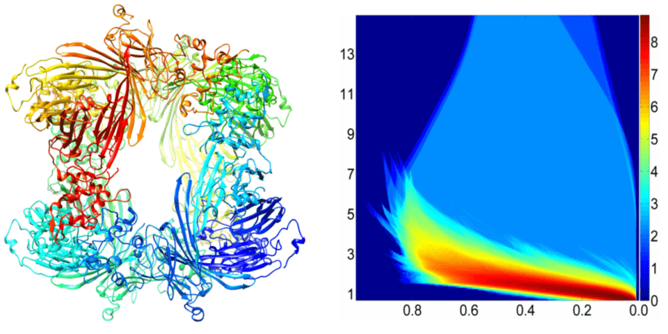 |
There have been considerable interest in developing multidimensional persistent homology or multidimensional persistence. Here, two classes of multidimensional persistence algorithms for biomolecular data are discussed. One class of multidimensional persistence is generated by repeated applications of 1D persistent homology to high-dimensional data, such as those from protein folding, molecular dynamics, geometric partial differential equations, etc. The resulting high-dimensional persistent homology is a pseudo-multidimensional persistence and has been applied to protein folding analysis to identify topological transitions [297].
Another class of multidimensional persistence is created by the geometric representation given in Eq. (131). As is an independent variable that can modify the geometry and topology of the underlying data set, one can carry out filtration with respect to both the original density and the resolution to obtain genuine 2D persistent homology [297]. Indeed, each value leads to a family of new simplicial complexes. Similarly, for an -dimensional persistent homology, -independent variables should be introduced for the filtration. Higher dimensional persistence has been demonstrated for macromolecular data [297]. Resolution induced persistent homology and multidimensional persistence have been applied to many biomolecular systems, including protein flexibility analysis, protein folding characterization, topological denoising, noise removal from cryo-EM data, and analysis of fullerene molecules. An example of multidimensional persistent homology is depicted in Fig. 12. Clearly, the maps of multidimensional persistent homology given in Fig. 12 can be employed for deep learning, which is an open field.
Basically each independent parameter that regulates the filtration process contributes a genuine persistent dimension. When the dimension is higher than two, the result representation is no longer straightforward. Additionally, how to make use of multidimensional persistence in realistic applications is also an interesting problem.
3.3 Differential geometry theory of surfaces
Differential geometry has fruitful applications in physics, particularly, general relativity and has found its success in biomolecular systems as well [21, 284, 62]. As stated earlier, in biophysical modeling, surface representation is a crucial subject and commonly used surface definitions lead to geometric singularities. Gaussian surface and general FRI rigidity surface are based on simple geometric ideas. In contrast, differential geometry theory of surfaces gives rise to natural description macromolecular surfaces [21, 284, 62]. This approach becomes powerful when it is combined with variation calculus for biomolecular modeling as shown in Section 3.4. In this section, a brief introduction is given about the differential geometry theory of surfaces using the notations and definitions from Ref. [167].
Surface elements and immersion
For an open set , a parametrized surface element is an immersion . Here is also known as a parametrization. One can call the elements of as parameters and their images under as points. If the rank of map is maximal, is an immersion. The point where the rank is not maximal, it is called a singular point or singularity [167].
One can use the following notations for a parametrized surface element . is the tangent space of at , ; is the tangent space of at , ; is the tangent space of at , ; and is the normal space of at . The element of is called tangent vector and the element of is the normal vector [167].
First fundamental form
The first fundamental form is the inner product between two tangent vectors in tangent planes , i.e., . For coordinate systems , the first fundamental form can be described by the following tensor matrix [167]
Also one can have the line element
and the surface area
where is the determinant.
Gauss map
Since each plane is essentially determined by its normal vector, the curvature of the surface can be studied by the variation of the normal vector, i.e., Gauss map. For a surface element , the Gauss map is and is defined by the formula [167]
where denotes the unite sphere .
Weingarten map
Let be a surface element with Gauss map , and for every the image plane of the linear map is parallel to the tangent plane . By canonically identifying , one can have at every point as the map . Moreover, by restricting to the image, one may view the map as a linear isomorphism . In this sense the inverse mapping is well-defined and is also an isomorphism. The map defined point-wisely by
is called a Weingarten map or the shape operator of . This map is independent of the parametrization of , and it is self-adjoint with respect to the first fundamental form I.
Second and third fundamental form
Let be a surface element with Gauss map . With the shape operator and tangent vectors and , the second fundamental is given by
and the third fundamental form is
and are symmetric bilinear forms on for every . The three fundamental forms , and have a relation as,
In coordinates , three fundamental forms can be expressed as
| (134) | |||
| (135) | |||
| (136) |
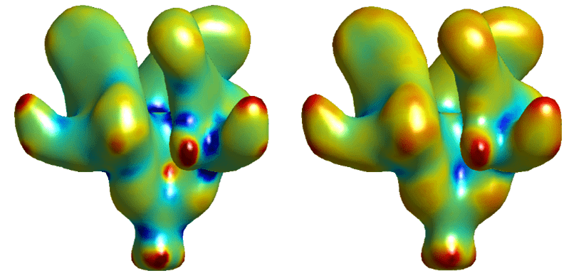 |
Principal curvature
For a unite tangent vector and , it is a principal curvature direction for if has a stationary value for , or is an eigenvalue of the Weingarten map . Further, the corresponding eigenvalue is the principal curvature.
Gaussian and mean curvature
Gaussian curvature is the determinate and mean curvature is the average value . Gaussian and mean curvatures can be expressed in local coordinates as
| (137) | |||
| (138) |
Gaussian and mean curvatures of an HIV virus fragment are illustrated in Fig. 13. In biophysics, the region with negative Gaussian is often associated membrane-protein interaction sites, while the region with negative mean curvatures on a protein surface is commonly regarded as a potential-ligand or protein-drug bonding site [291].
3.4 Differential geometry modeling and computation
With the advance of experimental technology, more than a hundred thousand of 3D macromolecular structural data has been accumulated. Differential geometry based surface modeling and curvature measurement are of essential importance to the geometric description and feature recognition of these 3D structural data [285, 302]. In this section, a brief review is given to the differential geometry based modeling of macromolecular surfaces. Two algorithms for curvature calculations are also discussed.
3.4.1 Minimal molecular surface
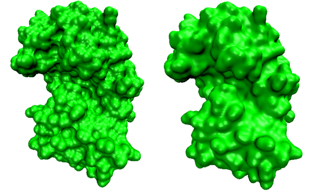 |
Minimal molecular surface (MMS) was introduced to construct surfaces free of geometric singularities via the variational principle [20, 21]. In this approach, a hypersurface is defined to represent the biomolecular surface. Basically, one assigns each point with coordinate a value , which represents the domain information. It can be viewed as a characteristic function of the macromolecular domain. By using geometric measure theory, the surface energy functional can be expressed as [284]
| (139) |
where is the surface tension. As it is convenient for us to set up the total free functional as a 3D integral in , one can make use of the concept of a mean surface area [284, 64] and the coarea formula [102] on a smooth surface
| (140) |
Here the value of hypersurface function is distributed between 0 and 1, represents the inverse function of and is defined as the whole domain. The variation of Eq. (139) with respect to leads to the vanishing of surface-tension weighted mean curvature, . The energy minimization of Eq. (139) can be realized by the introduction of an artificial time to obtain a generalized Laplace-Beltrami equation
| (141) |
The final MMS, which is free of geometric singularity, is obtained by extracting an iso-surface from the steady state hypersurface function. During each iteration, one can keep the value of in the van der Waals surface enclosed domain unchanged. Figure 14 illustrates the difference between the solvent excluded surface and the minimal molecular surface of protein 1PPL.
In the earlier works, sophisticated computational algorithms have been developed to accelerate the construction of MMSs for large biomolecules [19, 62, 63, 104]. Differential geometry based molecular surface modeling was extended to solvation analysis [284, 62], including level set approaches [65, 66], and ion channel transport [284, 288, 286, 57]. Numerical aspects were examined in the literature [311, 312]. Since these approaches work very well for biomolecular structure, function and dynamics, they will attract much attention in the future. However, a more detailed discussion of these issues is beyond the scope of the present review.
3.4.2 Scalar field curvature evaluation
For a given set of volumetric biomolecular data, the efficient and accurate computation of curvatures is needed. The evaluation of curvature properties from iso-surface embedded volumetric data has been studied in geometric modeling, although the related techniques have not received much attention in computational biophysics. In this section, two popular algorithms are reviewed. Many other elegant methods, including isophote surface based curvature evaluation [269], Sander-Zucker approach [242], Direct surface mapping based approach [257], piece-wise linear manifold techniques [257], etc, are often used in the computer science community.
Algorithm I
Essentially, the first and second fundamental forms in the differential geometry are involved in the definition and evaluation of the curvatures. a brief discussion of the mathematical background is given [254, 21].
The surface of interest can be extracted from a level set with iso-value , i.e., . One can assume to be non-degenerate, i.e., the norm of the gradient is non-zero at . Without loss of generality, one can assume that the projection onto is non-zero as well. Then the implicit function theorem states that locally, there exists a function , which parametrizes the surface as . One can express the iso-value relation as . The differentiation with respect to and variables leads to two more equations
where and can be given by
One can define and below to be the coefficients in the first and second fundamental forms. For simplicity, one can hide parameter labels. Their values for surface function can be given as
The Gaussian curvature can be expressed as the ratio of the determinants of the second and first fundamental forms,
Similarly, the mean curvature is given as the average second derivative with respect to the normal direction,
| (142) |
Note that curvature expressions become analytical when the implicit surface is given by the FRI rigidity density, Eq. (131).
Algorithm II
An alternative algorithm for the curvature extraction from volumetric data is the Hessian matrix method [159]. For volumetric data , one defines the surface gradient and surface norm .
| (143) | |||
| (144) |
Here denoting the transpose. One further calculates the matrix , which can be expressed as
| (145) | |||
where is the identity matrix, matrix and Hessian matrix is given by
| (149) |
Geometrically, project onto a one-dimensional span of . Here further projects onto the orthogonal space complement to the span of , which is the tangent plane.
In general, both and are symmetric but is not. If lies in the tangent plane, and . Therefore, for and in the tangent plane, one has
| (150) |
The restriction of to the tangent plane is symmetric and thus there exists an orthonormal basis for the tangent plane in which is a 2*2 diagonal matrix. This basis can be easily extended to an orthonormal basis for all . In this basis, the derivative of the surface normal is given by
| (154) |
The diagonal term in the bottom row is zero because no change in normal can lead to a change in length. Motion along and results in the change of along the same directions, with a ratio of and respectively. Here and are the principal curvature directions, while and are the principal curvatures. When there is a change in normal , it tilts according to and .
One can further multiply by to diagonalize the matrix
| (161) |
The surface curvature measurements are based on geometry tensor . In a volumetric data set or a scalar field, is known in terms of the Cartesian basis . Matrix invariants provide the leverage to extract the desired curvature values and from , regardless of the coordinate frame of the principal curvature direction. The trace of is . The Frobenius norm of , notated and defined as , is . Then and are found with the quadratic formula.
The two principal curvatures can be evaluated by the following procedure.
-
1.
Calculate matrix
-
2.
Evaluate matrix ,
(162) -
3.
Compute the trace and Frobenius norm of matrix ;
(163) (164) (165) (166)
When the two principal curvatures are available, the Gaussian curvature and mean curvature can be obtained as
| (167) | |||
| (168) |
Essentially, the Hessian matrix method generates the same results as Algorithm I derived from the first and second fundamental form. However, for FRI rigidity density given in Eq. (131), Algorithm I is preferred as it is analytical and without matrix diagonalization.
3.4.3 Analytical minimal molecular surface
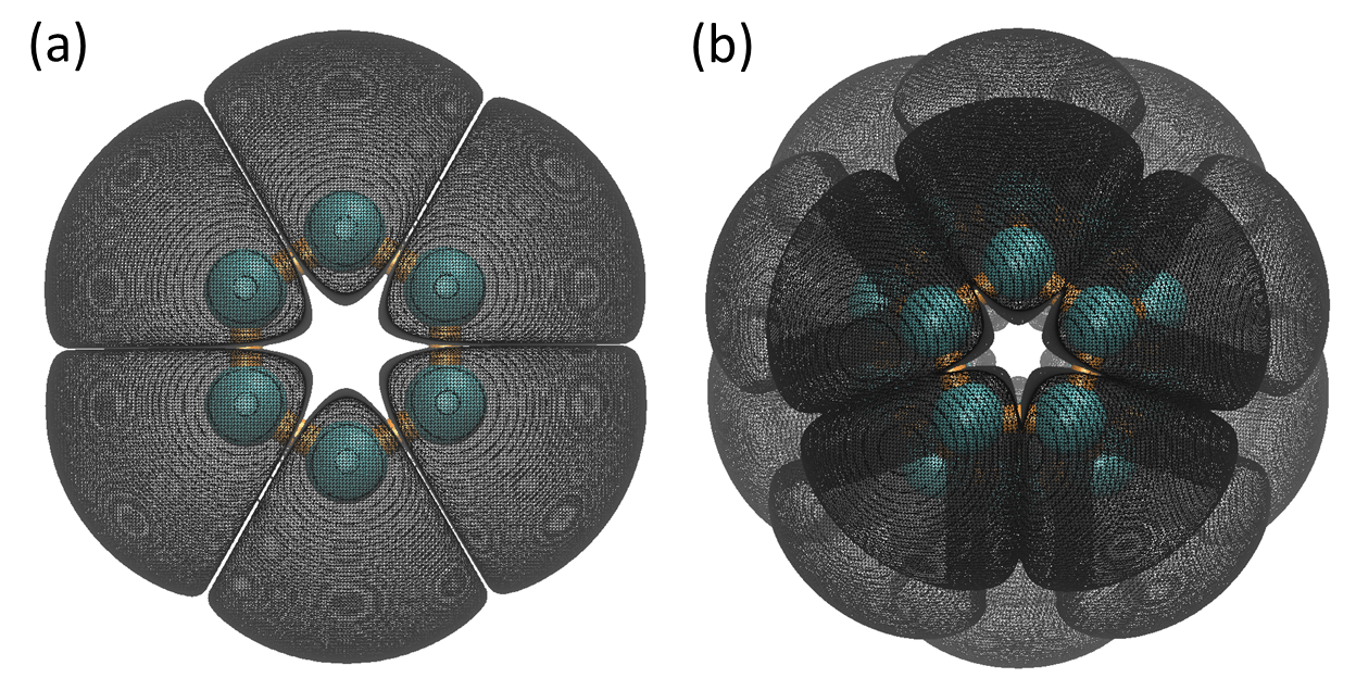 |
 |
Minimal surfaces are ubiquitous in nature and are a fascinating topic for centuries. The Euler-Lagrange minimization of the hypersurface was proposed to generate minimal molecular surface (MMS) for biomolecules [21]. In general, MMS is a result of vanishing mean curvature and is free of geometric singularity. It is a powerful concept in biophysical modeling [21, 284]. Nevertheless, it is still much more computationally expensive to construct MMS than to generate SES. In this work, a new minimal molecular surface, called analytical minimal molecular surface (AMMS) is proposed. In this approach, the rigidity density representation of biomolecular data by Eq. (131) is employed. Then the mean curvature can be analytically computed by using Eq. (142). Finally, AMMS can be constructed by setting the isosurface of the mean curvature to zero (or practically, a number very close zero).
One can consider the hexagonal ring and a fullerene C20 molecule in the study of AMMS. The coordinates used for particles in the hexagonal ring are (0.000, 1.403, 0.000; -1.215, 0.701, 0.000; -1.215, -0.701, 0.000; 0.000, -1.403, 0.000; 1.215, -0.701, 0.000; and 1.215, 0.701, 0.000). The fullerene C20 molecule has a highly symmetric cage structure made of 12 pentagons. Its atomic coordinates are (1.569, -0.657, -0.936; 1.767, 0.643, -0.472; 0.470, -0.665, -1.793; 0.012, 0.648, -1.826; 0.793, 1.467, -1.028; -0.487, -1.482, -1.216; -1.564, -0.657, -0.895; -1.269, 0.649, -1.277; -0.002, -1.962, -0.007; -0.770, -1.453, 1.036; -1.758, -0.638, 0.474; 1.288, -1.450, 0.163; 1.290, -0.660, 1.305; 0.012, -0.646, 1.853; 1.583, 0.645, 0.898; 0.485, 1.438, 1.194; -0.503, 0.647, 1.775; -1.606, 0.672, 0.923; -1.296, 1.489, -0.166; -0.010, 1.973, -0.006). The fullerene rigidity density is given by Eq. (131). In both cases, the parameters and are set to and , respectively.
When the mean curvature isovalue equals to zero, the resulting surfaces are usually composed by several non-intersecting surfaces perpendicular to atomic bonds near the BCPs. When loosing the condition a little bit by setting the isovalue to 0.001 (or some other very small positive value), a better AMMS can be generated. Figure 16 depicts the AMMS for the hexagonal ring and C20 molecule. It is found that for the hexagonal ring, each atom is enclosed by a surface segment. These segments are tightly close to each other and only connect near BCPs. For the C20 molecules, the surface appears to be better connected. However, a careful examination reveals the separation as well. Obviously, these surfaces are not minimal surfaces.
To generate better understanding of the minimal molecular surface generation, one can explore several values for the hexagonal ring. Figure 16 illustrates the results. From (a) to (d), is chosen as 0.5, 0.6, 0.7 and 0.8, respectively. All the isosurfaces are extracted at mean curvature equals to 0.001. It is seen that a relatively large value produces a better minimal surface. The gaps between atomic segments are gradually narrow as the increase of value and finally disappear, which gives rise to a good quality AMMS.
3.5 Scalar and vector field topology
Topological approaches have become an integral part in data analysis, visualization, and mathematical modeling for volumetric as well as for vector valued data sets. In fact, the results of scalar and vector field topology coincide each other for gradient vector fields, although the respective mathematical approaches are originated from different fields. Vector field topology is usually developed for analyzing the streamlines of fluid flow generated by the velocity vector during the flow evolution. These techniques can be applied to macromolecular analysis for dealing with cryo-EM data and improving the structural construction.
Three dimensional scalar field data, particular the molecular structural data are widely available from many data sources. To decipher useful information from these data requires highly efficient methods and algorithms. Mathematical approaches, including topological tools, such as fundamental groups, homology theory, contour tress, Reeb graphs [93], Morse theory [132, 199], etc, have a great potential for revealing the intrinsic connectivity or structure-function relationship. Topological data analysis (TDA) has gained much attention in the past decade. The traditional atoms in molecules (AIM) analysis [11, 12] can be viewed as an application of TDA to electron density analysis. Historically, the topological study of scalar fields, particularly electron densities, has advanced the understanding of molecules and their chemical and biological functions tremendously. The theory of AIM developed by Bader and his coworkers has provided an elegant and feasible approach to define atoms in molecule. AIM can be generalized into a more general theory called quantum chemical topology (QCT), which applies the topological analysis in AIM to the study of other physically meaningful scalar fields [229]. Mathematically, this topological analysis used in AIM is known as Morse theory. In general, all scalar fields used in QCT are some kind of Morse functions and more can be proposed as long as they satisfy the Mores function constraints. On the other hand, geometric modelings and analysis contribute a lot to the molecular visualization and structure-function relationship. Various surface definitions from geometric modelings facilitate the visualization and characterization of molecules. Geometric analysis has contributed enormously to the understanding of structure-function relationship. Among the geometric analysis, surface curvature analysis has provide great insights into solvation analysis, protein-protein interaction, drug design, etc. [291, 209]
In the QCT theory, researchers explore atomic or molecular properties through the analysis of various properties at critical points. In geometric analysis, curvature estimation is normally done on a special surface, such as SES, SAS, Gaussian surface, FRI rigidity surface, etc. Even though these two approaches are very efficient and powerful in capturing and characterizing atomic and molecular information, enormous information embedded in the electron density scalar field has not been fully utilized. Question is how to improve geometric and topological analysis for biomolecules. Note that in persistent homology analysis, physical properties are analyzed by a systematic filtration process. Therefore, for a given scalar field, a better understanding can be achieved if the topological analysis includes not only some critical points or a special iso-surface, but also a series of iso-surfaces derived from a systematic evolution of iso-values. Further, more information can be extracted if one considers not only simply scalar fields, such as electron density, electron density Laplacian, etc, but also geometric properties like individual eigenvalues and various curvatures. These approaches are developed in the present work.
In the following, a brief review of some very basic concepts in AIM is given. Their connection with mathematical theories is discussed. Then, some new approaches are presented. Examples of applications are provided.
3.5.1 Critical points and their classification
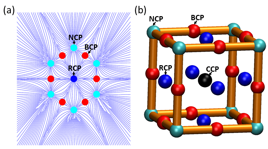 |
Three dimensional molecular electron density data can be collected through experimental tools, like electron microscopy, cryo-electron microscope, etc and also from theoretical models in quantum mechanics. One can denote electron density function and its domain . A quantitative method to analyze the topology of is to consider its first derivative, i.e., gradient . At certain points, called critical points (CPs), this gradient vanishes. The characteristics of these points is determined by the second derivatives, which form the Hessian of electron density .
By diagonalization of the Hessian matrix, one can obtain three eigenvalues . Their sum equals to the Laplacian of electron density . That is, locally [12]
| (169) |
It can be noticed that the Hessian matrix is symmetric, therefore all the eigenvalues are real. Based on the positive and negative sign of these three eigenvalues, one defines rank and signature to characterize critical points. The rank of a CP is the number of non-zero eigenvalues, and a signature is the algebraic sum of the signs (+1 or -1) of the eigenvalues. In general, a CP is non-degenerate, meaning its three eigenvalues are non-zero (Rank). A degenerate critical point is unstable in the sense that even a small change in the function will cause it either to vanish or bifurcate into a non-degenerate CP.
Based on the rank and signature value, non-degenerate CPs in three dimensional scalar field can be classified into four basic types, namely, nucleic critical point (NCP), bond critical point (BCP), ring critical point (RCP) and cage critical point (CCP). Table 1 lists these values. An illustration of these four types of CPs can be found in Figure 17. An NCP is a nucleic center of an atom. It is represented by a cyan-color point. A BCP is a bond center between two atoms and is marked with a red-color point. A RCP is usually found at the center of a ring structure and is colored in blue. A CCP is known as a cage critical point, and can only be found in the center of a cage structure. In general, an NCP is a local maximal point. BCP and RCP both are saddle points. A CCP is a local minimal point. In Sections 3.6.1 and 3.6.2, it will be demonstrated that these properties can be used to systematically characterize and analyze the eigenvalue and curvature isosuface information.
Topologically, the general structure connectivity can be characterized by the number of CPs. This is stated in the famous Poincaré-Hopf theorem as following:
| (170) |
where , , and are numbers of NCPs, BCPs, RCPs and NCPs, respectively. Here is the Euler characteristic. The Poincaré index is defined on a vector field. In the AIM theory, one studies the gradient vector field of electron density. Poincaré indices for NCP, BCP, RCP and NCP are and , respectively. More properties of this gradient vector field are discussed in the following section.
Equation (170) can also be derived from simplicial complex analysis [296]. Essentially, NCP can be viewed as a point (0-simplex); BCP corresponds to a straight line (1-simplex); RCP is for a polygon (2-simplex); CCP is then for a polyhedron (3-simplex). In this manner, Euler characteristic can be directly employed and the above equation can be obtained as well.
| Rank | Signature | Poincaré index | Simplex | Property | |
|---|---|---|---|---|---|
| NCP | 3 | 1 | 0-simplex | local maxima | |
| BCP | 3 | 1-simplex | saddle | ||
| RCP | 3 | 1 | 1 | 2-simplex | saddle |
| CCP | 3 | 3 | 3-simplex | local minima |
3.5.2 Vector field topology
The gradient on the entire domain forms a vector field. The topological property of this vector field can be explored by tools borrowed from dynamic systems or the mathematical analysis of fluid flows. These tools include integral line, separatrix, and Poincaré index, etc.
An integral line is a curve of a function that satisfies . Its origin and destination can be defined as
and
Integral lines satisfy the following conditions:
-
1.
Two integral lines are either disjoint or the same.
-
2.
Integral lines cover all the manifold.
-
3.
The limits, org and dest, are critical points.
In general, integral lines represent the gradient flow between critical points. All the integral lines that share the same form a region called atomic basin. The whole electron density domain is subdivided into many atomic basins. The interface between these attraction basins is called inter-atomic surface (IAS). Mathematical, IAS is the separatrix of a gradient vector field. Each attraction basin includes one and only one atom. This is known as the quantum topological definition of an atom in a molecule.
IAS also satisfies the zero-flux condition . Here is a gradient vector and is the normal vector to the IAS.
Morse theory
It should be noticed that basic concepts like critical point, degeneracy, critical point classification, basin, etc. are the essential part of a mathematical tool called Morse theory [132, 199]. In general, the atom in molecular method can be viewed as an application of Morse theory in molecular density field analysis. More specifically, various critical points, including NCP, BCP, RCP, and CCP, are the counterparts of peak point, saddle-1 point, saddle-2 point and valley point, respectively. The separatrix of a gradient vector field just provides a Morse decomposition of the underlying molecular scale field manifold [132, 199].
3.5.3 Topological characterization of chemical bonds
Chemical properties of molecular systems are profoundly determined or influenced by their atomic covalent bonds and noncovalent interactions. Usually, covalent bonds are much stronger and determine the structural integrity of a molecule. Noncovalent interactions are comparably weak but play important roles in macromolecular assembly, protein folding, macromolecular function, etc. Traditionally, the Laplacian of electron density can be used to interpret noncovalent interactions of a molecular system. Recently, signed electron density and reduced gradient, two scalar fields derived from electron density, have drawn much attention in quantum chemistry since they enable a qualitative visualization of noncovalent interactions even in complex molecular systems [150, 76, 75, 120, 128]. These approaches are reviewed below.
The Laplacian of electron density
The Laplacian of electron density can be used to indicate the electron density concentration and depletion. Essentially, the density is locally concentrated where , and locally depleted where . In this manner, if one defines the function , the maximum in denotes the maximum in the concentration of the density. The minimum in implies a depletion of density. The Laplacian of the electronic charge distribution, , demonstrates the presence of local concentrations of charge in the valence shell of an atom in a molecule. These local maxima faithfully duplicate in number, location, and size of the spatially localized electron pairs of the valence shell electron pair repulsion (VSEPR) model. Thus the Laplacian of the charge density provides a physical basis for the Lewis and VSEPR models [13].
Identifying noncovalent interactions
The study of the Hessian matrix and its three eigenvalues has yielded many intriguing results [12]. It has been found that all eigenvalues (, and ) are negative in the vicinity of the nuclei centers. Away from these centers, the largest eigenvalue becomes positive, and varies along the internuclear axis representing covalent bonds. It is also found that and describe the density variation orthogonal to this internuclear axis. More specially, is always negative even it is away from the nuclei. While can be either positive, meaning attractive interactions concentrating electron charge perpendicular to the bond, or negative, meaning repulsive interactions causing density depletion. Using this localized information, the signed electron density is defined as
| (171) |
The signed electron density additionally enables the differentiation of attracting and repulsive interactions.
To further reveal weak noncovalent interactions, the reduced gradient is introduced as following [120, 128]
| (172) |
The reduced density gradient describes the deviation in atomic densities due to interactions and has found interesting applications in in analyzing biomolecular structure and function [150, 120, 128].
3.6 Geometric-topological (Geo-Topo) fingerprints of scalar fields
3.6.1 Geo-Topo fingerprints of Hessian matrix eigenvalue maps
The QCT and AIM analyses of molecules are limited to special locations and given iso-surfaces. In this work, the Hessian eigenvalue analysis of a molecular density is considered, not only for a few critical points, but also for the whole domain. Additionally, the topology over a series of isosurfaces derived from a systematic evolution of isovalues are analyzed. More specifically, in the topological persistence of Hessian matrix eigenvalues, the topological properties of a series of isosurfaces, generated by varying the isovalue from the smallest to the largest, are systematically studied. Therefore, the isosurface value behaves like a filtration parameter. The topological transitions in this series of isosurfaces are emphasized. Another very important aspect of this analysis is to analyze the behavior of isosurfaces through its relation with the four types of CPs. It should also be noticed that these methods are greatly different from other interatomic surface methods [222, 228], as these models focus on a special surface.
 |
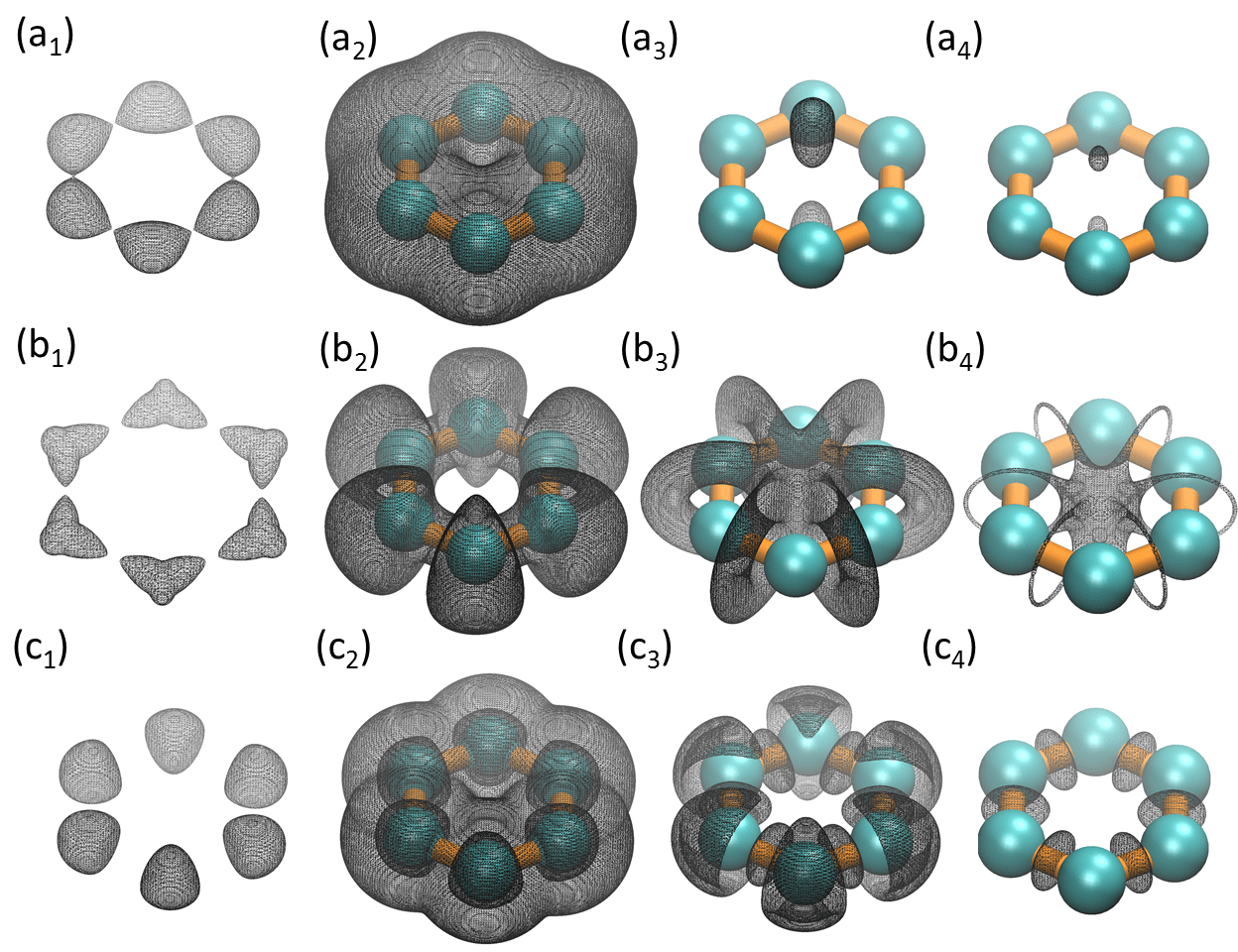 |
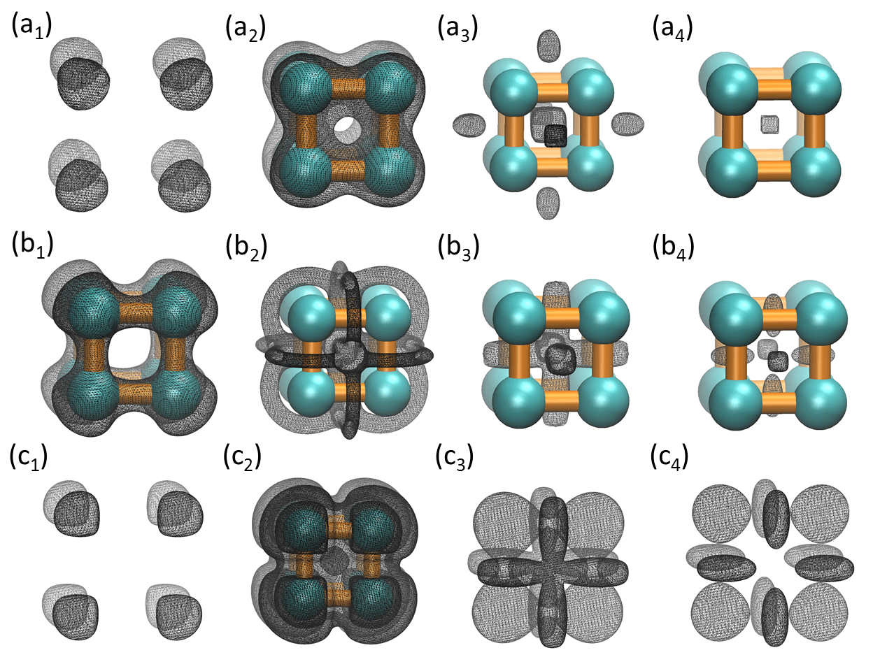 |
To illustrate the idea, two toy models, i.e., the hexagonal ring discussed earlier and a cubic structure are considered. The coordinates for the cubic structure are set to (1.245, 0.537, -0.073; 0.924, -0.995, 0.024; -0.123, -0.704, 1.155; 0.199, 0.828, 1.058; 0.123, 0.704, -1.155; -0.924, 0.995, -0.024; -1.245, -0.537, 0.073; and -0.199, -0.828, -1.058). The discrete to continuum mapping, Eq. (131), is used to generate rigidity density. The parameters and are chosen as Å and for all particles. In this approach, Hessian matrix is evaluated at each point of the computational domain and its eigenvalue is obtained everywhere as well, which forms an eigenvalue scalar field.
To have a general idea of the basic distribution of eigenvalues, one can study the Hessian matrix eigenvalue behavior of the hexagonal ring in a two-dimensional plane , as all its particles are located within this special plane. Results are illustrated in Fig. 18. Three eigenvalues, i.e., , and , are demonstrated in subfigure (a), (b) and (c), respectively. It can be seen that for regions near NCPs, all three eigenvalues are negative. For regions near BCPs, is always negative. While is negative in the very closed neighborhood and gradual increases to be positive further away. Finally, is always positive. For regions near RCPs, all three eigenvalues are positive. These results are consistent with findings in AIM [12].
To obtain more geometric insights, particularly eigenvalue behaviors on all four types of CPs, the cubic structure is considered. In this case, the CCP can be identified as having the positive and .
As stated above, this approach is to extract a series of isosufaces of Hessian matrix through a filtration process. One can carefully observe these eigenvalue isosurface patterns to detect topological transitions. The geometric information is further analyzed and its relation with the four types of CPs is summarized into several unique characteristics, called Geo-Topo fingerprints. Using hexagonal ring and cubic structure models, the Geo-Topo fingerprints for eigenvalues are revealed.
Figures 20 and 20 demonstrate four unique patters for each eigenvalue. The subscripts to indicate four representative eigenvalue isovalues, from the small to the large. The notations , and represent eigenvalue , and , respectively. As stated above, the filtration process delivers a series of isosurfaces. To avoid confusion, the filtration process always goes from the smallest value to the largest one. Only four representative isosurfaces that capture some unique topological features within the eigenvalue isosurface series, are selected. Through the comparison of these patterns, some common features can be extracted.
-
1.
For all the three eigenvalues, regions around NCPs are always concentrated with negative values.
-
2.
For , as the filtration goes, negative isosurfaces first appears near NCPs. Then they enlarge to incorporate regions near BCPs, being still negative. The regions near RCPs can be analyzed from two different perspectives: i.e., along the ring plane and perpendicular to the ring plane. Along the ring plane, values gradually increase to about 0 as the eigenvalue isosurface propagate to RCPs. There is a sudden topological transition for the isosuface when its value passes through 0. When becoming positive, eigenvalue isosurfaces form ellipsoids surfaces perpendicular to the ring plane near all RCPs. These ellipsoids usually appear in pairs and symmetric to each other along the ring plane as indicated in Fig. 20 . Finally, positive isosurface appears near the regions of CCPs.
-
3.
For , as the filtration process goes, negative isosurfaces first appear near NCPs and then enlarge to incorporate regions near BCPs just like . However, positive isosurfaces appear much earlier. They occupy the regions around RCPs and CCPs. More interestingly, they form a loop around each bond near its BCP. To be more precise, these loops are perpendicular to atom bonds at BCPs and atom bonds pass through them at their centers. The isosurfaces gradually shrink and reduce to regions around RCPs as their values grows.
-
4.
For , its negative isosurfaces concentrate only in regions around NCPs. Positive isosurfaces form a sphere slice around each atom. Isosurface with relative small positive value also appears around the RCPs and CCPs. As the filtration goes further, positive isosurfaces concentrate around regions near BCPs.
3.6.2 Geo-Topo fingerprints of curvature maps
In this work, the topological analysis of curvature maps are developed. One can still consider the hexagonal ring and the cubic structure discussed in the last section. The discrete to continuum mapping is carried out to generate the FRI rigidity density. Then curvatures are evaluated at all of the molecular FRI rigidity isosurfaces (or every point in the computational domain) to form a curvature map. At each curvature isovalue, topological analysis is applied. Here, topological analysis is twofold. One type of topological analysis is to identify topological critical points (i.e., 0-simplex, 1-simplex, 2-simplex, etc.). The other type is to carry out persistent homology analysis to track topological invariants during the density filtration of the curvature map.
Gaussian and mean curvature maps
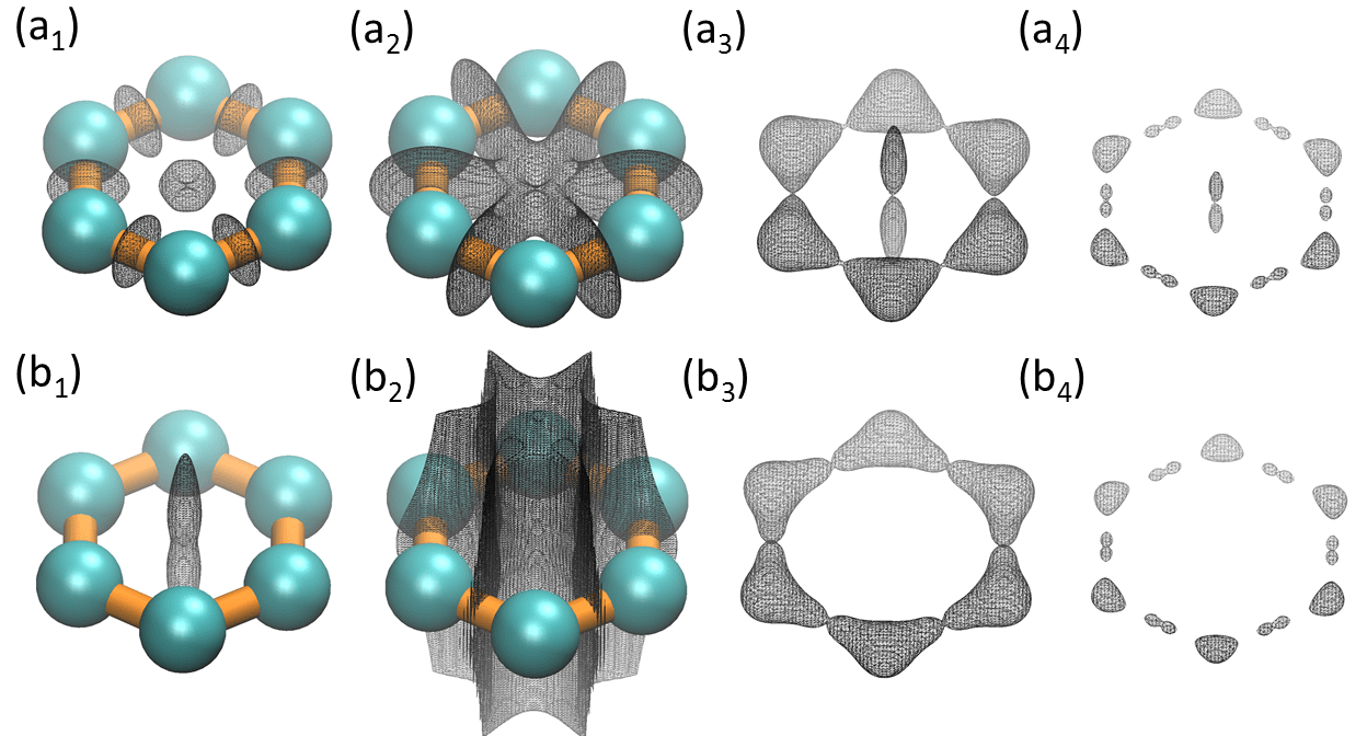 |
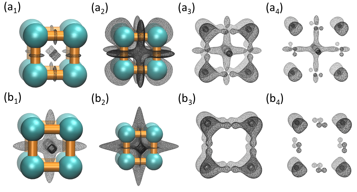 |
It is seen from the above analysis that with four types of CPs, Geo-Topo fingerprints are extracted and the behaviors of the Hessian matrix eigenvalue isosurfaces can been characterized very well. However, before the employment of this technique in Gaussian and mean curvature isosurface analysis, it is helpful to review a little bit more the four types of CPs. As stated in Section 3.5.1, NCPs and CCPs are locally maximal and locally minimal, respectively. Actually, for NCPs, all three eigenvalues have negative signs, while for CCPs, all three eigenvalues have positive signs. BCPs and RCPs are saddle points, which means that their eigenvalues have both positive and negative values. Geometrically, the curvature is isotropic near NCPs and CCPs, but anisotropic near BCPs and RCPs. So in this analysis, the isosurfaces near BCPs and RCPs can be divided into two types, namely, A-type or V-type. A-type isosurface means it is along atomic bonds or ring planes. V-type isosurface means it is vertical or perpendicular to the atomic bonds or ring planes. For instance, the isosurface near the RCP in Fig. 22 () is an A-type, but it becomes a V-type in Fig. 22 (). Another important property is that A-type and V-type isosurfaces are usually with different signs. That is, if A-type of isosurface is obtained from a positive isovlaue, the associated V-type isosurface can only be obtained from a negative isosurface. With this notation, one can extract some Geo-Topo fingerprints for Gaussian and mean curvatures. The basic results are summarized as below.
-
1.
For a Gaussian curvature field, it has negative A-type isosurfaces near RCPs and negative V-type isosurfaces near BCPs. In contrast, positive isosurfaces enclose regions near BCPs and CCPs. Positive V-type isosurfaces can be found in RCPs and positive A-type isosurfaces are found near BCPs. Finally, positive isosurfaces are found in NCPs.
-
2.
For a mean curvature field, it has negative isosurfaces near RCPs and CCPs. Negative V-type isosurfaces can be found near BCPs. Positive isosurfaces are found in NCPs. Finally, A-type positive isosurfaces can be found near BCPs.
Maximal and minimal curvature maps
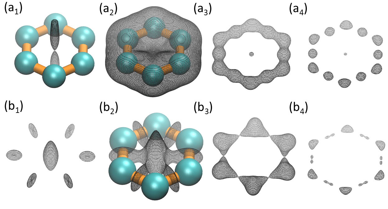 |
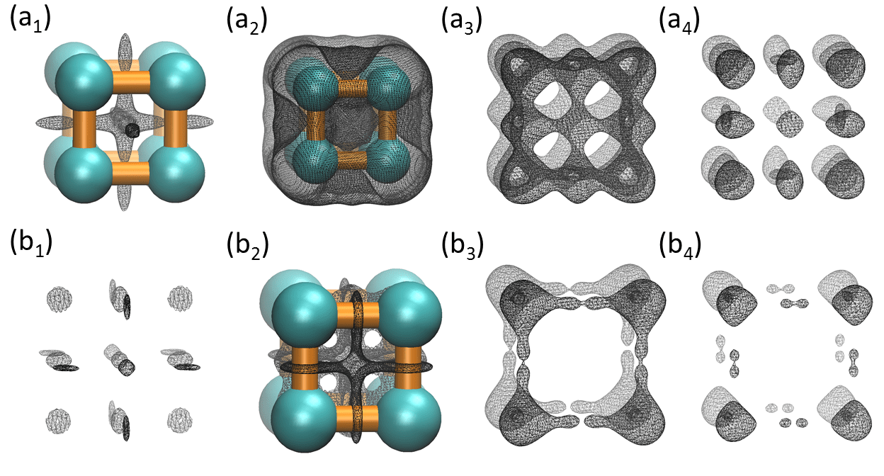 |
One can also carry out a throughout investigation of maximal and minimal curvature maps. Isosurfaces near NCPs, BCPs, RCPs and CCPs are analyzed in the same manner as they were done in the above two sections. Figures 24 and 24 illustrate results for the hexagonal ring and the cubic structure, respectively. Main results about their Geo-Topo fingerprints are summarized below:
-
1.
For maximal curvature map, it has negative isosurfaces near CCPs and negative V-type isosurfaces near RCPs. Positive isosurfaces enclose regions near BCPs and NCPs. Positive A-type isosurfaces can be found in RCPs.
-
2.
For minimal curvature map, it has negative isosurfaces near RCPs and negative V-type isosurfaces near BCPs. Negative isosurfaces also enclose region near CCPs. Positive isosurfaces are found in NCPs. Finally, A-type positive isosurfaces can be found near BCPs.
It can be noticed that all curvature representations, i.e., Gaussian curvature, mean curvature and two principal curvatures, are quite different from Hessian matrix eigenvalue distributions. In general, negative curvatures usually do not occur near NCPs, this is particularly true for maximal and mean curvatures. They are more likely to appear near BCPs, RCPs and CCPs. In contrast, positive isosurfaces are concentrated in the atomic basin (i.e., NCPs).
| NCP | BCP | RCP | CCP | |
|---|---|---|---|---|
| N | N | P | P | |
| N | N; P-Loop | P | P | |
| N | P | P | P | |
| P | A-type (P); V-type (N) | V-type (P); A-type (N) | P | |
| P | A-type (P); V-type(N) | N | N | |
| P | P | A-type (P); V-type (N) | N | |
| P | A-type (P); V-type (N) | N | N |
To have a more general understanding of the properties of eigenvalue and curvature maps, all of the above-mentioned results are summarized in Table 2. To show the consistency of results, one can take some simply tests. For instance, one can compare the results of and with . Since , their signs of isosurface at four types of CPs should match with each other. For NCP, and are both positive, and multiplying together yields a positive , exactly as it is found in the table. For all other three types of CPs, they all match very well.
3.7 Eigenvector field analysis
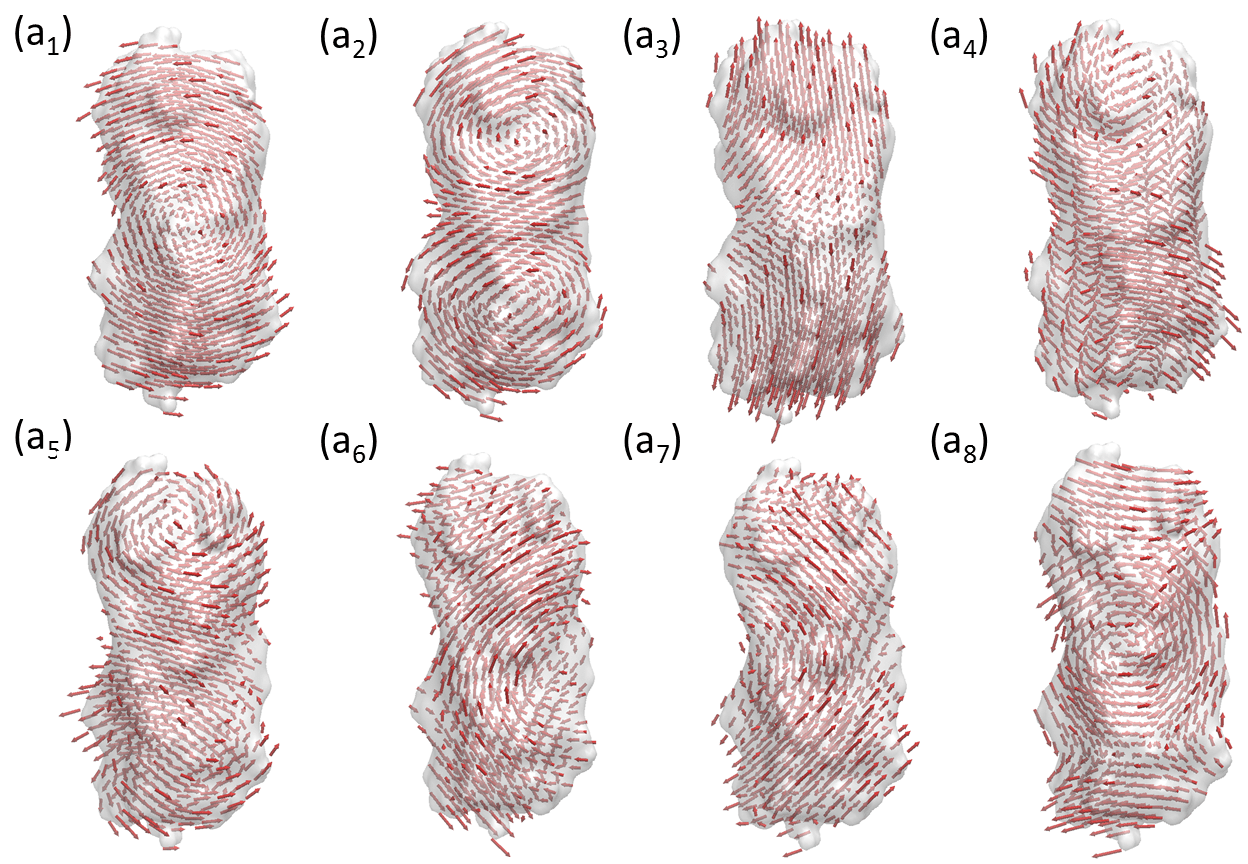 |
Tensor fields widely exist in natural world. For biomolecules, researchers are particular interested in their motions. As stated in the previous sections, various methods, including molecular dynamics, anisotropic network model (ANM), anisotropic FRI (aFRI), etc, have been developed to explore the dynamics of the biomolecular systems. However, all the above mentioned approaches rely on the discrete representation and can not be used in the study of motions of continuous biomolecular density function or data, particularly electron density data, such as cryo-electron microscopy (cryo-EM) maps. To analyze the motion of density profiles, a virtual particle model (VPM) is proposed to systematically explore the anisotropic motions of continuous density.
3.7.1 Virtual particle model
Both ANM and aFRI depend on their graph or discrete representation of biomolecules. It is not obvious how to construct a continuum model for a continuous density function to characterize its anisotropic motions. Previously, vector quantization (VQ) algorithm [127] is employed to decompose the electron density map of a biological molecule into a set of finite Voronoi cells. It is then combined with ANM to explore the dynamic of the cryo-EM data [263, 197].
In this section, virtual particles are introduced and defined for each small volume or element of a density data. To be more specific, the domain of the density function can be discretized into many elements. In general, the discretization can be non-uniformed and the elements may have different sizes. One can associate each element with a virtual particle, which is centered at the element center but having a continuous density profile. One can assume that all virtual particles are correlated with each other, but the correlations between them decrease with the distance or follow prescribed relations. The anisotropic motions of virtual particles are obtained. Similar to ANM and aFRI approaches, such anisotropic motions are evaluated from the eigenmodes of the anisotropic Kirchhoff matrix.
One can assume that the density function of interest is given by , which can be either a cryo-EM density map or a rigidity density computed from atomic coordinates by using the discrete to continuum mapping as shown in Section 3.1. One can consider two virtual particles centered at and and enclosed by the volume elements of and , respectively. A special scaling parameter is proposed as following:
| (173) |
where the coefficient is a normalization factor, is a FRI correlation function and is the characteristic length of elements. In contrast, the in the discrete to continuum mapping is the characteristic length of atomic distances.
Three realizations of VPM by constructing three anisotropic Kirchhoff matrices are proposed. First, one can modify ANM to construct a VPM anisotropic Kirchhoff matrix. For each matrix element , a local Hessian matrix for ANM in Eq. (34) is formed as
| (177) |
The diagonal elements are constructed following Eq. (35). The test indicates this ANM based VPM works very well. However, the demonstration of this test is skipped.
Additionally, one can modify the aFRI to generate two other realizations of VPM. It is very natural for one to derive the continuous aFRI by making use of the local anisotropic matrix defined in Eqs. (64) and (65). However, in general applications, the correlation can be more specifically defined to describe the interaction between each pair of virtual particles. To construct the final matrix, one can multiply the scaling parameter to the local flexibility Hessian matrix and , the corresponding two generalized Hessian matrices can be expressed as following:
| (178) | |||||
| (179) |
and
| (180) | |||||
| (181) |
where denotes the adjoint of matrix. Here is a matrix with every element being one.
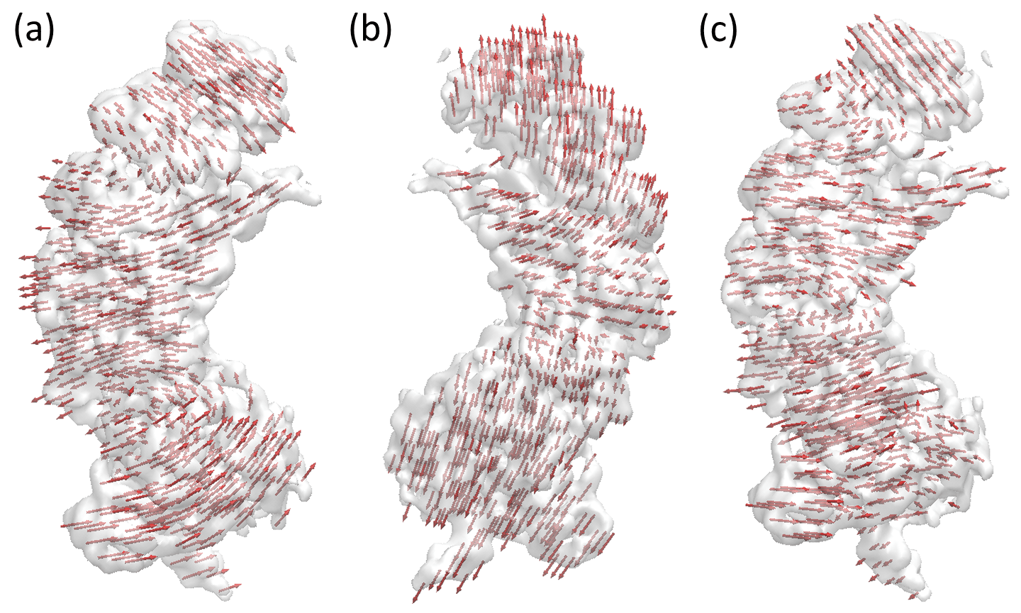 |
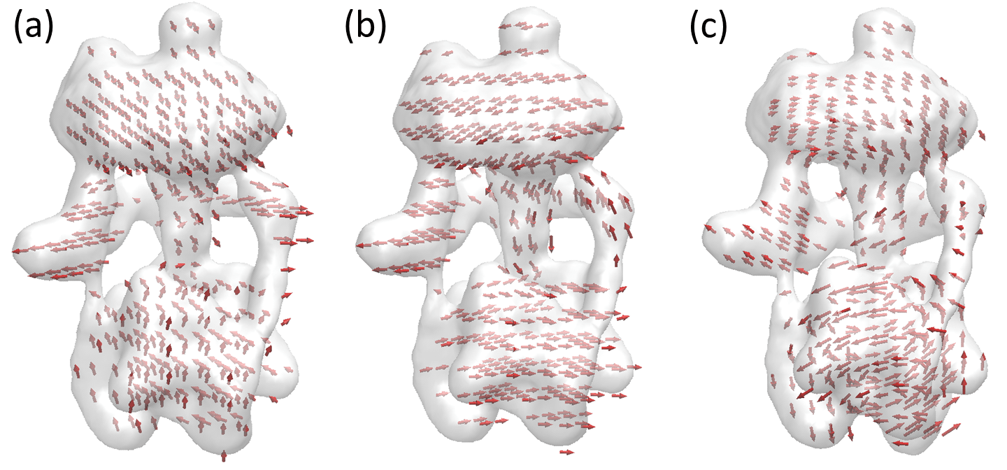 |
It should be noticed that clustering idea in the discrete aFRI matches with the idea of discretization of the continuous density function very well. However, only the simple global form is highlighted in Eqs. (178) and (180).
The proposed model is tested to evaluate the anisotropic motion of density volumetric data. One only needs to consider those density elements in the anisotropic motion analysis that their density values are larger than a threshold, which is the suggested value for biomolecular visualization. This truncation dramatically reduces the number of entries in final Kirchhoff matrix.
It has been verified that the VPM is able to recover ANM and aFRI when the elements are very small and each element contains at most one real particle (say a Cα). The demonstration of this result is omitted.
The proposed method is illustrated with three examples, i.e., protein 2XHF, cryo-EM maps EMD8295 and EMD3266. For protein 2XHF, the original data contain discrete atomic coordinates. The discrete to continuum mapping is used to generate rigidity density . In this transformation, the Lorentz kernel as in Eq. (39) is chosen with and Å. To construct VPM Hessian matrix, a threshold is chosen to be of the maximal density value. One can discretize the computational domain by using the element size of 3.0 Å. In the mode analysis, the second aFRI form is used. The Lorentz kernel in Eq. (39) is chosen with and Å. Mode 4 to mode 11 are illustrated in Fig. 25. The results are similar to those obtained with discrete aFRI [210].
For cryo-EM density map EMD8295, the data have a dimension of Å3. A mesh of 40*40*40 is used to discretize domain. The threshold of 0.08 is used to result in a total number of 639 nonzero elements in the matrix. Again the second aFRI form with the Lorentz kernel is used. The parameter used are and Å. Modes 4, 5 and 6 are illustrated in Fig. 26.
For EMD1590, the region is of the size Å3. A mesh of 25*25*25 is employed for the discretization. The biomolecular domain is chosen by using the threshold of 0.05 and there are 394 nonzero elements in the final matrix. The second aFRI form with the Lorentz kernel is used with and Å in matrix construction. Modes 4, 5 and 6 are illustrated in Fig. 27.
It can be noticed that VPM can be applied to other systems, such as stability analysis of cells, tissues, and some elastic systems with appropriate definitions of correlations functions. However, this aspect is beyond the scope of the present review.
3.7.2 Eigenvector analysis
Mathematically, vector field can be analyze by Poincaré index [227], winding number, Morse index [132, 199] and more interestingly, the Conley index [73, 200, 58, 192]. The essential idea of all these methods is to explore the behavior of the vectors around critical points.
To give a brief introduction of Conley index, one can consider a manifold and its associated vector field. If one expresses the vector field in terms of a differential equation . The solution can be expressed as a function . This solution is a flow that satisfies for all . One can also define the trajectory as . With this setting, an invariant set is . Two basic types of invariant sets are fixed points and periodic points, see Table 3. One can define an isolating neighborhood as for every , there exists , such that one has either or . The exit set of an isolating block is . The pair is called an index pair. The Conley index of an invariant set is the relative homology of the index pair , i.e., .
| Cases | Conely index |
|---|---|
| Attracting fixed point | (1 0 0) |
| Saddle fixed point | (0 1 0) |
| Repelling fixed point | (0 0 1) |
| attracting periodic orbit | (1 1 0) |
| Repelling periodic orbit | (0 1 1) |
It should be noticed that the VPM eigenvector field is different from the traditional vector field. For an individual eigenvector, one has local vectors associated all virtual particles. All these local vectors join together to form a unique global vector field, which is able to capture the collective motions of the biomolecule of interest. However, locally, various fixed points can also be identified as demonstrated in Figs. 25, 26 and 27. Rigorous analysis of these eigenvectors is still an open problem.
3.8 Demonstrations
In this section, the utility of Geo-Topo algorithms, namely, topological analysis of Hessian matrix eigenvalue and curvature maps is illustrated by a few case studies. Additionally, the persistent homology analysis of molecular Hessian matrix eigenvalue maps is also demonstrated.
3.8.1 Case studies
Three molecules, a fullerene, an alpha helix and beta sheet, to illustrate Geo-Topo methods are considered.
Fullerene C20
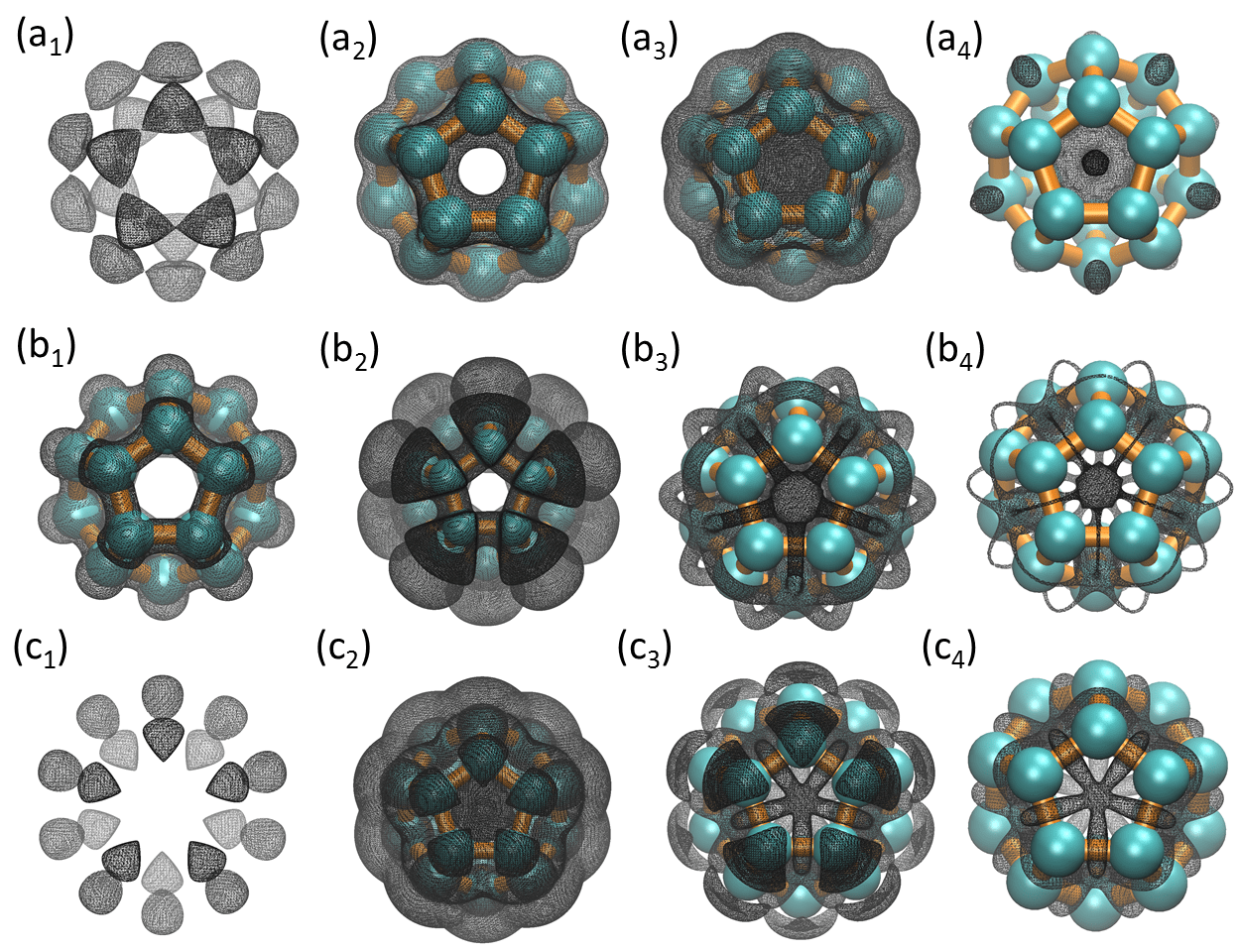 |
Using the proposed Geo-Topo fingerprint, one can study more complicated structures. The first one is the fullerene C20 considered in last section. Again, it rigidity density is described by Eq. (131) and parameter and are set to and , respectively.
First the Hessian matrix eigenvalue isosurfaces of C20 is studied. Figure 28 has illustrated four representative isosurfaces for each eigenvalue. Subscripts to indicate four isovalues from small to large. The notations to represent eigenvalues to . One can see that their isosurface behaviors are consistent with descriptions of the Geo-Topo fingerprints summarized in Table 2. State differently, the Geo-Topo fingerprints are able to capture the essential Geo-Topo properties of C20 isosurfaces.
More specifically, for , negative isosurfaces enclose all NCPs and BCPs. Anisotropic isosurfaces are found near RCPs with negative A-type and positive V-type behaviors. CCPs are enclosed with positive isosurfaces. For , negative isosurfaces enclose all NCPs and bond regions of BCPs. Positive loops can be found around BCP bonds. RCPs and CCPs are enclosed by positive isosurfaces. For , negative isosurfaces only enclose atomic basin of NCPs, whereas BCPs, RCPs and CCPs are all enclosed by positive isosurfaces.
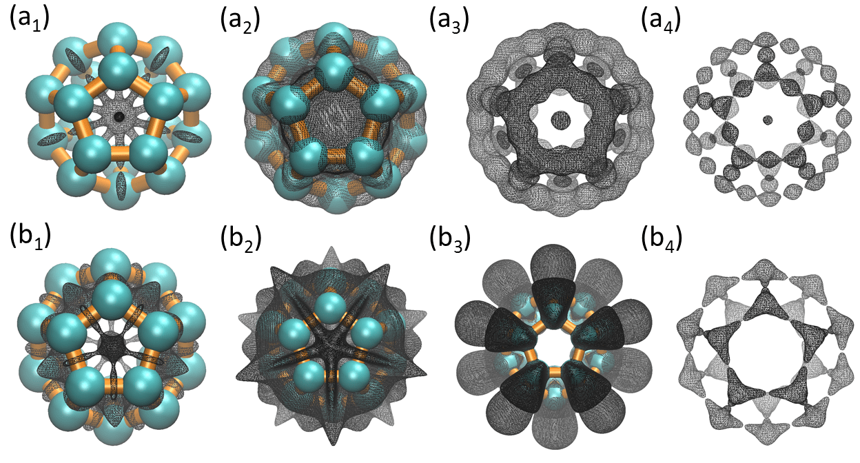 |
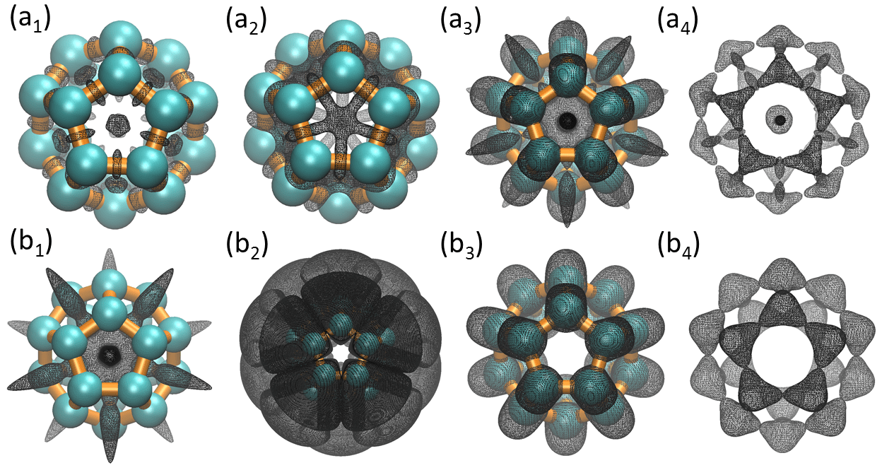 |
An -helix structure
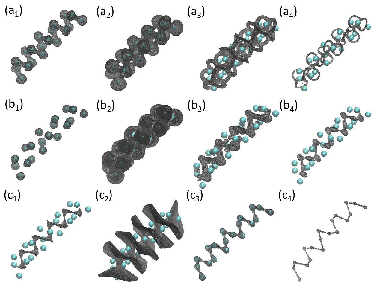 |
So far, all cases examined are about highly symmetric molecular structures. In this part, the Geo-Topo analysis is applied to irregular biomolecular structures, particularly protein structure. It is known that there are four distinct levels of protein structure, namely primary structure, which is a sequence of amino acids in the polypeptide chain; secondary structure, which is an -helix or a -strand; tertiary structure, which refers to the three-dimensional structure of a monomeric and multimeric protein molecule; and quaternary structure, which is the three-dimensional structure of a multi-subunit protein complex. The Geo-Topo analysis of proteins for protein secondary structures is demonstrated.
First, one can consider one of protein secondary structures, i.e., an -helix segment. This segment is extracted from protein with ID 1C26. The coarse-grained (CG) representation is employed and 19 Cα atoms from the th residue to th residue in chain A are used. The distance between two adjacent Cα atoms are about Å and the used in the CG rigidity density model is Å.
In stead of listing all results of eigenvalues and curvatures, only three geometric parameters of interest are examined, including eigenvalue , eigenvalue and mean curvature. For each of them, four representative isosurfaces are extracted. The results are illustrated in Fig. 31.
It can be found that results are very consistent with the Geo-Topo fingerprints. For , positive loops around atomic bonds are found. For , large eigenvalues are still concentrated in regions near BCPs. For mean curvature, one can still find positive A-type and negative V-type isosurfaces near BCPs. Also, large positive values are concentrates around NCPs and indicate the topological connectivity at Figs. 31(a1), (c3) and (c4).
It also should be noticed that unlike the regular symmetric structures studied in previous sections, RCPs and CCPs are more complicated in -helix structure. Moreover, since the characteristic distance is chosen as Å, bond effect (or topological connectivity) is observed not only between adjacent two atoms but also between atoms in close distance, as indicated in Fig. 31. At meantime, strongest topological connectivity (largest isovalues) is as usual found between adjacent two atoms as indicated in Fig. 31. Interestingly, in the case of , positive isosurface forms a strip that is parallel to the backbone of the -helix as demonstrated in Fig. 31. The largest isovalues are concentrated around BCPs not between adjacent two atoms, but neighboring two atoms in adjacent two circles as illustrated in Fig. 31. However, if characteristic distance was chosen as Å, the largest isovalues of would move to BCP regions between adjacent two atoms. This is due to the multiscale nature of the model. Further detailed discussion of this multiscale property is beyond the scope of this paper.
A -sheet structure
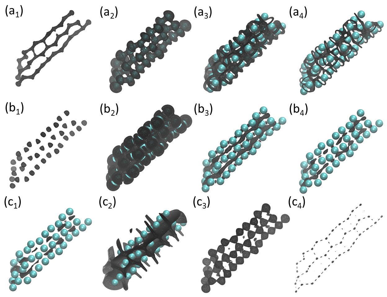 |
Another important protein secondary structure is -sheet. In this part, three adjacent -strands from protein with ID 4UW4 are considered. Again, the CG representation is used and three stands include residues from to , to and to in chain A. Just as the analysis in the -helix structure, the characteristic distance is chosen as Å. One can examine the second eigenvalue , the third eigenvalue and mean curvature in the Geo-Topo analysis. For each of them, four representative isosurfaces are extracted. The results are illustrated in Fig. 32.
Just as the -helix case, results for -sheet are also very consistent with the Geo-Topo fingerprints. Positive loops around BCPs for can be observed. For , small negative isosurfaces indicate NCPs. Large eigenvalues are concentrated in regions near BCPs. Positive A-type and negative V-type isosurfaces near BCPs are found in mean curvature. Large positive values are concentrates around NCPs. Further, as the characteristic distance is chosen as Å, the bond connection between the stands or sheets are amplified. It can be clearly observed in Fig. 32 .
3.8.2 Persistent homology for scalar field analysis
 |
Finally, the topological persistence in the scalar fields generated by Hessian matrix eigenvalues and curvatures is studied. The hexagonal ring is used for persistent homology analysis. The barcodes for eigenvalues , and are demonstrated in Figs. 33 (a)-(c). In each subfigure, results for , and are presented. The filtration goes from the smallest value to the largest in the persistent homology analysis.
For , at very small values, it has bars, i.e., independent components. Topologically, it represents small negative values concentrating around NCPs. As the filtration progresses, a loop is formed, which leads to a bar in . Further down the filtration process, isosurface begins to shrink to two balls perpendicular to the RCP. This contributes two bars in .
For , again bars are found in the earliest stage of the filtration. Here independent components quickly combine to form a loop as filtration progresses. More interesting, individual loops around BCPs form when filtration value is around the range from 0 to 1. Finally, isosurfaces shrink into the RCP.
For , smallest negative values are concentrated around NCPs and contribute bars at the earliest stage of the filtration. Once the isosurface value becomes positive, a new component emerges due to the generation of a new isosurface at the boundary region. This new isosurface also contributes to a long persisting bar. Further down the filtration, more bars occur, each representing a very narrow ring region around atom bonds. These regions are relatively small and quickly disappear. After that, “hat” regions attaching to the original NCP isosurfaces emerge, contributing to small loops. They quickly become detached and shrink away. At same time, isosurfaces form near the BCP and gradually disappear. Together they contribute 12 independent bars.
4 Concluding remarks
Every field in natural science, engineering, medicine, finance and social sciences becomes quantitative when it is getting mature. Mathematics is essential for all quantitative fields. Being regarded the last scientific forefront, biological sciences, particularly molecular biology and structural biology, have accumulated gigantic among of data in terms of biomolecular structures, activity relations and genetic sequences in the past few decades and are transforming from qualitative and phenomenological to quantitative and predictive. Such a transformation offers unprecedented opportunities for mathematically driven advances in biological sciences [287].
Geometry, topology, and graph theory are some of the core mathematics and have been naturally playing a unique role in molecular biology and molecular biophysics. In this paper, we present a brief review of geometric, topological, and graph theory apparatuses that are important to the contemporary molecular biology and biophysics. We first discuss the discrete methods and models, including graph theory, Gaussian network models, anisotropic network model, normal mode analysis, flexibility-rigidity index, molecular nonlinear dynamics, spectral graph theory and persistent homology for biomolecular modeling and analysis. Additionally, we describe continuous algorithms and theories, including, discrete to continuum mapping, multidimensional persistent homology for volumetric data sets, geometric modeling of biomolecules, differential geometry theory of surfaces, curvature analysis, atoms in molecule theory, and quantum chemical topology theory. Attention is paid to the connections between existing biophysical approaches and standard mathematical subjects, such as, Morse theory, Poincaré Hopf index, differential topology, etc. Open problems and potential new directions are point out in discussions.
Two new models, namely the analytical minimal molecular surface and virtual particle model, and two new methods, i.e., Hessian eigenvalue map and curvature map, are introduced for biomolecular modeling and analysis. These new approaches were inspirited by the subject under review during our preparation of this review. For simplicity, only the proof-of-principle applications are demonstrated for all methods, models, theories and algorithms covered in this review.
Selected mathematical topics in geometry, topology, and graph theory are based on our limited knowledge and understanding of mathematics and molecular biophysics. Many subjects in geometry, topology, and graph theory that have found much success in molecular biology and biophysics have not been covered in this review. One of these subjects is knot theory, particularly the DNA knot theory, which is also a very important ingredient of topological modeling of biomolecules [272, 226, 112, 18, 259, 83, 8, 40, 84, 33, 244]. Knot theory is an area of geometric topology that deals with knots and links. Mathematically, a knot is an embedding of circles or its homeomorphisms in the three-dimensional (3D) Euclidean space, . Physically, DNA, as a genetic material, exists usually in two very long strands that intertwine to form chromatins, bind with histones to build nucleosomes, tie into knots, and are subjected to successive coiling before package into chromosomes. The loss of knots in chromosomes can cause Angelman and Prader-Willi syndromes. DNA knot theory has been a very important topic in applied topology. However, in this review, focus is given to geometric and topological methods and models for atomistic biomolecular data. Therefore, DNA knot theory is not covered.
Another relevant subject that have not been considered in this review is biomolecular interaction network models, including protein interaction networks, metabolic networks and transcriptional regulatory networks. Obviously, part of the mathematical foundation of these models is graph theory. Protein interaction networks are designed to study protein-protein interactions [234, 122]. Normally, in these models, proteins are represented as nodes and physical interaction between them are represented by edges. To make the network more reliable, data from different sources are combined together and different rules are applied for the identification of protein interactions. A metabolic network considers all metabolic and physical processes happened within a cell [149, 218]. This network comprises the chemical reactions of metabolism, the metabolic pathways, as well as the regulatory interactions that guide these reactions. Transcriptional regulatory networks describe the regulatory interactions between genes [175, 241]. In this network, each gene is represented by a node and the regulation relations are represented by edges. The exclusion of this subject is also due to our focus on atomistic biomolecular data.
Topological graph theory concerns immersions of graphs as well as the embedding of graphs in surfaces, spatial embeddings of graphs, and graphs as topological spaces. It has had much success in the mathematical modeling of DNA recombination, DNA-RNA interactions, protein folding and protein-protein interactions [7]. The exclusion of this subject is due to our insufficient knowledge and understanding. For the same reason, fascinating applications of combinatorics, algebra and tiling theory in the modeling of viral capsid self assembly [151, 267, 7, 133, 136, 240] have not been covered in our review.
A continuous, differentiable curve can be embedded in a three-dimensional Euclidean space and its kinematic properties, such as the derivatives of tangent, normal, and binormal unit vectors, can be described by Frenet-Serret formulas in differential geometry [77]. Discrete Frenet-Serret frame offers an efficient description of amino-acid and/or nucleic acid chains [232, 145]. We believe that discrete Frenet-Serret frame can be easily applied to RNA chains, microtubules, nucleosomes, chromatins, active chromosomes and metaphase chromosomes.
An emergent approach is to combine machine learning with geometry, topology and/or graph theory for analyzing biomolecular data [43, 164]. Machine learning is a cutting edge computer science and statistical tool originally developed for pattern recognition and artificial intelligence. Its combination with mathematical apparatuses leads to extremely powerful approaches to massive biomolecular data challenges, such as the blind predictions of solvation free energies [276, 277] and protein-ligand binding affinity prediction [278]. For example, topological learning algorithm that utilizes exclusively persistent homology and machine learning for protein-ligand binding affinity predictions outperforms all the existing eminent methods in computational biophysics over massive binding data sets [42]. However, this subject is at its early stage and is evolving too fast to have a conclusive review at present.
It is worth mentioning that the geometric, topological and graph theory apparatuses discussed in this review can be employed in conjugation with partial differential equation (PDE), which is widely used in computational biophysics, to model biomolecular systems. Certainly geometric modeling is often a prerequisite in the PDE models of electrostatics, solvation, ion channels, membrane-protein interactions, and biomolecular elasticity [284]. As mentioned in Section 2.1.6, the graph cut problem can be formulated as a free energy minimization. Such a formulation makes it possible to combine spectral graph theory with PDE approaches for a wide range of biophysical modeling for biomolecular systems, including solvation, ion channel, biomembrane, protein-ligand binding, protein-protein interaction and protein-nucleic acid interaction. Finally, connection between algebraic topology and differential geometry, including Laplace-Beltrami operator, has been made [275]. Essentially, one defines an object function to optimize certain biophysical properties, which leads to a Laplace-Beltrami operator that generates a multiscale representation of the initial data and offers an object-oriented filtration process for persistent homology. The resulting differential geometry based object-oriented persistent homology is able to preserve desirable geometric features in the evolutionary filtration and enhances the corresponding topological persistence. However, how to design object-oriented persistent homology to automatically extract desirable features in the original biomolecular data during the filtration process is still an open problem.
Indeed, the application aspects of geometry, topology and graph theory have become a driven force for the development of abstract geometry, topology, homology, graph theory in recent years. It is expected that a versatile variety of pure mathematics concepts, methods and techniques will find their cutting edges in the transcend description of biomolecular structure, function, dynamics and transport. This health interaction between mathematics and molecular bioscience will benefit both fields and attract young researchers for generations to come.
Acknowledgments
This work was supported in part by a Start-Up Grant from The Nanyang Technological University (KLX), NSF IIS- 1302285 (GWW), NSF DMS-1160352 (GWW), NIH R01GM-090208 (GWW) and MSU Center for Mathematical Molecular Biosciences Initiative (GWW).
References
- [1] P. K. Agarwal, H. Edelsbrunner, J. Harer, and Y. Wang. Extreme elevation on a 2-manifold. Discrete and Computational Geometry (DCG), 36(4):553–572, 2006.
- [2] D. Aldous and J. Fill. Reversible Markov chains and random walks on graphs, 2002.
- [3] N. Alexandrov and I. Shindyalov. PDP: protein domain parser. Bioinformatics, 19(3):429–430, 2003.
- [4] M. P. Allen and D. J. Tildesley. Computer Simulation of Liquids. Oxford: Clarendon Press, 1987.
- [5] A. Amadei, A. Linssen, and H. JC Berendsen. Essential dynamics of proteins. Proteins: Structure, Function, and Bioinformatics, 17(4):412–425, 1993.
- [6] C. B. Anfinsen. Einfluss der configuration auf die wirkung den. Science, 181:223 – 230, 1973.
- [7] A. Angeleska, N. Jonoska, and M. Saito. Dna rearrangement through assembly graphs. Discrete and Applied Mathematics, 157:3020–3037, 2009.
- [8] J. Arsuaga, M. Vázquez, S. Trigueros, D. W. Sumners, and J. Roca. Knotting probability of DNA molecules confined in restricted volumes: DNA knotting in phage capsids. Proceedings of the National Academy of Sciences, 99(8):5373–5377, 2002.
- [9] A. R. Atilgan, S. R. Durrell, R. L. Jernigan, M. C. Demirel, O. Keskin, and I. Bahar. Anisotropy of fluctuation dynamics of proteins with an elastic network model. Biophys. J., 80:505 – 515, 2001.
- [10] A. Azran and Z. Ghahramani. A new approach to data driven clustering. In Proceedings of the 23rd international conference on Machine learning, pages 57–64. ACM, 2006.
- [11] R. F. Bader. Atoms in molecules. Accounts of Chemical Research, 18(1):9–15, 1985.
- [12] R. F. Bader. Atoms in molecules. Wiley Online Library, 1990.
- [13] R. F. Bader, R. J. Gillespie, and P. J. MacDougall. A physical basis for the VSEPR model of molecular geometry. Journal of the American Chemical Society, 110(22):7329–7336, 1988.
- [14] I. Bahar, A. R. Atilgan, M. C. Demirel, and B. Erman. Vibrational dynamics of proteins: Significance of slow and fast modes in relation to function and stability. Phys. Rev. Lett, 80:2733 – 2736, 1998.
- [15] I. Bahar, A. R. Atilgan, and B. Erman. Direct evaluation of thermal fluctuations in proteins using a single-parameter harmonic potential. Folding and Design, 2:173 – 181, 1997.
- [16] N. A. Baker. Improving implicit solvent simulations: a Poisson-centric view. Current Opinion in Structural Biology, 15(2):137–43, 2005.
- [17] N. A. Baker, D. Sept, S. Joseph, M. J. Holst, and J. A. McCammon. Electrostatics of nanosystems: Application to microtubules and the ribosome. Proceedings of the National Academy of Sciences of the United States of America, 98(18):10037–10041, 2001.
- [18] A. D. Bates and A. Maxwell. DNA topology. Oxford University Press, USA, 2005.
- [19] P. W. Bates, Z. Chen, Y. H. Sun, G. W. Wei, and S. Zhao. Geometric and potential driving formation and evolution of biomolecular surfaces. J. Math. Biol., 59:193–231, 2009.
- [20] P. W. Bates, G. W. Wei, and S. Zhao. The minimal molecular surface. arXiv:q-bio/0610038v1, [q-bio.BM], 2006.
- [21] P. W. Bates, G. W. Wei, and Shan Zhao. Minimal molecular surfaces and their applications. Journal of Computational Chemistry, 29(3):380–91, 2008.
- [22] U. Bauer, M. Kerber, and J. Reininghaus. Distributed computation of persistent homology. Proceedings of the Sixteenth Workshop on Algorithm Engineering and Experiments (ALENEX), 2014.
- [23] K. Beketayev, G. H. Weber, M. Haranczyk, P.T. Bremer, M. Hlawitschka, and B. Hamann. Topology-based visualization of transformation pathways in complex chemical systems. In Computer Graphics Forum, volume 30, pages 663–672. Wiley Online Library, 2011.
- [24] M. Belkin. Problems of learning on manifolds. PhD thesis, The University of Chicago, 2003.
- [25] M. Belkin and P. Niyogi. Towards a theoretical foundation for Laplacian-based manifold methods. In International Conference on Computational Learning Theory, pages 486–500. Springer, 2005.
- [26] Paul Bendich, Herbert Edelsbrunner, and Michael Kerber. Computing robustness and persistence for images. IEEE Transactions on Visualization and Computer Graphics, 16:1251–1260, 2010.
- [27] Paul Bendich and John Harer. Persistent intersection homology. Foundations of Computational Mathematics (FOCM), 11(3):305–336, 2011.
- [28] Janine Bennett, Fabien Vivodtzev, and Valerio Pascucci, editors. Topological and statistical methods for complex data: Tackling large-scale, high-dimensional and multivariate data spaces. Mathematics and Visualization. Springer-Verlag Berlin Heidelberg, 2015.
- [29] C.A.S. Bergstrom, M. Strafford, L. Lazorova, A. Avdeef, K. Luthman, and P. Artursson. Absorption classification of oral drugs based on molecular surface properties. J. Medicinal Chem., 46:558–570, 2003.
- [30] S. Biasotti, L. De Floriani, B. Falcidieno, P. Frosini, D. Giorgi, C. Landi, L. Papaleo, and M. Spagnuolo. Describing shapes by geometrical-topological properties of real functions. ACM Computing Surveys, 40(4):12, 2008.
- [31] F. Biegler-König and J. Schönbohm. Update of the AIM2000-program for atoms in molecules. Journal of computational chemistry, 23(15):1489–1494, 2002.
- [32] J. Blinn. A generalization of algebraic surface drawing. ACM Transactions on Graphics, 1(3):235–256, 1982.
- [33] R. Brasher, R. G. Scharein, and M. Vazquez. New biologically motivated knot table. Biochemical Society Transactions, 41:606–611, 2013.
- [34] P. T. Bremer, V. Pascucci I. Hotz, and R. Peikert, editors. Topological methods in data analysis and visualization III: Theory, algorithms and applications. Mathematics and Visualization. Springer International Publishing, 2014.
- [35] B. Brooks and M. Karplus. Harmonic dynamics of proteins: normal modes and fluctuations in bovine pancreatic trypsin inhibitor. Proceedings of the National Academy of Sciences, 80(21):6571–6575, 1983.
- [36] B. R. Brooks, R. E. Bruccoleri, B. D. Olafson, D.J. States, S. Swaminathan, and M. Karplus. Charmm: A program for macromolecular energy, minimization, and dynamics calculations. J. Comput. Chem., 4:187–217, 1983.
- [37] B. R. Brooks, D. Janežič, and M. Karplus. Harmonic analysis of large systems. I. Methodology. Journal of computational chemistry, 16(12):1522–1542, 1995.
- [38] Peter Bubenik. Statistical topological data analysis using persistence landscapes. Journal of Machine Learning Research, 16(1):77–102, 2015.
- [39] Peter Bubenik and Peter T. Kim. A statistical approach to persistent homology. Homology, Homotopy and Applications, 19:337–362, 2007.
- [40] D. Buck and E. Flapan. Predicting knot or catenane type of site-specific recombination products. Journal of molecular biology, 374(5):1186–1199, 2007.
- [41] Z. X. Cang and G. W. Wei. Feature functional theory-mutation predictor (FFT-MP) for the blind prediction of protein mutation energy changes. Submitted, 2016.
- [42] Z. X. Cang and G. W. Wei. Persistent topology based scoring function (T-Score) for the blind prediction of protein-ligand binding affinities . Submitted, 2016.
- [43] Zixuan Cang, Lin Mu, Kedi Wu, Kris Opron, Kelin Xia, and Guo-Wei Wei. A topological approach to protein classificationy. Molecular based Mathematical Biologys, 3:140–162, 2015.
- [44] G. Carlsson. Topology and data. Am. Math. Soc, 46(2):255–308, 2009.
- [45] G. Carlsson, T. Ishkhanov, V. Silva, and A. Zomorodian. On the local behavior of spaces of natural images. International Journal of Computer Vision, 76(1):1–12, 2008.
- [46] G. Carlsson and A. Zomorodian. The theory of multidimensional persistence. Discrete Computational Geometry, 42(1):71–93, 2009.
- [47] G. Carlsson, A. Zomorodian, A. Collins, and L. J. Guibas. Persistence barcodes for shapes. International Journal of Shape Modeling, 11(2):149–187, 2005.
- [48] Gunnar Carlsson and Vin De Silva. Zigzag persistence. Foundations of computational mathematics, 10(4):367–405, 2010.
- [49] Gunnar Carlsson, Vin de Silva, and Dmitriy Morozov. Zigzag persistent homology and real-valued functions. In Proc. 25th Annu. ACM Sympos. Comput. Geom., pages 247–256, 2009.
- [50] S. L. Chan and E. O. Purisima. Molecular surface generation using marching tetrahedra. J. Computat. Chem., 11:1268–1277, 1998.
- [51] H. W. Chang, S. Bacallado, V. S. Pande, and G. E. Carlsson. Persistent topology and metastable state in conformational dynamics. PLos ONE, 8(4):e58699, 2013.
- [52] Frédéric Chazal, David Cohen-Steiner, Marc Glisse, Leonidas J. Guibas, and Steve Oudot. Proximity of persistence modules and their diagrams. In Proc. 25th ACM Sympos. on Comput. Geom., pages 237–246, 2009.
- [53] Frédéric Chazal, Leonidas J. Guibas, Steve Y. Oudot, and Primoz Skraba. Persistence-based clustering in riemannian manifolds. In Proceedings of the 27th annual ACM symposium on Computational geometry, SoCG ’11, pages 97–106, 2011.
- [54] Duan Chen, Zhan Chen, Changjun Chen, W. H. Geng, and G. W. Wei. MIBPB: A software package for electrostatic analysis. J. Comput. Chem., 32:657 – 670, 2011.
- [55] Duan Chen, Zhan Chen, and G. W. Wei. Quantum dynamics in continuum for proton transport II: Variational solvent-solute interface. International Journal for Numerical Methods in Biomedical Engineering, 28:25 – 51, 2012.
- [56] Duan Chen and G. W. Wei. Quantum dynamics in continuum for proton transport—Generalized correlation. J Chem. Phys., 136:134109, 2012.
- [57] Duan Chen and G. W. Wei. Quantum dynamics in continuum for proton transport I: Basic formulation. Commun. Comput. Phys., 13:285–324, 2013.
- [58] Guoning Chen, Qingqing Deng, Andrzej Szymczak, Robert S Laramee, and Eugene Zhang. Morse set classification and hierarchical refinement using conley index. IEEE transactions on visualization and computer graphics, 18(5):767–782, 2012.
- [59] Minxin Chen and Benzhuo Lu. Tmsmesh: A robust method for molecular surface mesh generation using a trace technique. J Chem. Theory and Comput., 7:203–212, 2011.
- [60] Minxin Chen, Bin Tu, and Benzhuo Lu. Triangulated manifold meshing method preserving molecular surface topology. J. Mole. Graph. Model., 38:411–418, 2012.
- [61] Wenyu Chen, Jianmin Zheng, and Yiyu Cai. Kernel modeling for molecular surfaces using a uniform solution. Computer Aided Design, 42:267–278, 2010.
- [62] Z. Chen, N. A. Baker, and G. W. Wei. Differential geometry based solvation models I: Eulerian formulation. J. Comput. Phys., 229:8231–8258, 2010.
- [63] Z. Chen, N. A. Baker, and G. W. Wei. Differential geometry based solvation models II: Lagrangian formulation. J. Math. Biol., 63:1139– 1200, 2011.
- [64] Z. Chen and G. W. Wei. Differential geometry based solvation models III: Quantum formulation. J. Chem. Phys., 135:194108, 2011.
- [65] L. T. Cheng, Joachim Dzubiella, Andrew J. McCammon, and B. Li. Application of the level-set method to the implicit solvation of nonpolar molecules. Journal of Chemical Physics, 127(8), 2007.
- [66] Li-Tien Cheng, Yang Xie, Joachim Dzubiella, J. Andrew McCammon, Jianwei Che, and Bo Li. Coupling the level-set method with molecular mechanics for variational implicit solvation of nonpolar molecules. J. Chem. Theory Comput., 5:257–266, 2009.
- [67] F. Chiti and C. M. Dobson. Protein misfolding, functional amyloid, and human disease. Annu. Rev. Biochem., 75:333 – 366, 2006.
- [68] F. Chung. Spectral graph theory. American Mathematical Society, 1997.
- [69] J. Cioslowski and G. H. Liu. Topology of electron-electron interactions in atoms and molecules. II. the correlation cage. The Journal of chemical physics, 110(4):1882–1887, 1999.
- [70] David Cohen-Steiner, Herbert Edelsbrunner, and John Harer. Stability of persistence diagrams. Discrete & Computational Geometry, 37(1):103–120, 2007.
- [71] David Cohen-Steiner, Herbert Edelsbrunner, and John Harer. Extending persistence using poincaré and lefschetz duality. Foundations of Computational Mathematics, 9(1):79–103, 2009.
- [72] David Cohen-Steiner, Herbert Edelsbrunner, John Harer, and Dmitriy Morozov. Persistent homology for kernels, images, and cokernels. In Proceedings of the Twentieth Annual ACM-SIAM Symposium on Discrete Algorithms, SODA 09, pages 1011–1020, 2009.
- [73] Charles Conley. Isolated invariant sets and the Morse index. CBMS Regional Conference Series in Mathematics, 38. American Mathematical Society,Providence, R.I, 1978.
- [74] M. L. Connolly. Depth buffer algorithms for molecular modeling. J. Mol. Graphics, 3:19–24, 1985.
- [75] Julia Contreras-García, Erin R Johnson, Shahar Keinan, Robin Chaudret, Jean-Philip Piquemal, David N Beratan, and Weitao Yang. Nciplot: a program for plotting noncovalent interaction regions. Journal of chemical theory and computation, 7(3):625–632, 2011.
- [76] Julia Contreras-García, Weitao Yang, and Erin R Johnson. Analysis of hydrogen-bond interaction potentials from the electron density: integration of noncovalent interaction regions. The Journal of Physical Chemistry A, 115(45):12983–12990, 2011.
- [77] Hugh C Crenshaw and Leah Edelstein-Keshet. Orientation by helical motion—ii. changing the direction of the axis of motion. Bulletin of mathematical biology, 55(1):213–230, 1993.
- [78] P.B. Crowley and A. Golovin. Cation-pi interactions in protein-protein interfaces. Proteins - Struct. Func. Bioinf., 59:231–239, 2005.
- [79] Q. Cui. Combining implicit solvation models with hybrid quantum mechanical/molecular mechanical methods: A critical test with glycine. Journal of Chemical Physics, 117(10):4720, 2002.
- [80] Q. Cui and I. Bahar. Normal mode analysis: theory and applications to biological and chemical systems. Chapman and Hall/CRC, 2010.
- [81] Q. Cui, G. J. Li, J. Ma, and M. Karplus. A normal mode analysis of structural plasticity in the biomolecular motor f(1)-atpase. J. Mol. Biol., 340(2):345 – 372, 2004.
- [82] Y. Dabaghian, F. Memoli, L. Frank, and G. Carlsson. A topological paradigm for hippocampal spatial map formation using persistent homology. PLoS Comput Biol, 8(8):e1002581, 08 2012.
- [83] I. K. Darcy. Biological distances on DNA knots and links: applications to XER recombination. Journal of Knot Theory and its Ramifications, 10(02):269–294, 2001.
- [84] I. K. Darcy and M. Vazquez. Determining the topology of stable protein-DNA complexes. Biochemical Society Transactions, 41:601–605, 2013.
- [85] S. J. Darnell, L. LeGault, and J. C. Mitchell. KFC server: interactive forecasting of protein interaction hot spots. Nucleic Acids Research, 36:W265–W269, 2008.
- [86] Bhaskar DasGupta and Jie Liang. Models and Algorithms for Biomolecules and Molecular Networks. John Wiley & Sons, 2016.
- [87] Vin de Silva, Dmitriy Morozov, and Mikael Vejdemo-Johansson. Persistent cohomology and circular coordinates. Discrete and Comput. Geom., 45:737–759, 2011.
- [88] S. Decherchi and W. Rocchia. A general and robust ray-casting-based algorithm for triangulating surfaces at the nanoscale. PLoS ONE, 8:e59744, 2013.
- [89] O. N. A. Demerdash, M. D. Daily, and J. C. Mitchell. Structure-based predictive models for allosteric hot spots. PLOS Computational Biology, 5:e1000531, 2009.
- [90] Omar N. A. Demerdash and Julie C. Mitchell. Density-cluster NMA: A new protein decomposition technique for coarse-grained normal mode analysis. Proteins:Structure Function and Bioinformatics, 80(7):1766–1779, JUL 2012.
- [91] T. K. Dey, K. Y. Li, J. Sun, and C. S. David. Computing geometry aware handle and tunnel loops in 3d models. ACM Trans. Graph., 27, 2008.
- [92] Tamal K Dey, Fengtao Fan, and Yusu Wang. Computing topological persistence for simplicial maps. In Proc. 30th Annu. Sympos. Comput. Geom. (SoCG), pages 345–354, 2014.
- [93] Tamal K. Dey and Y. S. Wang. Reeb graphs: Approximation and persistence. Discrete and Computational Geometry, 49(1):46–73, 2013.
- [94] Barbara Di Fabio and Claudia Landi. A mayer-vietoris formula for persistent homology with an application to shape recognition in the presence of occlusions. Foundations of Computational Mathematics, 11:499–527, 2011.
- [95] C. H. Q. Ding, X. F. He, H. Y. Zha, M. Gu, and H. D. Simon. A min-max cut algorithm for graph partitioning and data clustering. In Data Mining, 2001. ICDM 2001, Proceedings IEEE International Conference on, pages 107–114. IEEE, 2001.
- [96] A.I. Dragan, C.M. Read, E.N. Makeyeva, E.I. Milgotina, M.E.A. Churchill, C. Crane-Robinson, and P.L. Privalov. Dna binding and bending by hmg boxes: Energetic determinants of specificity. J. Mol. Biol., 343:371–393, 2004.
- [97] B. S. Duncan and A. J. Olson. Shape analysis of molecular surfaces. Biopolymers, 33:231–238, 1993.
- [98] H. Edelsbrunner and J. Harer. Persistent homology-a survey. Contemporary mathematics, 453:257–282, 2008.
- [99] H. Edelsbrunner, D. Letscher, and A. Zomorodian. Topological persistence and simplification. Discrete Comput. Geom., 28:511–533, 2002.
- [100] H. Edelsbrunner and E. P. Mucke. Three-dimensional alpha shapes. Physical Review Letters, 13:43–72, 1994.
- [101] Herbert Edelsbrunner and John Harer. Computational topology: an introduction. American Mathematical Soc., 2010.
- [102] H. Federer. Curvature Measures. Trans. Amer. Math. Soc., 93:418–491, 1959.
- [103] X. Feng, K. L. Xia, Y. Y. Tong, and G. W. Wei. Multiscale geometric modeling of macromolecules II: lagrangian representation. Journal of Computational Chemistry, 34:2100–2120, 2013.
- [104] Xin Feng, Kelin Xia, Yiying Tong, and Guo-Wei Wei. Geometric modeling of subcellular structures, organelles and large multiprotein complexes. International Journal for Numerical Methods in Biomedical Engineering, 28:1198–1223, 2012.
- [105] D. Fera, N. Kim, N. Shiffeldrim, J. Zorn, U. Laserson, H. H. Gan, and T. Schlick. RAG: RNA-as-graphs web resource. BMC bioinformatics, 5(1):1, 2004.
- [106] P. J. Flory. Statistical thermodynamics of random networks. Proc. Roy. Soc. Lond. A,, 351:351 – 378, 1976.
- [107] S. Fortunato. Community detection in graphs. Physics reports, 486(3):75–174, 2010.
- [108] Patrizio Frosini. A distance for similarity classes of submanifolds of a Euclidean space. BUllentin of Australian Mathematical Society, 42(3):407–416, 1990.
- [109] Patrizio Frosini and Claudia Landi. Size theory as a topological tool for computer vision. Pattern Recognition and Image Analysis, 9(4):596–603, 1999.
- [110] Patrizio Frosini and Claudia Landi. Persistent betti numbers for a noise tolerant shape-based approach to image retrieval. Pattern Recognition Letters, 34:863–872, 2013.
- [111] Issei Fujishiro, Yuriko Takeshima, Taeko Azuma, and Shigeo Takahashi. Volume data mining using 3d field topology analysis. IEEE Computer Graphics and Applications, 20(5):46–51, 2000.
- [112] F. B. Fuller. The writhing number of a space curve. Proceedings of the National Academy of Sciences, 68(4):815–819, 1971.
- [113] M. Gameiro, Y. Hiraoka, S. Izumi, M. Kramar, K. Mischaikow, and V. Nanda. Topological measurement of protein compressibility via persistence diagrams. Japan Journal of Industrial and Applied Mathematics, 32:1–17, 2014.
- [114] H. H. Gan, D. Fera, J. Zorn, N. Shiffeldrim, M. Tang, U. Laserson, N. Kim, and T. Schlick. RAG: RNA-as-graphs database—concepts, analysis, and features. Bioinformatics, 20(8):1285–1291, 2004.
- [115] A. E. García. Large-amplitude nonlinear motions in proteins. Physical review letters, 68(17):2696, 1992.
- [116] W. Geng and G. W. Wei. Multiscale molecular dynamics using the matched interface and boundary method. J Comput. Phys., 230(2):435–457, 2011.
- [117] Weihua Geng, Sining Yu, and G. W. Wei. Treatment of charge singularities in implicit solvent models. Journal of Chemical Physics, 127:114106, 2007.
- [118] Z. Nevin Gerek and S. Banu Ozkan. A flexible docking scheme to explore the binding selectivity of pdz domains. Protein Science, 19:914–928, 2010.
- [119] R. Ghrist. Barcodes: The persistent topology of data. Bull. Amer. Math. Soc., 45:61–75, 2008.
- [120] N. Gillet, R. Chaudret, J. Contreras-Garcı˝́a, W. T. Yang, B. Silvi, and J. P. Piquemal. Coupling quantum interpretative techniques: another look at chemical mechanisms in organic reactions. Journal of chemical theory and computation, 8(11):3993–3997, 2012.
- [121] E. Giné and V. Koltchinskii. Empirical graph Laplacian approximation of Laplace Beltrami operators: Large sample results. In High dimensional probability, pages 238–259. Institute of Mathematical Statistics, 2006.
- [122] L. Giot, J. S. Bader, C. Brouwer, A. Chaudhuri, B. Kuang, Y. Li, Y.L. Hao, C.E. Ooi, B. Godwin, E. Vitols, et al. A protein interaction map of drosophila melanogaster. science, 302(5651):1727–1736, 2003.
- [123] N. Go, T. Noguti, and T. Nishikawa. Dynamics of a small globular protein in terms of low-frequency vibrational modes. Proc. Natl. Acad. Sci., 80:3696 – 3700, 1983.
- [124] J. A. Grant and B. T. Pickup. A gaussian description of molecular shape. Journal of Physical Chemistry, 99:3503–3510, 1995.
- [125] J. A. Grant, B. T. Pickup, M. T. Sykes, C. A. Kitchen, and A. Nicholls. The Gaussian Generalized Born model: application to small molecules. Physical Chemistry Chemical Physics, 9:4913–22, 2007.
- [126] J. Andrew Grant, Barry T. Pickup, and Anthony Nicholls. A smooth permittivity function for Poisson-Boltzmann solvation methods. Journal of Computational Chemistry, 22(6):608–640, 2001.
- [127] R. Gray. Vector quantization. IEEE Assp Magazine, 1(2):4–29, 1984.
- [128] D. Günther, A. Jacobson, J. Reininghaus, H. P. Seidel, O. Sorkine-Hornung, and T. Weinkauf. Fast and memory-efficient topological denoising of 2D and 3D scalar fields. IEEE Transactions on Visualization and Computer Graphics, 20:12, 2014.
- [129] J. T. Guo, D. Xu, D. Kim, and Y. Xu. Improving the performance of DomainParser for structural domain partition using neural network. Nucleic Acids Research, 31(3):944–952, 2003.
- [130] L. Hagen and A. B. Kahng. New spectral methods for ratio cut partitioning and clustering. IEEE transactions on computer-aided design of integrated circuits and systems, 11(9):1074–1085, 1992.
- [131] B. Halle. Flexibility and packing in proteins. PNAS, 99:1274–1279, 2002.
- [132] Shaun Harker, Konstantin Mischaikow, Marian Mrozek, and Vidit Nanda. Discrete morse theoretic algorithms for computing homology of complexes and maps. Foundations of Computational Mathematics, pages 1–34, 2013.
- [133] S. C. Harvey, Y. Zeng, and C. E. Heitsch. The icosahedral rna virus as a grotto: organizing the genome into stalagmites and stalactite. J Biol Phys, Chapter 7:163–172, 2013.
- [134] S. Hayward and B. L. De Groot. Normal modes and essential dynamics. Molecular Modeling of Proteins, pages 89–106, 2008.
- [135] M. Hein, J. Y. Audibert, and U. Von Luxburg. From graphs to manifolds–weak and strong pointwise consistency of graph Laplacian. In International Conference on Computational Learning Theory, pages 470–485. Springer, 2005.
- [136] C. Heitsch and S. Poznanovic. Combinatorial insights into rna secondary structure, in N. Jonoska and M. Saito, editors. Discrete and Topological Models in Molecular Biology, Chapter 7:145–166, 2014.
- [137] G. Henkelman, A. Arnaldsson, and H. Jónsson. A fast and robust algorithm for Bader decomposition of charge density. Computational Materials Science, 36(3):354–360, 2006.
- [138] K. Hinsen. Analysis of domain motions by approximate normal mode calculations. Proteins, 33:417 – 429, 1998.
- [139] K. Hinsen. Structural flexibility in proteins: impact of the crystal environment. Bioinformatics, 24:521 – 528, 2008.
- [140] L. Holm and C. Sander. Mapping the protein universe. Science, 273(5275):595, 1996.
- [141] Michael Holst. The Poisson-Boltzmann equation: Analysis and multilevel numerical solution. PhD thesis, California Institute of Technology, 1994.
- [142] D. Horak, S Maletic, and M. Rajkovic. Persistent homology of complex networks. Journal of Statistical Mechanics: Theory and Experiment, 2009(03):P03034, 2009.
- [143] G. Hu, J. H. Yang, and W. J. Liu. Instability and controllability of linearly coupled oscillators: Eigenvalue analysis. Phys. Rev. E, 58:4440– 4453, 1998.
- [144] H. Y. Hu, T. Laurent, M. A. Porter, and A. L. Bertozzi. A method based on total variation for network modularity optimization using the MBO scheme. SIAM Journal on Applied Mathematics, 73(6):2224–2246, 2013.
- [145] Shuangwei Hu, Martin Lundgren, and Antti J Niemi. Discrete frenet frame, inflection point solitons, and curve visualization with applications to folded proteins. Physical Review E, 83(6):061908, 2011.
- [146] YunKyong Hyon, Bob Eisenberg, and Chun Liu. A mathematical model of the hard sphere repulsion in ionic solutions. Communications in Mathematical Sciences, 9:459–475, 2010.
- [147] R.M. Jackson and M.J. Sternberg. Dna binding and bending by hmg boxes: Energetic determinants of specificity. J. Mol. Biol., 250:258–275, 1995.
- [148] A. K. Jain. Data clustering: 50 years beyond k-means. Pattern recognition letters, 31(8):651–666, 2010.
- [149] H. Jeong, B. Tombor, R. Albert, Z. N. Oltvai, and A. L. Barabási. The large-scale organization of metabolic networks. Nature, 407(6804):651–654, 2000.
- [150] E. R. Johnson, S. Keinan, P. Mori-Sanchez, J. Contreras-Garcia, A. J. Cohen, and W.T. Yang. Revealing noncovalent interactions. Journal of the American Chemical Society, 132(18):6498–6506, 2010.
- [151] N. Jonoska and G. McColm. Complexity classes for self-assembling flexible tiles. Theoretical Computer Science, 410:332–346, 2009.
- [152] T. Kaczynski, K. Mischaikow, and M. Mrozek. Computational homology. Springer-Verlag, 2004.
- [153] Tomasz Kaczynski, Konstantin Mischaikow, and Marian Mrozek. Computational Homology, volume 157 of Applied Mathematical Sciences. Springer-Verlag, New York, 2004.
- [154] P. M. Kasson, A. Zomorodian, S. Park, N. Singhal, L. J. Guibas, and V. S. Pande. Persistent voids a new structural metric for membrane fusion. Bioinformatics, 23:1753–1759, 2007.
- [155] T. A. Keith and R. F. Bader. Topological analysis of magnetically induced molecular current distributions. The Journal of chemical physics, 99(5):3669–3682, 1993.
- [156] T. A. Keith, R. F. Bader, and Y. Aray. Structural homeomorphism between the electron density and the virial field. International journal of quantum chemistry, 57(2):183–198, 1996.
- [157] O. Keskin, I. Bahar, D. Flatow, D. G. Covell, and R. L. Jernigan. Molecular mechanisms of chaperonin groel-groes function. Biochem., 41:491 – 501, 2002.
- [158] N. Kim, N. Shiffeldrim, H. H. Gan, and T. Schlick. Candidates for novel RNA topologies. Journal of molecular biology, 341(5):1129–1144, 2004.
- [159] G. Kindlmann, R. Whitaker, T. Tasdizen, and T. Möller. Curvature-based transfer functions for direct volume rendering: methods and applications. Proc. IEEE Visualization, 2003.
- [160] A. Kitao, F. Hirata, and N. Gō. The effects of solvent on the conformation and the collective motions of protein: normal mode analysis and molecular dynamics simulations of melittin in water and in vacuum. Chemical physics, 158(2-3):447–472, 1991.
- [161] M. Kohout, K. Pernal, F. R. Wagner, and Y. Grin. Electron localizability indicator for correlated wavefunctions. I. Parallel-spin pairs. Theoretical Chemistry Accounts, 112(5-6):453–459, 2004.
- [162] W. L. Koltun. Precision space-filling atomic models. Biopolymers, 3:667–679, 1965.
- [163] D. A. Kondrashov, A. W. Van Wynsberghe, R. M. Bannen, Q. Cui, and Jr. G. N. Phillips. Protein structural variation in computational models and crystallographic data. Structure, 15:169 – 177, 2007.
- [164] Violeta Kovacev-Nikolic, Peter Bubenik, Dragan Nikolić, and Giseon Heo. Using persistent homology and dynamical distances to analyze protein binding. Stat. Appl. Genet. Mol. Biol., 15(1):19–38, 2016.
- [165] Bala Krishnamoorthy, Scott Provan, and Alexander Tropsha. A topological characterization of protein structure. In Data Mining in Biomedicine, Springer Optimization and Its Applications, pages 431–455, 2007.
- [166] L.A. Kuhn, M. A. Siani, M. E. Pique, C. L. Fisher, E. D. Getzoff, and J. A. Tainer. The interdependence of protein surface topography and bound water molecules revealed by surface accessibility and fractal density measures. J. Mol. Biol., 228:13–22, 1992.
- [167] W. Kühnel. Differential Geometry: Curves-Surfaces-Manifolds, Student Math. American mathematical Society, 2015.
- [168] S. Kundu, J. S. Melton, D. C. Sorensen, and Jr. G. N. Phillips. Dynamics of proteins in crystals: comparison of experiment with simple models. Biophys. J., 83:723 – 732, 2002.
- [169] S. Kundu, D. C. Sorensen, and G. N. Phillips. Automatic domain decomposition of proteins by a Gaussian network model. Proteins: Structure, Function, and Bioinformatics, 57(4):725–733, 2004.
- [170] Z. Zhang L. Li, C. Li and Emil Alexov. On the dielectric ”constant” of proteins: Smooth dielectric function for macromolecular modeling and its implementation in DelPhi. J. Chem. Theory Comput., 9:2126–2136, 2013.
- [171] S. S. Lafon. Diffusion maps and geometric harmonics. PhD thesis, Yale University, 2004.
- [172] M. Leboeuf, A. M. Köster, K. Jug, and D. R. Salahub. Topological analysis of the molecular electrostatic potential. The Journal of chemical physics, 111(11):4893–4905, 1999.
- [173] B. Lee and F. M. Richards. The interpretation of protein structures: estimation of static accessibility. J Mol Biol, 55(3):379–400, 1971.
- [174] H Lee, H. Kang, M. K. Chung, B. Kim, and D. S. Lee. Persistent brain network homology from the perspective of dendrogram. Medical Imaging, IEEE Transactions on, 31(12):2267–2277, Dec 2012.
- [175] T. I. Lee, N. J. Rinaldi, F. Robert, D. T. Odom, Z. Bar-Joseph, G. K. Gerber, N. M. Hannett, C. T. Harbison, C. M. Thompson, I. Simon, et al. Transcriptional regulatory networks in saccharomyces cerevisiae. science, 298(5594):799–804, 2002.
- [176] M. Levitt, C. Sander, and P. S. Stern. The normal modes of a protein: Native bovine pancreatic trypsin inhibitor. International Journal of Quantum Chemistry, 24(S10):181–199, 1983.
- [177] M. Levitt, C. Sander, and P. S. Stern. Protein normal-mode dynamics: Trypsin inhibitor, crambin, ribonuclease and lysozyme. J. Mol. Biol., 181(3):423 – 447, 1985.
- [178] D. W. Li and R. Brüschweiler. All-atom contact model for understanding protein dynamics from crystallographic b-factors. Biophysical journal, 96(8):3074–3081, 2009.
- [179] G. H. Li and Q. Cui. A coarse-grained normal mode approach for macromolecules: an efficient implementation and application to Ca(2+)-ATPase. Bipohys. J., 83:2457 – 2474, 2002.
- [180] J. Li, P. Mach, and P. Koehl. Measuring the shapes of macromolecules - and why it matters. Comput Struct Biotechnol J., 8:e201309001, 2013.
- [181] Lin Li, Chuan Li, and Emil Alexov. On the modeling of polar component of solvation energy using smooth gaussian-based dielectric function. Journal of Theoretical and Computational Chemistry, 13:10.1142/S0219633614400021, 2014.
- [182] V.J. LiCata and N.M. Allewell. Functionally linked hydration changes in escherichia coli aspartate transcarbamylase and its catalytic subunit. Biochemistry, 36:10161–10167, 1997.
- [183] C. P. Lin, S. W. Huang, Y. L. Lai, S. C. Yen, C. H. Shih, C. H. Lu, C. C. Huang, and J. K. Hwang. Deriving protein dynamical properties from weighted protein contact number. Proteins: Structure, Function, and Bioinformatics, 72(3):929–935, 2008.
- [184] Beibei Liu, Bao Wang, Rundong Zhao, Yiying Tong, and Guo Wei Wei. ESES: software for Eulerian solvent excluded surface. Preprint, 2015.
- [185] Xu Liu, Zheng Xie, and Dongyun Yi. A fast algorithm for constructing topological structure in large data. Homology, Homotopy and Applications, 14:221–238, 2012.
- [186] D. R. Livesay, S. Dallakyan, G. G. Wood, and D. J. Jacobs. A flexible approach for understanding protein stability. FEBS Letters, 576:468–476, 2004.
- [187] J. R. López-Blanco, O. Miyashita, F. Tama, and P. Chacón. Normal mode analysis techniques in structural biology. eLS, 2014.
- [188] E. N. Lorenz. Deterministic nonperiodic flow. Journal of the Atmospheric Sciences, 20:130– 141, 1963.
- [189] L. Lovász. Random walks on graphs. Combinatorics, Paul erdos is eighty, 2:1–46, 1993.
- [190] J. P. Ma. Usefulness and limitations of normal mode analysis in modeling dynamics of biomolecular complexes. Structure, 13:373 – 180, 2005.
- [191] P. Mach and P. Koehl. Geometric measures of large biomolecules: Surface, volume, and pockets. J. Comp. Chem., 32:3023–3038, 2011.
- [192] Ciprian Manolescu. The conley index, gauge theory, and triangulations. Journal of Fixed Point Theory and Applications, 13(2):431–457, 2013.
- [193] J. A. McCammon, B. R. Gelin, and M. Karplus. Dynamics of folded proteins. Nature, 267:585–590, 1977.
- [194] M. Meila and J. B. Shi. A random walks view of spectral segmentation. In In Tenth International Workshop on Artificial Intelligence and Statistics AISTATS, 2001.
- [195] E. Merkurjev, T. Kostic, and A. L. Bertozzi. An MBO scheme on graphs for classification and image processing. SIAM Journal on Imaging Sciences, 6(4):1903–1930, 2013.
- [196] P. G. Mezey. Catchment region partitioning of energy hypersurfaces,I. Theoretica chimica acta, 58(4):309–330, 1981.
- [197] D. M. Ming, Y. F. Kong, M. A. Lambert, Z. Huang, and J. P. Ma. How to describe protein motion without amino acid sequence and atomic coordinates. Proceedings of the National Academy of Sciences, 99(13):8620–8625, 2002.
- [198] K. Mischaikow, M Mrozek, J. Reiss, and A. Szymczak. Construction of symbolic dynamics from experimental time series. Physical Review Letters, 82:1144–1147, 1999.
- [199] K. Mischaikow and V. Nanda. Morse theory for filtrations and efficient computation of persistent homology. Discrete and Computational Geometry, 50(2):330–353, 2013.
- [200] Konstantin Mischaikow and Marian Mrozek. Conley index. Chapter 9 in Handbook of Dynamical Systems, vol 2, pp 393-460. Elsevier, 2002.
- [201] B. Mohar. Some applications of Laplace eigenvalues of graphs. In Graph symmetry, pages 225–275. Springer, 1997.
- [202] B. Mohar, Y. Alavi, G. Chartrand, and O. R. Oellermann. The laplacian spectrum of graphs. Graph theory, combinatorics, and applications, 2(871-898):12, 1991.
- [203] A. G. Murzin, S. E. Brenner, T. Hubbard, and C. Chothia. SCOP: a structural classification of proteins database for the investigation of sequences and structures. Journal of molecular biology, 247(4):536–540, 1995.
- [204] Vidit Nanda. Perseus: the persistent homology software. Software available at http://www.sas.upenn.edu/~vnanda/perseus.
- [205] V. Natarajan, P. Koehl, Y. Wang, and B. Hamann. Visual analysis of biomolecular surfaces. In L. Linsen, H. Hagen, and B. Hamann, editors, Mathematical Methods for Visualization in Medicine and Life Science, pages 237–256. Springer Verlag, 2008.
- [206] M. E. J. Newman. Modularity and community structure in networks. Proceedings of the national academy of sciences, 103(23):8577–8582, 2006.
- [207] M. E. J. Newman and M. Girvan. Finding and evaluating community structure in networks. Physical review E, 69(2):026113, 2004.
- [208] A. Y. Ng, M. I. Jordan, and Y. Weiss. On spectral clustering: Analysis and an algorithm. Advances in neural information processing systems, 2:849–856, 2002.
- [209] Duc D Nguyen and G. W. Wei. The impact of surface area, volume, curvature and lennard-jones potential to solvation modeling. Journal of Computational Chemistry, submitted 2016.
- [210] Duc D Nguyen, K. L. Xia, and G. W. Wei. Generalized flexibility-rigidity index. Journal of Chemical Physics, 144:234106, 2016.
- [211] P. Niyogi, S. Smale, and S. Weinberger. A topological view of unsupervised learning from noisy data. SIAM Journal on Computing, 40:646–663, 2011.
- [212] J. N. Onuchic, Z. Luthey-Schulten, and P. G. Wolynes. Theory of protein folding: The energy landscape perspective. Annu. Rev. Phys. Chem, 48:545–600, 1997.
- [213] K. Opron, K. L. Xia, and G. W. Wei. Fast and anisotropic flexibility-rigidity index for protein flexibility and fluctuation analysis. Journal of Chemical Physics, 140:234105, 2014.
- [214] Kristopher Opron, K. L. Xia, Z. Burton, and G. W. Wei. Flexibility-rigidity index for protein-nucleic acid flexibility and fluctuation analysis. Journal of Computational Chemistry, 37:1283–1295, 2016.
- [215] Kristopher Opron, K. L. Xia, and G. W. Wei. Communication: Capturing protein multiscale thermal fluctuations. Journal of Chemical Physics, 142(211101), 2015.
- [216] C. A. Orengo, A. D. Michie, S. Jones, D. T. Jones, M. B. Swindells, and J. M. Thornton. CATH–a hierarchic classification of protein domain structures. Structure, 5(8):1093–1109, 1997.
- [217] Steve Y. Oudot and Donald R. Sheehy. Zigzag Zoology: Rips Zigzags for Homology Inference. In Proc. 29th Annual Symposium on Computational Geometry, pages 387–396, June 2013.
- [218] R. Overbeek, N. Larsen, G. D. Pusch, M. D’Souza, E. Selkov Jr, N. Kyrpides, M. Fonstein, N. Maltsev, and E. Selkov. WIT: integrated system for high-throughput genome sequence analysis and metabolic reconstruction. Nucleic Acids Research, 28(1):123–125, 2000.
- [219] D. Pachauri, C. Hinrichs, M.K. Chung, S.C. Johnson, and V. Singh. Topology-based kernels with application to inference problems in alzheimer’s disease. Medical Imaging, IEEE Transactions on, 30(10):1760–1770, Oct 2011.
- [220] J. K. Park, Robert Jernigan, and Zhijun Wu. Coarse grained normal mode analysis vs. refined gaussian network model for protein residue-level structural fluctuations. Bulletin of Mathematical Biology, 75:124 –160, 2013.
- [221] L. M. Pecora, T. L. Carroll, G. A. Johnson, and D. J. Mar. Fundamentals of synchronization in chaotic systems, concepts and applications. Chaos, 7:520– 543, 1997.
- [222] A. M. Pendás and V. Luaña. Curvature of interatomic surfaces. i. fundamentals. The Journal of chemical physics, 119(15):7633–7642, 2003.
- [223] J. A. Perea, A. Deckard, S. B. Haase, and J. Harer. Sw1pers: Sliding windows and 1-persistence scoring; discovering periodicity in gene expression time series data. BMC Bioinformatics, 16:257, 2015.
- [224] J. A. Perea and J. Harer. Sliding windows and persistence: An application of topological methods to signal analysis. Foundations of Computational Mathematics, 15:799–838, 2015.
- [225] D. Petrey and B. Honig. GRASP2: Visualization, surface properties, and electrostatics of macromolecular structures and sequences. Methods in Enzymology, 374:492–509, 2003.
- [226] W. F. Pohl. DNA and differential geometry. The Mathematical Intelligencer, 3(1):20–27, 1980.
- [227] J. H. Poincaré. Sur le probleme des trois corps et les équations de la dynamique. divergence des s ries de m. lindstedt. Acta Mathematica, 13:A3–A270, 1890.
- [228] P. L. Popelier. On the differential geometry of interatomic surfaces. Canadian journal of chemistry, 74(6):829–838, 1996.
- [229] P. L. Popelier. Quantum chemical topology: on bonds and potentials. In Intermolecular forces and clusters I, pages 1–56. Springer, 2005.
- [230] P. L. Popelier, F. M. Aicken, and S. E. O’Brien. Atoms in molecules. Chemical Modelling: Applications and Theory, 1:143–198, 2000.
- [231] J. R. Quine, S. Achuthan, T. Asbury, R. Bertram, M.S. Chapman, J. Hu, and T.A. Cross. Intensity and mosaic spread analysis from PISEMA tensors in solid-state NMR. Journal of Magnetic Resonance, 179(2):190–198, 2006.
- [232] JR Quine, Timothy A Cross, Michael S Chapman, and Richard Bertram. Mathematical aspects of protein structure determination with nmr orientational restraints. Bulletin of mathematical biology, 66(6):1705–1730, 2004.
- [233] A. J. Rader, D. H. Vlad, and I. Bahar. Maturation dynamics of bacteriophage hk97 capsid. Structure, 13:413 – 421, 2005.
- [234] J. C. Rain, L. Selig, H. De Reuse, V. Battaglia, C. Reverdy, S. Simon, G. Lenzen, F. Petel, J. Wojcik, V. Schächter, Y. Chemama, A. Labigne, and P. Legrain. The protein-protein interaction map of Helicobacter pylori. Nature, 409(6817):211–215, 2001.
- [235] T.M. Raschke, J. Tsai, and M. Levitt. Quantification of the hydrophobic interaction by simulations of the aggregation of small hydrophobic solutes in water. Proc. Natl. Acad. Sci. USA, 98:5965–5969, 2001.
- [236] F. M. Richards. Areas, volumes, packing, and protein structure. Annual Review of Biophysics and Bioengineering, 6(1):151–176, 1977.
- [237] Bastian Rieck, Hubert Mara, and Heike Leitte. Multivariate data analysis using persistence-based filtering and topological signatures. IEEE Transactions on Visualization and Computer Graphics, 18:2382–2391, 2012.
- [238] Vanessa Robins. Towards computing homology from finite approximations. In Topology Proceedings, volume 24, pages 503–532, 1999.
- [239] W. Rocchia, S. Sridharan, A. Nicholls, E Alexov, A Chiabrera, and B. Honig. Rapid grid-based construction of the molecular surface and the use of induced surface charge to calculate reaction field energies: Applications to the molecular systems and geometric objects. Journal of Computational Chemistry, 23:128 – 137, 2002.
- [240] F. Sadre-Marandi, J. Liu, S. Tavener, and C. Chen. Generating vectors for the lattice structures of tubular and conical viral capsids. Mol. Based Math. Biol., 2:128–140, 2014.
- [241] H. Salgado, A. Santos-Zavaleta, S. Gama-Castro, M. Peralta-Gil, M. I. Peñaloza-Spínola, A. Martínez-Antonio, P. D. Karp, and J. Collado-Vides. The comprehensive updated regulatory network of escherichia coli k-12. BMC bioinformatics, 7(1):1, 2006.
- [242] P. T. Sander and S. W. Zucker. Inferring surface trace and differential structure from 3D images. IEEE Transactions on Pattern Analysis and Machine Intelligence, 12(9):833–854, 1990.
- [243] M. F. Sanner, A. J. Olson, and J. C. Spehner. Reduced surface: An efficient way to compute molecular surfaces. Biopolymers, 38:305–320, 1996.
- [244] T. Schlick and W. K. Olson. Trefoil knotting revealed by molecular dynamics simulations of supercoiled DNA. Science, 257(5073):1110–1115, 1992.
- [245] H. L. Schmider and A. D. Becke. Chemical content of the kinetic energy density. Journal of Molecular Structure: THEOCHEM, 527(1):51–61, 2000.
- [246] M Schroder and R. J. Kaufman. The mammalian unfolded protein response. Annual Review of Biochemistry, 74:739 – 789, 2005.
- [247] J. B. Shi and J. Malik. Normalized cuts and image segmentation. IEEE Transactions on pattern analysis and machine intelligence, 22(8):888–905, 2000.
- [248] X. Shi and P. Koehl. Geometry and topology for modeling biomolecular surfaces. Far East J. Applied Math., 50:1–34, 2011.
- [249] V. D. Silva and R Ghrist. Blind swarms for coverage in 2-d. In In Proceedings of Robotics: Science and Systems, page 01, 2005.
- [250] B. Silvi and A. Savin. Classification of chemical bonds based on topological analysis of electron localization functions. Nature, 371(6499):683–686, 1994.
- [251] G. Singh, F. Mémoli, and G. E. Carlsson. Topological methods for the analysis of high dimensional data sets and 3D object recognition. In SPBG, pages 91–100, 2007.
- [252] G. Singh, F. Memoli, T. Ishkhanov, G. Sapiro, G. Carlsson, and D. L. Ringach. Topological analysis of population activity in visual cortex. Journal of Vision, 8(8), 2008.
- [253] L. Skjaerven, S. M. Hollup, and N. Reuter. Normal mode analysis for proteins. Journal of Molecular Structure: Theochem., 898:42 – 48, 2009.
- [254] O. Soldea, G. Elber, and E. Rivlin. Global segmentation and curvature analysis of volumetric data sets using trivariate b-spline functions. IEEE Trans. on PAMI, 28(2):265 – 278, 2006.
- [255] G. Song and R. L. Jernigan. vgnm: a better model for understanding the dynamics of proteins in crystals. J. Mol. Biol., 369(3):880 – 893, 2007.
- [256] R. S. Spolar and M. T. Record Jr. Coupling of local folding to site-specific binding of proteins to dna. Science, 263:777–784, 1994.
- [257] E. M. Stokely and S. Y. Wu. Surface parametrization and curvature measurement of arbitrary 3-D objects: five practical methods. IEEE Transactions on pattern analysis and machine Intelligence, 14(8):833–840, 1992.
- [258] D. Strombom. Persistent Homology in the cubical setting. Master’s Thesis, Lulea University of Technology, 2007.
- [259] D. W. Sumners. Knot theory and DNA. In Proceedings of Symposia in Applied Mathematics, volume 45, pages 39–72, 1992.
- [260] F. Tama and C. K. Brooks III. Diversity and identity of mechanical properties of icosahedral viral capsids studied with elastic network normal mode analysis. J. Mol. Biol., 345:299 – 314, 2005.
- [261] F. Tama and Y. H. Sanejouand. Conformational change of proteins arising from normal mode calculations. Protein Eng., 14:1 – 6, 2001.
- [262] F. Tama, M. Valle, J. Frank, and C. K. Brooks III. Dynamic reorganization of the functionally active ribosome explored by normal mode analysis and cryo-electron microscopy. Proc. Natl Acad. Sci., 100(16):9319 – 9323, 2003.
- [263] F. Tama, W. Wriggers, and C. L. Brooks. Exploring global distortions of biological macromolecules and assemblies from low-resolution structural information and elastic network theory. Journal of molecular biology, 321(2):297–305, 2002.
- [264] M. Tasumi, H. Takenchi, S. Ataka, A. M. Dwidedi, and S. Krimm. Normal vibrations of proteins: Glucagon. Biopolymers, 21:711 – 714, 1982.
- [265] Andrew Tausz, Mikael Vejdemo-Johansson, and Henry Adams. Javaplex: A research software package for persistent (co)homology. Software available at http://code.google.com/p/javaplex, 2011.
- [266] M. M. Tirion. Large amplitude elastic motions in proteins from a single-parameter, atomic analysis. Phys. Rev. Lett., 77:1905 – 1908, 1996.
- [267] R. Twarock and N. Jonoska. Blueprints for dodecahedral dna cages. Journal of Physics A: Mathematical and Theoretical, 41:304043 –304057, 2008.
- [268] V. Uversky and A. K. Dunker. Controlled chaos. Sceince, 322:1340 – 1341, 2008.
- [269] P. W. Verbeek and L. J. Van Vliet. Curvature and bending energy in digitized 2D and 3D images. In 8th Scandinavian Conference on Image Analysis, Tromso, Norway, 1993.
- [270] S. Veretnik and I. Shindyalov. Computational methods for domain partitioning of protein structures. In Computational Methods for Protein Structure Prediction and Modeling, pages 125–145. Springer New York, 2007.
- [271] N. Volkmann. Methods for segmentation and interpretation of electron tomographic reconstructions. In Methods Enzymol, volume 483, pages 31–46, 2010.
- [272] A. Vologodskii. Topology and Physics of Circular DNA. CRC Press, 1992.
- [273] U. Von Luxburg. A tutorial on spectral clustering. Statistics and computing, 17(4):395–416, 2007.
- [274] B. Wang and G. W. Wei. Parameter optimization in differential geometry based solvation models. Journal Chemical Physics, 143:134119, 2015.
- [275] B. Wang and G. W. Wei. Object-oriented persistent homology. Journal of Computational Physics, 305:276–299, 2016.
- [276] Bao Wang, Chengzhang Wang, and G. W. Wei. Feature functional theory - solvation predictor (fft-sp) for the blind prediction of solvation free energy. Journal of Chemical Information and Modeling, submitted 2016.
- [277] Bao Wang, Zhixiong Zhao, and G. W. Wei. Automatic parametrization of non-polar implicit solvent models for the blind prediction of solvation free energies. Journal of Chemical Physics, 145:124110, 2016.
- [278] Bao Wang, Zhixiong Zhao, and G. W. Wei. Feature functional theory - binding predictor (fft-bp) for the blind prediction of binding free energy. Journal of Chemical Thoery and Computation, submitted 2016.
- [279] Bei Wang, Brian Summa, Valerio Pascucci, and M. Vejdemo-Johansson. Branching and circular features in high dimensional data. IEEE Transactions on Visualization and Computer Graphics, 17:1902–1911, 2011.
- [280] Lin Wang, Lin Li, and Emil Alexov. pKa predictions for proteins, RNAs and DNAs with the Gaussian dielectric function using DelPhiPKa. Proteins, 83:2186–2197, 2015.
- [281] Y. Wang, A. J. Rader, I. Bahar, and R. L. Jernigan. Global ribosome motions revealed with elastic network model. J. Struct. Biol., 147:302 – 314, 2004.
- [282] A. Warshel and M. Levitt. Theoretical studies of enzymic reactions: Dielectric, electrostatic and steric stabilization of the carbonium ion in the reaction of lysozyme. Journal of Molecular Biology, 103:227–249, 1976.
- [283] G. W. Wei. Wavelets generated by using discrete singular convolution kernels. Journal of Physics A: Mathematical and General, 33:8577 – 8596, 2000.
- [284] G. W. Wei. Differential geometry based multiscale models. Bulletin of Mathematical Biology, 72:1562 – 1622, 2010.
- [285] G. W. Wei, Y. H. Sun, Y. C. Zhou, and M. Feig. Molecular multiresolution surfaces. arXiv:math-ph/0511001v1, pages 1 – 11, 2005.
- [286] Guo Wei Wei. Multiscale, multiphysics and multidomain models I: Basic theory. Journal of Theoretical and Computational Chemistry, 12(8):1341006, 2013.
- [287] Guo Wei Wei. Mathematical molecular bioscience and biophysics. SIAM News, 49(7), September 2016.
- [288] Guo-Wei Wei, Qiong Zheng, Zhan Chen, and Kelin Xia. Variational multiscale models for charge transport. SIAM Review, 54(4):699 – 754, 2012.
- [289] S. H. White and W. C. Wimley. Membrane protein folding and stability: Physical principles. Annual Review of Biophysics and Biomolecular Structure, 28:319–365, 1999.
- [290] David Whitley. Analysing molecular surface properties, in Drug design strategies: computational techniques and applications, ed. Lee Banting and Tim Clark. Royal Society of Chemistry, 2012.
- [291] K. L. Xia, X. Feng, Y. Y. Tong, and G. W. Wei. Multiscale geometric modeling of macromolecules i: Cartesian representation. Journal of Computational Physics, 275:912–936, 2014.
- [292] K. L. Xia, X. Feng, Y. Y. Tong, and G. W. Wei. Persistent homology for the quantitative prediction of fullerene stability. Journal of Computational Chemsitry, 36:408–422, 2015.
- [293] K. L. Xia, K. Opron, and G. W. Wei. Multiscale multiphysics and multidomain models — Flexibility and rigidity. Journal of Chemical Physics, 139:194109, 2013.
- [294] K. L. Xia, K. Opron, and G. W. Wei. Multiscale Gaussian network model (mGNM) and multiscale anisotropic network model (mANM). Journal of Chemical Physics, 143:204106, 2015.
- [295] K. L. Xia and G. W. Wei. Molecular nonlinear dynamics and protein thermal uncertainty quantification. Chaos, 24:013103, 2014.
- [296] K. L. Xia and G. W. Wei. Persistent homology analysis of protein structure, flexibility and folding. International Journal for Numerical Methods in Biomedical Engineerings, 30:814–844, 2014.
- [297] K. L. Xia and G. W. Wei. Multidimensional persistence in biomolecular data. Journal Computational Chemistry, 36:1502–1520, 2015.
- [298] K. L. Xia and G. W. Wei. Persistent topology for cryo-EM data analysis. International Journal for Numerical Methods in Biomedical Engineering, 31:e02719, 2015.
- [299] K. L. Xia, Z. X. Zhao, and G. W. Wei. Multiresolution persistent homology for excessively large biomolecular datasets. Journal of Chemical Physics, 143:134103, 2015.
- [300] K. L. Xia, Z. X. Zhao, and G. W. Wei. Multiresolution topological simplification. Journal Computational Biology, 22:1–5, 2015.
- [301] C. Xu, D. Tobi, and I. Bahar. Allosteric changes in protein structure computed by a simple mechanical model: hemoglobin t ¡–¿ r2 transition. J. Mol. Biol., 333:153 – 168, 2003.
- [302] Guoliang Xu, Qing Pan, and Chandrajit L. Bajaj. Discrete surface modeling using partial differential equations. Computer Aided Geometric Design, 23(2):125–145, 2006.
- [303] L. W. Yang and C. P. Chng. Coarse-grained models reveal functional dynamics–I. elastic network models–theories, comparisons and perspectives. Bioinformatics and Biology Insights, 2:25 – 45, 2008.
- [304] Lee-Wei Yang, A Rader, Xiong Liu, Cristopher Jursa, Shann Chen, Hassan Karimi, and Ivet Bahar. oGNM: online computation of structural dynamics using the gaussian network model. Nucleic Acids Research, 34(Web Server issue):W24–W31, 2006.
- [305] Y. Yao, J. Sun, X. H. Huang, G. R. Bowman, G. Singh, M. Lesnick, L. J. Guibas, V. S. Pande, and G. Carlsson. Topological methods for exploring low-density states in biomolecular folding pathways. The Journal of Chemical Physics, 130:144115, 2009.
- [306] S. N. Yu and G. W. Wei. Three-dimensional matched interface and boundary (MIB) method for treating geometric singularities. J. Comput. Phys., 227:602–632, 2007.
- [307] Z. Yu, M. Holst, T. Hayashi, C. L. Bajaj, M. H. Ellisman, J. A. McCammon, and M. Hoshijima. Three-dimensional geometric modeling of membrane-bound organelles in ventricular myocytes: Bridging the gap between microscopic imaging and mathematical simulation. Journal of Structural Biology, 164:304–313, 2008.
- [308] Z. Y. Yu, M. Holst, Y. Cheng, and J. A. McCammon. Feature-preserving adaptive mesh generation for molecular shape modeling and simulation. Journal of Molecular Graphics and Modeling, 26:1370–1380, 2008.
- [309] Lihi Zelnik-manor and Pietro Perona. Self-tuning spectral clustering. In Advances in Neural Information Processing Systems 17, pages 1601–1608. MIT Press, 2004.
- [310] Y. Zhang, H. Yu, J. H. Qin, and B. C. Lin. A microfluidic dna computing processor for gene expression analysis and gene drug synthesisn. Biomicrofluidics, 3(044105), 2009.
- [311] Shan Zhao. Pseudo-time-coupled nonlinear models for biomolecular surface representation and solvation analysis. International Journal for Numerical Methods in Biomedical Engineering, 27:1964–1981, 2011.
- [312] Shan Zhao. Operator splitting ADI schemes for pseudo-time coupled nonlinear solvation simulations. Journal of Computational Physics, 257:1000 – 1021, 2014.
- [313] Q. Zheng, S. Y. Yang, and G. W. Wei. Molecular surface generation using PDE transform. International Journal for Numerical Methods in Biomedical Engineering, 28:291–316, 2012.
- [314] Qiong Zheng, Duan Chen, and G. W. Wei. Second-order Poisson-Nernst-Planck solver for ion transport. Journal of Comput. Phys., 230:5239 – 5262, 2011.
- [315] Qiong Zheng and G. W. Wei. Poisson-Boltzmann-Nernst-Planck model. Journal of Chemical Physics, 134:194101, 2011.
- [316] W. Zheng, B. R. Brooks, and D. Thirumalai. Allosteric transitions in the chaperonin groel are captured by a dominant normal mode that is most robust to sequence variations. Biophys. J., 93:2289 – 2299, 2007.
- [317] W. J. Zheng and S. Doniach. A comparative study of motor-protein motions by using a simple elastic-network model. Proc. Natl. Acad. Sci. USA., 100(23):13253 – 13258, 2003.
- [318] Y. C. Zhou, M. J. Holst, and J. A. McCammon. A nonlinear elasticity model of macromolecular conformational change induced by electrostatic forces. Journal of Mathematical Analysis and Applications, 340:135–164, 2008.
- [319] A. Zomorodian and G. Carlsson. Computing persistent homology. Discrete Comput. Geom., 33:249–274, 2005.