Towards monitoring critical microscopic parameters for electropermeabilization
Abstract.
Electropermeabilization is a clinical technique in cancer treatment to locally stimulate the cell metabolism. It is based on electrical fields that change the properties of the cell membrane. With that, cancer treatment can reach the cell more easily. Electropermeabilization occurs only with accurate dosage of the electrical field. For applications, a monitoring for the amount of electropermeabilization is needed. It is a first step to image the macroscopic electrical field during the process. Nevertheless, this is not complete, because electropermeabilization depends on critical individual properties of the cells such as their curvature. From the macroscopic field, one cannot directly infer that microscopic state. In this article, we study effective parameters in a homogenization model as the next step to monitor the microscopic properties in clinical practice. We start from a physiological cell model for electropermeabilization and analyze its well-posedness. For a dynamical homogenization scheme, we prove convergence and then analyze the effective parameters, which can be found by macroscopic imaging methods. We demonstrate numerically the sensitivity of these effective parameters to critical microscopic parameters governing electropermeabilization. This opens the door to solve the inverse problem of rreconstructing these parameters.
Mathematics Subject Classification (MSC2000): 35B30, 35R30.
Keywords: electropermeabilization, cell membrane, homogenization, sensitivity of effective parameters.
1. Introduction
The technique of electropermeabilization (formerly referred to as electroporation) is employed to make the chemotherapeutical treatment of cancer more efficient and avoid side-effects. Instead of spreading out drugs over the whole body, electropermeabilization makes it possible to focus drug application on special areas. The mechanism of electropermeabilization relies on careful exposition of biological tissue to electrical fields: this changes the membrane properties of the cells such that treatment can enter more easily just at precisely defined areas of the tissue [6, 12].
The local change in microscopic tissue properties, which electropermeabilization effects, occurs only with field strengths above a certain threshold. On the other hand, too strong fields result in cell death. One therefore thinks of electropermeabilization occurring within a certain threshold of intensity of the local electric field [4].
For treatment planning in electropermeabilization, one is interested in the percentage of electroporated cells over the whole tissue to form decisions in the short term how to gear treatment [10, 4, 12].
One would like supervise the electropermeabilization using measurements of the electric field distribution with image modalities like in [10]. In that work, measurements of magnetic resonance electrical impedance tomography [19] have been employed to find the electrical field distribution. A threshold is then applied to find the electroporated cells.
Yet this approach is only the first step in a larger program:
-
•
the electrical field distribution reconstructed by an imaging modality is a macroscopic quantity;
-
•
the thresholding hypothesis is a simplification and should be refined [4];
-
•
the minimum transmembrane voltage governing electropermeabilization is determined by specific cell characteristics like the curvature of the cell membrane [21].
One solution to find about microscopic parameters from measurements is to take general models and do a specific parameter fitting with preselected cells like in [4]. In clinical practice, though, a preselected cell population may be unavailable for the analysis.
In this paper, we tackle the next step in electropermeabilization monitoring and investigate the question to determine microscopic parameters from macroscopic measurements. The modelling used stems from general physiological tissue models for cells, asymptotically simplified by Neu and Krassowska [14]. Whereas the mathematical well-posedness of the model of that model is not available in the literature, there exists an investigation of well-posedness for a similar model in [8]. In this paper, demonstrate the local well-posedness of the asymptotic cell model of [14], as well as the absence of a blow up. A variant of the model is shown to be globally well-posed.
In order to describe the relation between macroscopic and microscopic quantities, we apply the homogenization scheme in [2] to the cell model of Neu and Krassowska [14]. This not only describes isotropic effective parameters such as classical theory [16], but includes also anisotropy. We provide a convergence analysis for the homogenized solution.
Then we study numerically the sensitivity of the effective parameters to:
-
•
the conductivities of the extra- and intracellular media;
-
•
the shape of the cell membrane;
-
•
the volume fraction of the cells;
-
•
the lattice structure of the cells.
We refer to research in [21, 7, 11, 15, 18], where these critical parameters for electropermeabilization have been investigated, partly from an empirical or computer simulation point of view.
The structure of the paper is as follows. In Section 2, we introduce the model of [14] on the cellular scale. In Section 3, we investigate its well-posedness properties. In Section 4, we perform the homogenization and show the convergence of the homogenized solution. In Section 5, we provide a sensitivity analysis of the effective parameters, showing dependence on microscopic properties, summarized in Table 2. A discussion and final remarks in Section 6 conclude the article.
2. Modelling electropermeabilization on the cellular scale
2.1. Membrane model
Let be a bounded domain representing the cell, and let be the membrane of the cell. Let
where (resp. ) is be the inner (resp. the outer) domain. Let be the conductivity of the cell domain , and be the conductivity outside the cells on .
Let be an imposed voltage on the boundary of . An electrostatic model for the electrical field on in the inner and outer domain is
| (1) | |||||
| (2) | |||||
| (3) |
Here and throughout this paper, denotes the normal derivative.
2.2. Electropermeabilization models
In addition to the membrane model, a time-varying conductivity for is taken account of. The general effect of electropermeabilization is described by relating and the membrane thickness to the transmembrane potential (TMP) jump in an ordinary differential equation (ODE) on :
| (4) |
Here, the vector is the outward normal to , is the normal derivative, the superscripts denote the limits for outside and inside , and is a positive constant.
The membrane conductivity in (4) is described by different models. In [7], Mir et al. propose a static model based on
| (5) |
for some constants and , and used the model (1)-(4) and (5) as a boundary-value problem for an elliptic equation with nonlinear transmission conditions at the membrane.
The classical and more involved model for due to Neu and Krassowska [14] is explained in the following. It assumes that is the sum of and an electropermeabilization current. The latter is proportional to the pore density , which in turn is governed by an ordinary differential equation:
| (6) | |||||
| (7) | |||||
| (8) |
where and are constants, is the minimum transmembrane voltage for electropermeabilization, and is the final time.
3. Wellposedness of the electropermeabilization model
In this section, we treat the classical electropermeabilization model model (1)-(4) and (6)-(9) and study it in the form of an ODE on the membrane .
As a preliminary step, let us prove the following representation of the pore density .
Lemma 1.
Proof.
Note that the solution to a linear inhomogeneous ordinary differential equation
| (13) |
is given by [1, Thm. 5.14]
| (14) |
where
Equation (8) is a special form of (13), and the coefficients and are
and
Inserting and into the general solution (14), we directly obtain the representation (11) in (i).
Using the norm , the boundedness property in (ii) is then immediate. ∎
Remark 1.
In practice, it is clear that the potential stays finite. One may therefore choose a real number and work instead of with the function
| (15) |
For , this cutoff preserves the pore density: . In Lemma 3, it is shown that the function , considered in , has a global Lipschitz property.
3.1. Reduction to an ordinary differential equation
Definition 1 (Stekhlov-Poincaré operators).
Let be the standard Sobolev space on of order . Let be given. Define solutions of Dirichlet boundary value problems and assign the Neumann data via the Stekhlov-Poincaré operators , : and : ,
where are solutions to
and
The following results hold.
Lemma 2.
Proof.
For establishing existence and uniqueness results (in Theorem 1), we use the following lemma on the Lipschitz property of the function introduced in Remark 1.
Lemma 3.
Proof.
Let , . One has the algebraic identity
| (19) |
Using the boundedness of , (19) shows that it suffices to prove that is global Lipschitz in .
Consider the explicit form of in (11). As , there exists a constant such that
| (20) |
Therefore, we have
| (21) |
and the global Lipschitz property of in holds. ∎
Using Lemma 3, we now come to the well-posedness results. For this end, we introduce the following auxiliary problem. As a variant to (4), we consider
| (4’) |
Using the same procedure as in Lemma 2, we find that the model (1)-(3),(4’) and (6)-(9) is equivalent to solving
| (22) | |||
Let us now state the well-posedness properties of our initial value problems on .
Theorem 1.
Proof.
(i): Let be a constant and consider the initial value problem (22). Fix a number .
Due to the global Lipschitz property of shown in Lemma 3, one can apply the fixed point argument in [8, Thm.10]) to conclude that there exists a unique solution solving (22).
If one additionally assumes that and , then one can likewise conclude . Then we have that . With such boundary regularity, we infer , similarly . Then . Using this argument once again, we have that .
(ii): We will now show that the solution to (22) found in point (i) solves locally the original problem (16). – Using the Sobolev embedding theorem one has that
Take a constant such that, for any , one has
Define
Then, for , one gets
But for , one has that and . Therefore, the expressions in (16) and (22) are the same, which implies that, locally, solves as well the original initial value problem (16).
(iii): Take two solutions to (16) in . Due to closedness of and continuity of the norm , there exists a such that for every , one has
But then the cutoff with respect to does not change the functions: and . Therefore, and also solve (22). But for that ODE, one has a global uniqueness property. Therefore on . ∎
We now give a more detailed analysis of the terms in equation (16) to show that a solution cannot blow up in finite time (see Theorem 2).
Note that for given by (6), there exists a such that one has for all that
| (23) |
This immediately follows from the expression of the membrane conductivity in (6) and the fact that both the pore density as well as in (15) are positive.
Theorem 2.
For a function which solves (16), it is impossible that
Proof.
Take as an indirect assumption a blow up-sequence with . Without loss of generalization, we may choose , where is a neighborhood of such that is nonzero on . Due to the -regularity property of and , the function
is then continuously differentiable.
The sequence having the Cauchy property, the slope of the secants satisfies
as well. We then will work with a sequence such that
| (24) |
chosen by the mean-value theorem.
4. Homogenization
Let be a bounded domain in , which carries a periodic structure made up by periodic open sets . The reference domain contains a cell inside with membrane , where is the intracellular domain and is the extracellular domain. The whole domain is thus composed of
where is the collection of extracellular domains, is the collection of intracellular domains and is the collection of membranes.
We write the thickness of the membrane of the cells in the form
where is the scale of the cell and is the reference cell membrane thickness for .
As in [3], we want to study behavior of the electrical field on this cell cluster and recover features of the microscopic cell model from tissue measurements. Considering the cell model in (1)-(4) and (6)-(9) for a domain , we first give the model equation for in :
| (26) | |||||
where and . The pore density is governed by (8).
Here, in the second equation on , the quantity is understood in the sense of the definition in (15), i.e., for a constant .
Given the physical observation that the voltage stays bounded, it is reasonable that for proper , the system (26) is an accurate model for the real potential. Given Lemma 2 and Theorem 1, it is also well-posed.
We want to explore the limit of the solution as . For this end, we start with an energy estimate on the solution which will be needed later when investigating the limit.
Proposition 1.
-
(i)
We have for in (26) the energy estimate
(27) -
(ii)
In particular, the estimate
(28) holds.
Proof.
Multiply (26) by , then integrate by parts to find
| (29) |
The statement is then derived from the fact that
and . ∎
For now, let us formally assume that the solution of (26) has the form
| (30) |
We will calculate the equation for in Subsection 4.1 and then prove rigorously that converges in an appropriate sense to in Subsection 4.2.
4.1. Formal calculation of the homogenization limit
To find the precise form of the terms in the ansatz (30), we can apply the arguments developed in [2]. For this end, it is required that for the membrane conductivity one has that
(see [2, Secs. 3.2 and 3.3]). This condition can be ensured for the model (6), together with (8): From (11), one can prove that , and therefore .
Before calculating the limit, we first give some definitions. Introduce the transform
where
by
with being the solution to the following system with boundary data :
We define next the cell problems and . For this, let be the -th unit vector in . Then the component satisfies
The component is defined by
| (31) |
By a calculation analogous to [2, Sec.3], one finds that the candidate in equation (30) satisfies
| (32) |
Here, the matrices , and are defined by
| (33) |
where and , with and given above.
4.2. Convergence
While in Subsection 4.1, we derived the formal limit (32) for the ansatz of the asymptotic expansion (30), we now state its convergence properties.
Theorem 3.
The proof relies on arguments developed in [2]. For the sake of a readability, we outline them in the appendix, and only prove here the crucial lemma needed for their adaption to our case.
Lemma 4.
For , there exists a constant such that
| (34) |
Proof.
We have
| (35) | |||||
| (36) |
By the explicit form of in (11) and , there exists a constant such that
| (37) |
and .
Together with the fact that , we can thus conclude that
The lemma then follows by the energy estimate (28). ∎
| Symbol | Value | Definition |
|---|---|---|
| 0.455 | intracellular conductivity | |
| 5 | extracellular conductivity | |
| computation domain size | ||
| cell radius | ||
| membrane thickness | ||
| 0.76 | pore radius | |
| 0.0746 | pore conductivity | |
| 0.258 | characteristic voltage of electropermeabilization | |
| electropermeabilization parameter | ||
| equilibrium pore density | ||
| membrane capacitance |
5. Numerical experiments
In the preceding section, we have modeled macroscopic processes as homogenized quantities with specific effective material parameters. In this section we show the sensitivity of the effective parameters to microscopic properties relevant in electropermeabilization.
We use FEM with mesh generator [17] to implement all the numerical simulations. We present the numerical experiments from two aspects: First we will simulate the single cell model (16) and show the electropermeabilization at cell level. Next we show how the microscopic parameters affect effective parameters and anisotropy properties in the homogenized model (32).
5.1. Electropermeabilization simulation for a single cell
We simulate the single cell model (16) in a square domain , the cell is a circular in the center of the square with cell radius . The parameter in (8) is given by
| (38) |
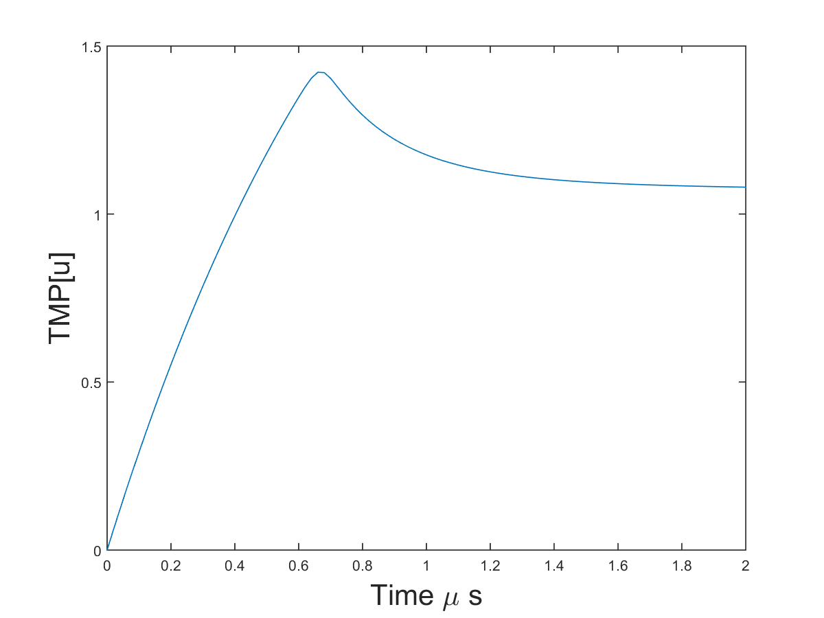
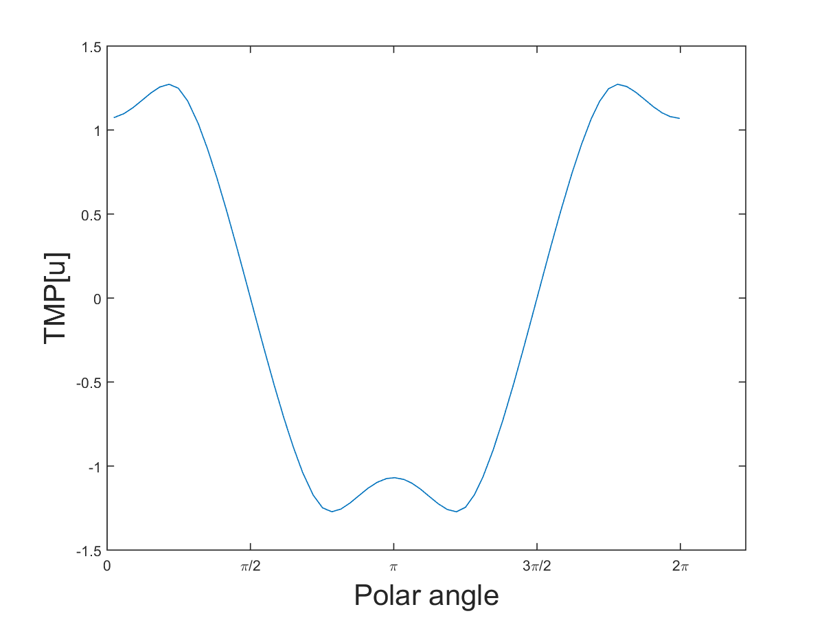
All the parameters are given in Table 1. Figure 1 shows the results for the time evolution and the voltage after .


5.2. Homogenization for electropermeabilization model
In this section, we show the sensitivity of the effective parameters , , and in (32) to
-
•
the conductivities and ;
-
•
the shape of the cell with membrane ;
-
•
the volume fraction ;
-
•
the lattice of the cells in the domain .
We perform four experiments, the results of which are found in Table 2.

Example 1. We fix the shape and size of the cell and change the ratio of the interior and exterior conductivities and .
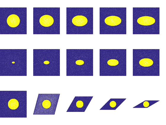
Example 2. In this example, we show how the shape of the cell membrane produces different effective anisotropy properties. We fix conductivities and the volume fraction of the cell, but take as cell shapes ellipses with different excentricity .
Example 3. We investigate the effect of different volume fractions of a cell with the same shape.
Example 4. In this example, we show how the angle of the lattice in which the cells are arranged affects the effective parameters.
For all these experiments, Table 2 presents the reactions of the effective conductivity and the effective anisotropy properties and to the microscopical change. One sees clearly that , as well as and react to a change of cell and conductivity parameters. Most of the sensitivity functions are in fact monotonic.
The best contrast is seen in:
-
•
the reaction of to the change in conductivity and to a change in the lattice angle ;
-
•
the reaction of both and to the cell shape.
The volume fraction alone does not show so much contrast in the anisotropy of the effective parameters.
Given the results of the sensitivity analysis, it is promising to infer shape parameters from macroscopic effective properties in electropermeabilization, as it was done in [3] from multifrequency admittivity measurements.
| effective conductivity | eigenvalues of | eigenvalues of . |
| Example 1: Difference in conductivity (ratio of interior and exterior conductivity). | ||
![[Uncaptioned image]](/html/1603.00764/assets/sigmaratio_sigma0.png) |
![[Uncaptioned image]](/html/1603.00764/assets/sigmaratio_A0.png) |
![[Uncaptioned image]](/html/1603.00764/assets/sigmaratio_A1.png) |
| Example 2: Difference in cell shape: change of the excentricity (see Fig. 2, 1st row). | ||
![[Uncaptioned image]](/html/1603.00764/assets/abratio_sigma0.png) |
![[Uncaptioned image]](/html/1603.00764/assets/abratio_A0.png) |
![[Uncaptioned image]](/html/1603.00764/assets/abratio_A1.png) |
| Example 3: Difference in volume fraction of the cells (see Fig. 2, 2nd row). | ||
![[Uncaptioned image]](/html/1603.00764/assets/fv_sigma0.png) |
![[Uncaptioned image]](/html/1603.00764/assets/fv_A0.png) |
![[Uncaptioned image]](/html/1603.00764/assets/fv_A1.png) |
| Example 4: Difference in angle of the lattice arrangement (see Fig. 2, 3rd row). | ||
![[Uncaptioned image]](/html/1603.00764/assets/Tratio_sigma0.png) |
![[Uncaptioned image]](/html/1603.00764/assets/Tratio_A0.png) |
![[Uncaptioned image]](/html/1603.00764/assets/Tratio_A1.png) |
6. Concluding remarks
We introduced a homogenization scheme relating critical microscopic and macroscopic quantities in electropermeabilization. The sensitivity analysis of the effective parameters showed this dependence and opens the door to solve the inverse problem to monitor those critical microscopic quantities in practice.
While setup optimization for electropermeabilization has been studied using computer simulations, for instance, in [12, 20, 5, 13, 11], from our approach comes an additional constraint: for mapping of the effective parameters and , two currents have to be applied which are nowhere parallel. An electrode configuration providing this allows for unique reconstruction [9].
Appendix A Convergence for homogenization
We give here the outline of the method used in [2]. It shows how Lemma 4 is used to prove Theorem 3 for our application.
Theorem 4.
Proof.
From the estimate (27) we get, extracting subsequences if needed
| (39) | |||||
Next, consider the weak formulation of system (26):
| (40) |
The general idea is to pass to the limit in this equation, and therefore to obtain the equation for . This is possible for special test functions .
Choose for the functions for , where is a smooth with compact support on , and is built by the cell functions and :
For this definition, given in [2, (5.1)] one has the weak formulation in [2, (5.2)-(5.4)].
By subtracting the weak equation (40) for and the equations [2, (5.2)-(5.4)], one can isolate the term :
| (41) |
with
| (42) | ||||
The limits of and are the same as in [2, p.18], whereas for the limit , one can show that by Lemma 4. One can take then the limit in (41) in order to obtain information on the specific form of the limit in (39). We get
| (43) |
with , , defined as in (33). Choosing in (40), combining with (43), and differentiating in gives then expressions which show that and that actually (32) is the correct equation of the limit .
∎
References
- [1] H. Amann. Ordinary differential equations. An introduction to nonlinear analysis. de Gruyter Studies in Mathematics. Walter de Gruyter, Berlin, New York, 1990.
- [2] M. Amar, D. Andreucci, P. Bisegna, and R. Gianni. Evolution and momory effects in the homogenization limit for electrical conduction in biological tissues. Math. Models Methods Appl. Sci., 14(09):1261–1295, 2004.
- [3] H. Ammari, J. Garnier, L. Giovangigli, W. Jing and J.K. Seo, Spectroscopic imaging of a dilute cell suspension, J. Math. Pure Appl., doi:10.1016/j.matpur.2015.11.009.
- [4] J. Dermol and D. Miklavčič. Predicting electroporation of cells in an inhomogeneous electric field based on mathematical modeling and experimental CHO-cell permeabilization to propidium iodide determination. Bioelectrochemistry, 100:52–61, 2014.
- [5] A. Golberg and B. Rubinsky. Towards electroporation based treatment planning considering electric field induced muscle contractions. Technol. Cancer. Res. Treat., 11(2):189–201, 2012.
- [6] A. Ivorra. Tissue electroporation as a bioelectric phenomenon: Basic concepts. In B. Rubinsky, editor, Irreversible Electroporation, Series in Biomedical Engineering, pages 23–61. Springer, Berlin, Heidelberg, 2010.
- [7] A. Ivorra, J. Villemejane, and L. M. Mir. Electrical modeling of the influence of medium conductivity on electroporation. Phys. Chem. Chem. Phys., 12:10055–10064, 2010.
- [8] O. Kavian, M. Leguèbe, C. Poignard, and L. Weynans. ”Classical” electropermeabilization modeling at the cell scale. J. Math. Biol., 68:235–265, 2014.
- [9] Y. J. Kim, O. Kwon, J. K. Seo, and E. J. Woo. Uniqueness and convergence of conductivity imge reconstruction in magnetic resonance electrical impedance tomography. Inverse Probl., 19:1213–1225, 2003.
- [10] M. Kranjc, B. Markelc, F. Bajd, M. Čemažar, I. Serša, T. Blagus, and D. Miklavčič. In situ monitoring of electric field distribution in mouse tumor during electroporation. Radiology, 274(1):115–123, 2015.
- [11] D. Miklavčič, K. Beravs, D. Šemrov, M. Čemačar, F. Demsar, and G. Serša. The importance of electric field distribution for effective in vivo electroporation of tissues. Biophys. J., 74:2152–5158, 1998.
- [12] D. Miklavčič, M. Snoj, A. Zupanic, B. Kos, M. Čemažar, M. Kropivnik, M. Bracko, T. Pecnik, E. Gadzijev, and G. Serša. Towards treatment planning and treatment of deep-seated solid tumors by electrochemotherapy. Biomed. Eng. Online, 9(10), 2010.
- [13] D. Miklavčič, D. Šemrov, H. Mekid, and L. M. Mir. A validated model of in vivo electric field distribution in tissues for electrochemotherapy and for DNA electrotransfer for gene therapy. Biochim. Biophys. Acta, 1523:73–83, 2000.
- [14] J. C. Neu and W. Krassowska. Asymptotic model of electroporation. Phys. Rev. E, 59(3):3471–3482, 1999.
- [15] M. Pavlin, N. Pavšelj, and D. Miklavčič. Dependence of induced transmembrane potential on cell density, arrangement and cell position inside a cell system. IEEE Trans. Biomed. Eng., 49(6):605–612, 2002.
- [16] M. Pavlin, T. Slivnik, and D. Miklavčič. Effective conductivitiy of cell suspensions. IEEE Trans. Biomed. Eng., 49(1):77–80, 2002.
- [17] P.-O. Persson and G. Strang. A simple mesh generator in MATLAB. SIAM Rev., 46(2):329–345, 2004.
- [18] G. Pucihar, T. Kotnik, B. Valič, and D. Miklavčič. Numerical determination of transmembrane voltage induced on irregularly shaped cells. Ann. Biomed. Eng., 34(4):642–652, 2006.
- [19] J.K. Seo and E.J. Woo, Magnetic resonance electrical impedance tomography (MREIT), SIAM Rev., 53 (2011), 40–68.
- [20] K. Sugibayashi, M. Yoshida, K. Mori, T. Watanabe, and T. Hasegawa. Electric field analysis on the improved skin concentration of benzoate by electroporation. Int. J. Pharm., 219:107–112, 2001.
- [21] L. Towhidi, Kotnik. T., G. Pucihar, S. M. P. Firoozabadi, H. Mozdarani, and D. Miklavčič. Variability of the minimal transmembrane voltage resulting in detectable membrane electroporation. Electromagn. Biol. Med., 27:372–385, 2008.