Measuring the defect structure orientation of a single NV- centre in diamond
Abstract
The negatively charged nitrogen-vacancy (NV-) centre in diamond has many exciting applications in quantum nano-metrology, including magnetometry, electrometry, thermometry and piezometry. Indeed, it is possible for a single NV- centre to measure the complete three-dimensional vector of the local electric field or the position of a single fundamental charge in ambient conditions. However, in order to achieve such vector measurements, near complete knowledge of the orientation of the centre’s defect structure is required. Here, we demonstrate an optically detected magnetic resonance (ODMR) technique employing rotations of static electric and magnetic fields that precisely determines the orientation of the centre’s major and minor trigonal symmetry axes. Thus, our technique is an enabler of the centre’s existing vector sensing applications and also motivates new applications in multi-axis rotation sensing, NV growth characterization and diamond crystallography.
pacs:
76.30.Mi; 76.70.Hb1 Introduction
The negatively charged nitrogen-vacancy (NV-) centre is a remarkable point defect in diamond [1] that is at the frontier of quantum technology. In particular, the NV- centre has many exciting applications as a highly sensitive quantum sensor in nano-metrology, including magnetometry [2, 3, 4, 5], electrometry [6, 7], thermometry [8, 9, 10, 11, 12], piezometry [13], and gyroscopy [14, 15]. High sensitivity is principally achieved by the long-lived coherence of the centre’s electron spin, which persists in ambient and extreme conditions [8, 16, 17]. Nanoscale sensing is enabled by the atomic size of the NV- centre, its bright fluorescence that allows single centres to be located with nano-resolution and its mechanism of optical spin-polarization/ readout that allows the magnetic resonance of its electron spin to be optically detected [1].
Single NV- centres have demonstrated the ability for three-dimensional vector sensing of electric fields [6] with single fundamental charge sensitivity [7]. NV- electrometry is achieved by observing the spin resonances as an applied magnetic field is rotated. Each vector component of the unknown electric field can be determined from its combined effect with the applied magnetic field, as long as the precise orientation of the magnetic field with respect to the defect structure of the NV- centre is known. The latter requirement has not yet been fulfilled and, as a consequence, complete NV- vector electrometry has not been possible. In principle, vector magnetic field sensing could also be achieved using a single NV- centre by an analogous technique, where an applied electric field is rotated in place of an applied magnetic field, but this is yet to be demonstrated. Whilst vector magnetic field sensing has been demonstrated using an ensemble of at least four differently orientated NV- centres [21, 22, 23, 24, 25, 26], a demonstration using a single NV- centre is likely to have several advantages due to the superior spin coherence of the single centre and its potential for greater spatial resolution. Here, we describe a technique for the measurement of the defect structure orientation of a single NV- centre relative to applied electric/ magnetic fields, which is key to the realization of high-sensitivity NV- vector sensing.
More generally, this technique will also prove useful for material science because it is prototypical for measuring the defect structure of spin defects with trigonal symmetry, such as (hh) or (kk) divacancies in silicon carbide [18]. The ability to precisely characterise which orientations of NV- centres are formed as the result of different fabrication methods will provide an invaluable tool to the study of the NV- growth mechanisms [19]. Such detailed knowledge of the growth mechanisms can be used to improve the fabrication of homogeneous ensembles of NV- centres, within which all centres are aligned and therefore can be more effectively employed in hybrid quantum devices (e.g. coupled superconducting resonators and NV- spin ensembles [20]). Measuring the structure of a single NV- centre also allows for local characterisation of the diamond lattice. In this way, the structure of nanodiamonds or polycrystalline sectors can be non-invasively determined.
The orientation measurement technique described here may be directly used to realize three-axis rotation sensing using a single NV- centre. Three-axis rotation sensing can be achieved in two different geometries (see figure 1): (1) the NV- diamond rotates with respect to a fixed apparatus of electric and magnetic fields or (2) the apparatus rotates with respect to a fixed NV- diamond. A possible example of the first geometry is the sensing of the rotational dynamics of a nanodiamond within a biological cell [27]. An example of the second geometry is a high sensitivity rotation sensor for a micro-mechanical system, where one element of the system is free to rotate with respect to another element that contains a NV- diamond. Notably, there has been related proposals of single-axis NV- gyroscopy using single centres [14] and ensembles [15]. These proposals differ from the one made here, as they are sensitive to the rotational frequency about a single rotational axis, not the rotational coordinates of a rotation about possibly all three rotational axes.

In this paper, we demonstrate such a technique for measuring the defect structure orientation of a single NV- centre. After further introduction to the properties of the NV- centre, we first derive the relationship between the spin resonances of a NV- centre in the presence of combined electric and magnetic fields and its defect structure. The orientation measurement technique arises naturally form the derivation and we subsequently demonstrate the technique by measuring the defect structure orientation of a single NV- centre. The latter measurement, in essence, mimics our second proposed geometry for rotation sensing. Based upon our demonstration, we finally examine the application of the technique to three-axis rotation sensing using single NV- centres.
2 Theory of the orientation measurement technique
The NV- centre is a point defect in diamond consisting of a substitutional nitrogen atom adjacent to a carbon vacancy trapping an additional electron (refer to figure 2a). The orientation of the centre’s defect structure is characterised by the directions of its major and minor symmetry axes. The trigonal structure of the centre has a major symmetry axis that is defined by the direction joining the nitrogen and vacancy. For a given crystal orientation, the centre’s [111] major symmetry axis has four possible alignments (see figure 2b). The centre’s minor symmetry axis is defined as being orthogonal to its major symmetry axis and also contained within one of the centre’s three reflection planes (eg. ), which corresponds to the direction joining a point on the centre’s major symmetry axis and one of the vacancy’s nearest neighbour carbon atoms. Considering an isolated single NV- centre, if the alignment of its major symmetry axis is known (i.e. via magnetic field alignment), then even with knowledge of the crystal orientation, the orientation of the centre’s defect structure is not fully determined.

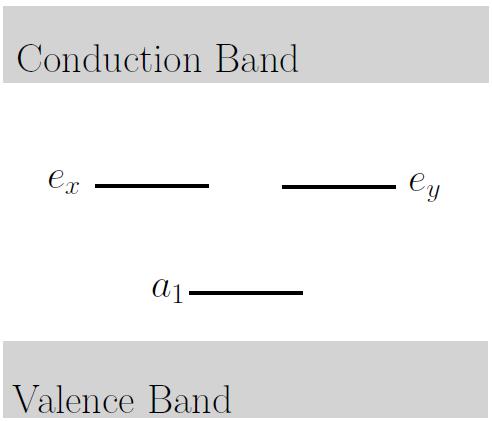
As depicted in figure 3a, the one-electron orbital level structure of the NV- centre contains three defect orbital levels (, and ). EPR observations and ab initio calculations indicate that these defect orbitals are highly localized to the centre [28, 29, 30, 31]. Figure 3b shows the centre’s many-electron electronic structure generated by the occupation of the three defect orbitals by four electrons [32, 33], including the zero phonon line (ZPL) energies of the optical (1.945 eV/637 nm) [34] and infrared (1.190 eV/1042 nm) [35, 36, 37] transitions. The energy separations of the spin triplet and singlet levels ( and ) are unknown.
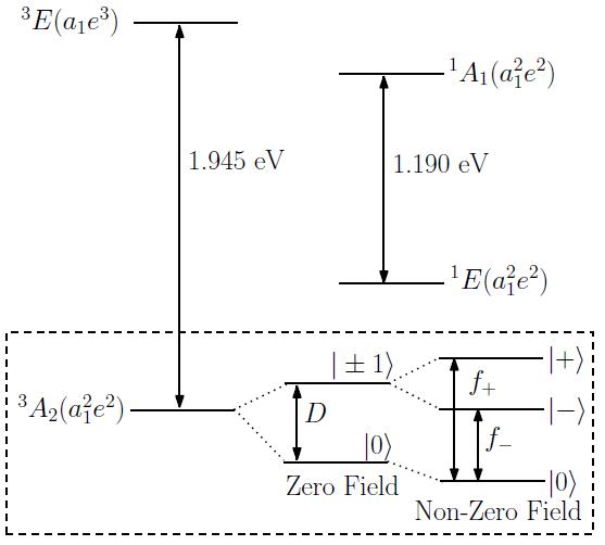
As depicted in the inset of figure 3b, the ground level exhibits a zero field fine structure splitting between the and spin sub-levels of GHz, which is principally due to first-order electron spin-spin interaction [38]. The spin quantization axis is thus defined by the trigonal unpaired electron spin density distribution of the ground level to be parallel to the centre’s major symmetry axis. Spin-orbit and spin-spin mixing of the and levels makes the fine structure susceptible to electric fields, yet does not significantly perturb the g-factor of the spin magnetic interaction from its free electron value [39].
A detailed derivation of the spin-Hamiltonian that describes the fine structure in the presence of electric and magnetic fields has been previously reported [39]. The spin-Hamiltonian derived in [39] is
| (1) | |||||
where are the dimensionless electron spin operators, , is the Bohr magneton, is the electron g-factor [29], is the Planck constant, and are the magnetic and electric fields, respectively, and kHz m/V and kHz m/V are the electric susceptibility parameters [40].
Although the spin-Hamiltonian derivation in [39] is detailed, there is limited discussion of the relationship between the spin-Hamiltonian and the defect structure of the NV- centre, in particular, the physical basis for the adopted coordinate system and the behaviour of the spin-Hamiltonian under coordinate transformations. Consequently, we present a brief review of the spin-Hamiltonian derivation in appendix A. The derivation proceeds by the application of perturbation theory to the fine structure interactions of the NV- centre and ends in the definition of each of the spin-Hamiltonian parameters in terms of integrals of the centre’s orbital wavefunctions. The key outcome of the review is the identification of the implicit coordinate system definition in (1) as the choice of the and coordinate axes as being directed along the centre’s major and minor symmetry axes, respectively, such that the electric susceptibility parameters are positive. As demonstrated in appendix B, a simple molecular orbital argument can be applied to the evaluation of the orbital integrals of the electric susceptibility parameters to show that they are positive when the coordinate axis is directed from the nitrogen towards the vacancy and when the coordinate axis is directed from the coordinate axis towards one of the vacancy’s three nearest-neighbour carbons (see figure 2a). Since there are three equivalent choices of the coordinate direction due to the centre’s trigonal symmetry, the spin-Hamiltonian (1) is invariant under rotations about the major symmetry axis.
Having established the relationship between the spin-Hamiltonian and the centre’s defect structure, we now discuss the orientation dependence of the centre’s observable spin resonances in the presence of electric and magnetic fields. The zero-field spin states (defined by spin-projection) are mixed in the presence of electric and magnetic fields to form a new state basis (refer to figure 3b) [39]. The frequencies of the spin transitions are [7]
| (2) |
where , , , , , , and . Note that the validity of the above expression is constrained by the conditions .
In the presence of either an electric or a magnetic field, the spin frequencies reduce to
| (3) |
where , , and . In each case, the spin frequencies depend on the alignment of the electric/magnetic field with the centre’s major symmetry axis (i.e. and ), but do not depend on the transverse orientation of the electric/ magnetic field (i.e. and ). This invariance to the transverse orientation of an individual field is due to the equal spin susceptibility to and field components, which is itself a consequence of the centre’s trigonal symmetry.
If the electric/ magnetic field is near alignment to the centre’s major symmetry axis, the spin frequencies obtain their maximum/ minimum values, such that
| (4) |
which may be used as a condition to identify the orientation of the symmetry axis. However, noting that , it can not be determined whether the magnetic field is parallel () or anti-parallel () with the coordinate axis. This is not the case for the electric field, where the observable sign of the common shift is directly dependent on whether the electric field is parallel or anti-parallel with the coordinate axis. Hence, whilst either electric or magnetic fields can be used to determine the orientation of the centre’s major symmetry axis, only an electric field can differentiate the precise direction of the centre’s coordinate axis.
This conclusion forms the basis of a technique for measuring the orientation of the coordinate axis (refer to figure 4). Since the magnetic susceptibility of the ground state spin is much greater than its electric susceptibility, the most sensitive method is to first rotate a known magnetic field until it is aligned with the centre’s major symmetry axis and then apply a parallel electric field to determine the direction of the coordinate axis. Although the electric field shift is typically small, as long as it can be detected, it will be sufficient because only its sign is relevant.

It is clear from Eq. (2), that the spin frequencies become dependent on the transverse orientations of electric and magnetic fields if they are simultaneously applied. This dependence obtains a maximum when the fields are transverse to the major symmetry axis
| (5) | |||||
where the approximation in the second line has been taken in the limit , which is relevant to typical experimental conditions [6, 7]. For an explanation of the transverse susceptibility see appendix A.
Since the second term of (5) leads to a splitting of the spin frequencies that is independent of the transverse field orientations, the variations of the spin frequencies as the fields are rotated may be individually observed and are governed by the third term, such that
| (6) |
Figure 5 depicts for the cases where the orientation of one field is fixed and the other is rotated. As discussed in the demonstrations of NV- vector electrometry [6] and single charge detection [7], the mixed argument of the cosine function implies that to determine the transverse orientation of an unknown electric field by observing as a transverse magnetic field is rotated, one requires knowledge of the orientation of the transverse magnetic field with respect to the centre’s coordinate system (i.e. ). The opposite is also true for the measurement of the transverse orientation of an unknown magnetic field via a rotation of a transverse electric field. This interdependence motivates the simultaneous rotation of transverse electric and magnetic fields as a means to determine the transverse orientation of the centre’s coordinate system/ defect structure.

Defining the combined electric-magnetic field angles and , the orientation dependence of the spin frequencies become
| (7) |
If is fixed whilst is varied by the fields being simultaneously rotated in the same direction at the same rate, the variation of (see figure 5) immediately reveals the trigonal structure of the NV- centre. Indeed, if the fields are parallel, such that , the three maxima of directly correspond to when the fields are directed along the three equivalent coordinate axes of the centre (i.e. the directions from the major symmetry axis to the vacancy’s three nearest-neighbour carbons).

This conclusion forms the basis of a technique for measuring the orientation of the coordinate axis (refer to figure 6). Having first determined the orientation of the centre’s major symmetry axis using the technique discussed previously, parallel electric and magnetic fields can be aligned such that they are transverse to the -axis. If the fields are rotated together as above, the direction of the coordinate axis can be identified as the field direction corresponding to one of the three maxima of .
3 Demonstration of the orientation measurement technique
In this section we present a proof-of-principle demonstration of the orientation measurement technique. We performed ODMR measurements on an as-grown single NV- centre in a bulk CVD diamond sample with a orientated surface. We independently verified the crystallographic directions by x-ray diffraction. However, note that this is no requirement for our method. Our experimental setup is sketched in figure 7a. Three orthogonal magnetic field coils allowed a magnetic field of G to be aligned and rotated in the transverse plane. A two-dimensional quadrupole electrode microstructure was engineered onto the diamond surface using gold lithography and similarly allowed an electric field to be aligned and rotated in the transverse plane. Microwaves were applied to the sample by two orthogonal wires underneath the diamond sample. Optical excitation with 532 nm light and red-shift fluorescence collection were achieved using a confocal microscope.

The dependence of on the orientation of the transverse electric and magnetic fields was observed using a modified spin-echo ODMR pulse sequence (see figure 7b). In the modified sequence, the last -pulse is phase-shifted by to the other two pulses to enable the measurement of the sign of the accumulated phase (i.e. the direction of the energy level shift). The microwave pulses were separated by a fixed delay time s, during which the transverse electric field was applied with a sinusoidal magnitude . The application of the electric field during the pulse sequence results in a net accumulated phase difference between and , which is converted into an optically detectable population difference between the spin states by the final -pulse.

Figure 8 depicts the observed behaviour of for the three distinct rotations of the transverse electric and magnetic fields. In each case, the measurements agree well with the theoretical prediction and any small differences can be explained by small deviations of the fields from transverse alignment as they are rotated. The strength of the measured electric field could be calculated to Vm-1 for the maximum applied voltage of V, which compares to a simulated electric field strength of Vm-1 for such a quadrupole structure, depending on the exact position of the NV centre within the structure. As discussed in the previous section, because the electric and magnetic fields are parallel (), the trigonal pattern of figure 8c directly corresponds to the orientation of the centre’s trigonal defect structure and would be shifted by for a differently oriented NV. EPR and crystallographic results concerning the direction of the minor symmetry axis coincide within the error margins.
4 Discussion of applications in multi-axis rotation sensing
The proposed concept of three-axis rotation sensing using a single NV- centre is to employ the orientation measurement technique to measure the orientations of the centre’s and coordinate axes at discrete intervals (refer to figure 9). From the discrete set of coordinate orientation measurements , the three-axis rotations that occurred during each time interval, such that and , can be reconstructed using
| (8) | |||||
where and are the axis and angle of the rotations. Note that the rotational dynamics are assumed to be slow enough such that there is no appreciable rotation during each orientation measurement. Given an apparatus where the direction of the parallel fields are precisely known with respect to a chosen reference frame, the accuracy of the orientation measurements will thus be determined by the sensitivity of the ODMR signal to misalignment of the fields.

The critical characteristic of our rotation sensing proposal is the average time required to perform an orientation measurement, which depends on the the average number of ODMR measurements for achieving a sufficient angle estimate for each axis and the relevant coherence time that determines the time of each ODMR measurement. NV- quantum sensing is performed via the measurement of spin frequencies using ODMR pulse sequences, such as the Ramsey, spin-echo and more advanced dynamical decoupling sequences. In essence, during a pulse sequence a coherent superposition of two spin states (e.g. and ) acquires a phase with a certain frequency. The latter is then sensitive to various quantities. For a single NV center and practical experimental parameters, the frequency sensitivity is
| (9) |
where given an average fluorescence photon count rate (under cw illumination with readout laser intensity) of kcounts, an ODMR contrast of and an exponential decay of the Ramsey or spin-echo-like signal over the phase accumulation time . For ms, this yields a frequency sensitivity of .
For an appropriate experimental setting we can convert this frequency sensitivity into an angle sensitivity for or . In particular, we might perform combined ac-magnetometry as depicted in fig. 9a (see also ref. [16]) and ac-electrometry as illustrated in figs. 7 and 9b. In that case, we divide the frequency sensitivity by the derivative of the frequency shift with respect to the angle or (assuming a superposition of spin states and ) at the corresponding working point
| (10) | |||
| (11) |
Using practical field magnitudes of G and Vm-1 we obtain
| (12) | |||
| (13) |
Even higher sensitivities might be achieved when using superposition states of and . In that case the derivatives in equations (10) and(11) do increase and we obtain for the sensitivities
| (14) | |||
| (15) |
In the following we describe a measurement scenario based on a rotating NV diamond and adjustable electric and magnetic fields in the lab frame (see fig. 1b). In the case of a slow rotation speed compared to the measurement rate, for both the estimation of the and the direction we perform each two measurements. Starting from the expected/ previous axis we apply magnetic fields with and two values which differ by (see fig. 9a). The result is an updated/ actual direction. For the estimation of the axis we apply parallel electric and magnetic fields transverse to the actual direction and with angles with respect to the expected axis (see fig. 9b). Finally, and directions are updated.
If we target at a similar accuracy for and we can neglect the time for the estimation measurement compared to the estimation measurement because of the large mismatch of the respective sensitivities. Therefore, the overall angle sensitivity for three-axis rotation sensing is equal to given the above assumptions. For a total three-axis rotation measurement time of s, this corresponds to an angular accuracy of 0.02 , which compares favourably to a previous single-axis rotational sensing demonstration where the rotational dynamics of a nanodiamond within a biological cell were deg/ hour [27]. Faster rotational dynamics can be captured at the expense of angular accuracy. For example, if a much smaller ODMR measurement time s is used, the total three-axis measurement rate is increased by three orders of magnitude at the expense of a 33 fold decrease in angular accuracy. Of course, in a real three-axis rotational sensing experiment, it is important to adjust the estimation rate to the rotational dynamics in order to exploit maximum possible sensitivities. The measurement sequence proposed here may also be improved or tailored to the specific sensing task in order to optimize sensitivity. Given these considerations, it is clear that since three-axis rotational sensing enables the rotational dynamics of a nanodiamond to be almost completely reconstructed, the technique proposed here may have significant applications in the study of the internal mechanics of biological cells.
5 Conclusion
In this paper, we have demonstrated an ODMR technique employing rotations of static electric and magnetic fields that precisely measures the orientation of the NV- centre’s defect structure. We developed our technique through a theoretical examination of the relationship between the centre’s observable spin resonances and its defect structure and showed that, by observing the variation of the spin resonances as parallel electric and magnetic fields are rotated, the fields can be aligned with specific directions within the centre’s defect structure. Whilst our technique is a vital enabler of the centre’s existing vector sensing applications, it also motivates new applications in multi-axis rotation sensing, NV growth characterization and diamond crystallography. Indeed, the application of our technique to multi-axis rotation sensing of nanodiamonds within biological cells has significant potential in the study of internal cellular mechanics.
Appendix A Spin-Hamiltonian derivation
In this appendix, we briefly review the detailed derivation of the ground state spin-Hamiltonian (1) presented in [39]. Through the application of perturbation theory, it was shown in [39] that spin-orbit and spin-spin coupling between the ground and excited levels results in the ground state spin becoming susceptible to electric fields, but does not result in its g-factor being significantly perturbed from the free electron value. Here, we present an abridged derivation that highlights the relationship between the spin-Hamiltonian and the defect structure of the NV- centre.
We begin with the centre’s electronic Hamiltonian
| (16) | |||||
where is the purely orbital Hamiltonian that includes electronic kinetic energy and electron-nucleus and electron-electron electrostatic interactions, is spin-orbit interaction, is spin-spin interaction, is the electric dipole interaction, and includes orbital and spin magnetic interactions. Note that , and are spherically symmetric and invariant to coordinate transformations and can each be written as linear combinations of products of orbital and spin operators [32].
The LS-coupling states corresponding to the triplet levels are [32]
| (17) |
where are orbital wavefunctions that transform as the row of the irreducible representation of the group and are the triplet spin states with . It is important to note that the orbital wavefunctions are defined by the centre’s defect structure. The labels and recognise the fact the orbital wavefunctions transform analogously to the coordinates under the operations, if the coordinates are defined such that the and coordinate directions are aligned with the centre’s major and (one of the three) minor symmetry axes. Hence, as in [39], it is natural to adopt this coordinate system that is aligned with the centre’s defect structure.
The LS-coupling states are eigenstates of and thus form the basis for the perturbative calculation of the fine structure interactions . Since it was shown in [39] that only interactions between the triplet levels perturb the ground state fine structure, the ground state spin-Hamiltonian may be immediately constructed to second-order in terms of orbital integrals as
| (18) | |||||
where eV is the energy separation of the triplet levels and it is to be understood that the spin operators of that remain after the evaluation of the orbital integrals act upon the triplet spin states . The above expression demonstrates that is symmetric, since its behaviour under a coordinate transformation will be determined by the behaviour of the symmetric orbital wavefunctions.
Evaluating the orbital integrals in the above yields the final expression of the spin-Hamiltonian (1). Introducing the centre’s defect orbitals (, and ), the explicit expressions for the electric susceptibility parameters in the adopted coordinate system are [39]
| (19) |
where
| (20) |
are transverse and axial electric dipole moments,
| (21) | |||||
is an orbital integral of spin-spin interaction, is the fundamental charge, is the permeability of free space, is the position of the electron, , and and are real spin-coupling coefficients formed from linear combinations of spin-orbit and spin-spin interaction integrals (refer to [32, 39] for further details).
The above demonstrates that the electric susceptibility parameters are products of the electric dipole and spin-spin interactions between the triplet levels. Consequently, the electric field interaction may be interpreted as resulting in the lowering of the symmetry of the ground state spin density. As discussed in section 2, whilst the adopted coordinate system is aligned with the centre’s symmetry axes, there is still freedom to choose the precise coordinate directions so that electric susceptibility parameters are positive. Since the spin-coupling coefficients are real, and thus , only the electric dipole determines the sign of . Both the spin-spin and electric dipole contributions to determine its sign. However, notably the spin-spin interaction is invariant to a coordinate inversion because it is quadratic in the coordinates. It is shown in appendix B that the electric susceptibility parameters are positive for the coordinate system definition depicted in figure 2a, where the coordinate axis is directed from the nitrogen towards the vacancy and when the coordinate axis is directed from the coordinate axis towards one of the vacancy’s three nearest-neighbour carbons.
Appendix B Molecular orbital calculation of the electric susceptibility parameters
In this appendix, the well established molecular model of the centre [32, 33] is drawn upon to perform a molecular orbital calculation of the sign of the electric susceptibility parameters. Similar calculations have been used to establish basic aspects of the temperature and pressure response of the ground state spin [12, 13]. The molecular model exploits the highly localized nature of the centre’s defect orbitals to approximate the orbitals as linear combinations of the dangling sp3 atomic orbitals (,,,) of the vacancy’s nearest neighbor nitrogen and carbon atoms (refer to figure 10a) [12]
| (22) |
where is a real linear coefficient and
| (23) |
are normalization constants.
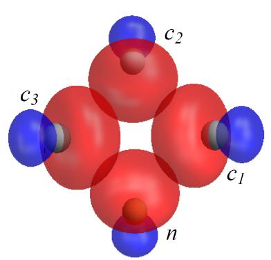
Expanding the electric dipole integrals (20) and spin-spin integral (21) using the above defect orbital definitions, applying symmetry operations and ignoring orbital overlaps, the integrals become
| (24) | |||||
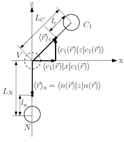
Using knowledge of the approximately tetrahedral nuclear geometry of the NV centre, the one-electron atomic orbital integrals within the electric dipole integrals can be evaluated. For the purpose of determining the sign of the electric dipole integrals, we can proceed with simple geometric arguments. For the carbon sp3 atomic orbitals, the expected position () of an electron occupying one of those orbitals occurs at a distance towards the vacancy away from the carbon [13]. Likewise, the expected position of an electron occupying the nitrogen orbital occurs at a distance towards the vacancy away from the nitrogen. The distances between the vacancy and the carbon and nitrogen atoms are and , respectively [31]. Consequently, through inspection of the geometry (refer to figure 10b), it is clear that both electric dipole integrals are positive.
Unlike the electric dipole integrals, the spin-spin integral contains two-electron direct integrals between densities of two carbon sp3 orbitals. For the purpose of determining the sign of the spin-spin integral, the difficulty of evaluating these direct integrals can be avoided and a semi-classical approximation can be performed instead, where the direct integrals between atomic orbitals are replaced by expressions containing the expected positions of the electrons occupying the orbitals [13]
| (25) |
Using these semi-classical expressions and the geometry of the centre (refer to figure 10c), the spin-spin integral becomes
| (26) |
which is clearly positive. Given that each of the electric dipole and spin-spin integrals are positive, it follows from (19) that the electric susceptibility parameters are also positive for the adopted coordinate system.
References
References
- [1] Doherty M W , Manson N B, Delaney P, Jelezko F and Hollenberg L C L 2013 Physics Reports 528 1
- [2] Hong S, Grinolds M S, Phama L M, Le Sagea D, Luana L, Walsworth R L and Yacoby A 2013 MRS Bull. 38 155
- [3] Grinolds M S, Hong S, Maletinsky P, Luan L, Lukin M D, Walsworth R L and Yacoby A 2013 Nature Physics 9 215
- [4] Mamin H J, Kim M, Sherwood M H, Rettner C T, Ohno K, Awschalom D D and Rugar D 2013 Science 339 557
- [5] Staudacher T, Shi F, Pezzagna S, Meijer J, Du J, Meriles C A, Reinhard F and Wrachtrup J 2013 Science 339 561
- [6] Dolde F, Fedder H, Doherty M W, Nöbauer T, Rempp F, Balasubramanian G, Wolf T, Reinhard F, Hollenberg L C L, Jelezko F and Wrachtrup J 2011 Nature Physics 7 459
- [7] Dolde F, Doherty M W, Michl J, Jakobi I, Naydenov B, Pezzagna S, Meijer J, Neumann P, Jelezko F, Manson N B and Wrachtrup J 2014 Phys. Rev. Lett. 112 097603
- [8] Toyli D M, Christle D J, Alkauskas A, Buckley B B, Van de Walle C G and Awschalom D D 2012 Phys. Rev. X 2 031001
- [9] Toyli D M, de las Casas C F, Christle D J, Dobrovitski V V and Awschalom D D 2013 PNAS 110 8417
- [10] Neumann P, Jakobi I, Dolde F, Burk C, Reuter R, Waldherr G, Honert J, Wolf T, Brunner A, Shim J H, Suter D, Sumiya H, Isoya J and Wrachtrup J 2013 Nano Lett. 13 2738
- [11] Kucsko G, Maurer P C, Yao N Y, Kubo M, Noh H J, Lo P K, Park H and Lukin M D 2013 Nature 500 54
- [12] Doherty M W, Acosta V M, Jarmola A, Barson M S J, Manson N B, Budker D and Hollenberg L C L 2013 arXiv:1310.7303
- [13] Doherty M W, Struzhkin V V, Simpson D A, McGuinness L P, Meng Y, Stacey A, Karle T J, Hemley R J, Manson N B, Hollenberg L C L and Prawer S 2014 Phys. Rev. Lett. 112 047601
- [14] Maclaurin D, Doherty M W, Hollenberg L C L and Martin A M 2012 Phys. Rev. Lett. 108 240403
- [15] Ledbetter M, Jensen K, Fischer R, Jarmola A and Budker D 2012 Phys. Rev. A 86 052116
- [16] Balasubramanian G R et al 2009 Nature Materials 8 383
- [17] Pham L M, Bar-Gill N, Belthangady C, Sage D L, Cappellaro P, Lukin M D, Yacoby A and Walsworth R L 2012 Phys. Rev. B 86 045214
- [18] Isoya J, Umeda T, Mizuochi N, Son N T, Janzen E and Ohshima T 2008 phys. stat. sol (b) 245 7
- [19] Michl J, Teraji T, Zaiser S, Jakobi I, Waldherr G, Dolde F, Neumann P, Doherty M W, Manson N B, Isoya J and Wrachtrup J 2014 arXiv:1401.4106
- [20] Amsüss R, Koller C, Nöbauer T, Putz S, Rotter S, Sandner K, Schneider S, Schramböck M, Steinhauser G, Ritsch H, Schmiedmayer J and Majer J 2011 Phys. Rev. Lett. 107 060502
- [21] Steinert S, Dolde F, Neumann P, Aird A, Naydenov B, Balasubramanian G, Jelezko F and Wrachtrup J 2010 Review of Scientific Instruments 81 043705
- [22] Steinert S, Ziem F, Hall L T, Zappe A, Schweikert M, Götz N, Aird A, Balasubramanian G, Hollenberg L C L and Wrachtrup J 2013 Nature Communications 4 1607
- [23] Le Sage D, Arai K, Glenn D R, DeVience D J, Pham L M, Rahn-Lee L, Lukin M D, Yacoby A, Komeili A and Walsworth R L 2013 Nature 496 486
- [24] Maertz B J, Wijnheijmer A P, Fuchs G D, Nowakowski M E and Awschalom D D 2010 Appl. Phys. Lett. 96 092504
- [25] Pham L M, Sage D L, Stanwix P L, Yeung T K, Glenn D, Trifonov A, Cappellaro P, Hemmer P R, Lukin M D, Park H, Yacoby A and Walsworth R L 2011 New J. Phys. 13 045021
- [26] Hall L T, Beart G C G, Thomas E A, Simpson D A, McGuinness L P, Cole J H, Manton J H, Scholten R E, Jelezko F, Wrachtrup J, Petrou S and Hollenberg L C L 2012 Scientific Reports 2 401
- [27] McGuinness L P, Yan Y, Stacey A, Simpson D A, Hall L T, Maclaurin D, Prawer S, Mulvaney P, Wrachtrup J, Caruso F, Scholten R E and Hollenberg L C L 2011 Nature Nanotech. 6 358
- [28] He X F, Manson N B and Fisk P T H 1993 Phys. Rev. B 47 8816
- [29] Felton S, Edmonds A M, Newton M E, Martineau P M, Fisher D, Twitchen D J and Baker J M 2009 Phys. Rev. B 79 075203
- [30] Larsson J A and Delaney P 2008 Phys. Rev. B 77 165201
- [31] Gali A, Fyta M and Kaxiras E 2008 Phys. Rev. B 77 155206
- [32] Doherty M W, Manson N B, Delaney P and Hollenberg L C L 2011 New J. Phys. 13 024019
- [33] Maze J R, Gali A, Togan E, Chu Y, Trifonov A, Kaxiras E and Lukin M D 2011 New J. Phys. 13 025205
- [34] Davies G and Hamer M F 1976 Proc. R. Soc. Lond. A 348 285
- [35] Rogers L J, Armstrong S, Sellars M J and Manson N B 2008 New J. Phys. 10 103024
- [36] Acosta V M, Jarmola A, Bauch E and Budker D 2010 Phys. Rev. B 82 201202(R)
- [37] Manson N B, Rogers L, Doherty M W and Hollenberg L C L 2010 arXiv:1011.2840v1
- [38] Loubser J H N and van Wyk J A 1978 Rep. Prog. Phys. 41 1202
- [39] Doherty M W, Dolde F, Fedder H, Jelezko F, Wrachtrup J, Manson N B and Hollenberg L C L 2012 Phys. Rev. B 85 205203
- [40] van Oort E and Glasbeek M 1990 Chem. Phys. Lett. 168 529
- [41] Neumann P, Beck J, Steiner M, Rempp F, Fedder H, Hemmer P R, Wrachtrup J and Jelezko F 2010 Science 329 542