High-spin low-spin transition
Abstract
Temperature dependent nuclear inelastic-scattering (NIS) of synchrotron radiation was applied to investigate both spin states of the spin-crossover complex [Fe(tpa)(NCS)2] (tpa=tris(2-pyridylmethyl)amine). A remarkable increase of the iron-ligand bond stretching upon spin crossover has unambiguously been identified by comparing the measured NIS spectra of with theoretical simulations based on density-functional calculations.
:
This is an unedited preprint. The original publication is available at
http://www.springerlink.com
http://www.doi.org/10.1023/A:1017056831002
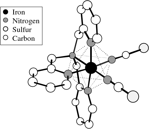
The iron(II) complex [Fe(tpa)(NCS)2] (Fig. 1, tpa = tris(2-pyridylmethyl) amine) belongs to the family of thermally driven spin-crossover complexes, which exhibit a transition from a low-spin (LS) to a high-spin (HS) state by increasing the temperature. These complexes are very promising materials for optical information storage [1].
IR measurements on several spin-crossover complexes with a central [FeN6] octahedron indicate a remarkable increase of the Fe-N bond stretching frequencies from about 25 to 30 meV in the HS state to about 50 to 60 meV in the LS state[2]. In this case nuclear inelastic scattering (NIS) turns out a very valuable alternative to IR and Raman spectroscopy because the Fe-N stretching modes can be definitely identified in the NIS spectra, whereas the IR and Raman spectra are rather complex in this frequency region [3] making an unambiguous assignment of these modes very difficult.
NIS spectra were recorded at the Nuclear Resonance Beamline ID 18 of the European Synchrotron Radiation Facility (ESRF) in Grenoble, France [4]. The 6 GeV electron storage ring was operated in 16 bunch mode and the purity of the filling (population of parasitic bunches compared to the single bunch) was better than . The incident beam was monochromatized by a double-crystal Si(111) premonochromator to the bandwidth of 2.5 eV at the energy of 14.413 keV. A further decrease of bandwidth down to 6 meV was obtained with a ”nested” high-resolution monochromator [5]. The sample was mounted in a closed-cycle cryostat to allow measurements at different temperatures. An avalanche photo-diode with mm2 area, 100 m active thickness, and a time resolution of less than 1 ns has been used as a detector [6] to count the 14.413 keV -quanta and, mainly, the K-fluorescence photons ( keV). The data were collected during several energy scans with 140 steps on average, each with 2 meV stepsize and 10 s measuring time. All individual scans were corrected for the approximately 9% decrease of beam intensity of the storage ring during the 1500 s required for each scan and added up afterwards.

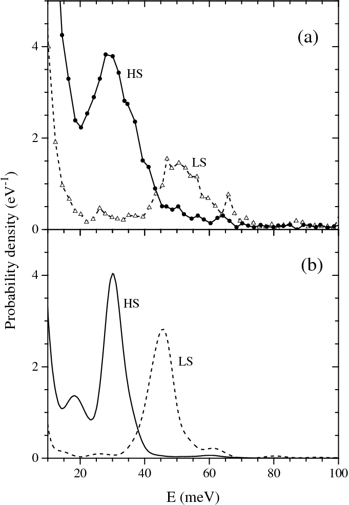
The NIS spectra of the HS and LS isomers of [Fe(tpa)(NCS)2] exhibit central peaks of 12 and 7 meV linewidth, respectively, and a pronounced inelastic peak at 30 meV in the HS state and at 50 meV in the LS state (Fig. 2)[7, 8, 9]. Comparing the intensity of the pronounced inelastic peaks in the HS and the LS spectrum it should be kept in mind, that the HS peak at 30 meV is located on the shoulder of the corresponding central peak. The linewidth of the LS peak observed at 50 meV is significantly larger than the linewidth of the corresponding central peak. The LS peak should, therefore, be regarded as a superposition of two or more individual peaks. The LS spectrum exhibits another, rather small peak at 66 meV, which is invisible in the HS spectrum.
The first momentum of the measured absorption probability density of the HS isomer (1.8 meV) is in reasonable agreement with the recoil energy = 1.96 meV of the free 57Fe nucleus, as expected according to Lipkin’s rule, [10] whereas the first momentum of the LS spectrum amounts to 4 meV, which is about twice as large as . This large measured is due to the increased attenuation of the incident radiation at nuclear resonance [11]. This phenomenon can be neglected if the elastic peak has a small weight . If however a large fraction of the spectrum belongs to the elastic peak the measured absorption probability density has to be corrected. For this reason the NIS spectrum of the LS isomer was normalized according to
| (1) |
The resolution function describes the energy distribution of the incident radiation and is assumed to be an even function.
For the HS and the LS isomers of [Fe(tpa)(NCS)2] electronic structure calculations were performed using the density functional theory (DFT) method B3LYP, [12] implemented in the gaussian94 program system [13] together with the split valence 3-21G* basis set [14]. The geometries were fully optimized applying the Berny algorithm to redundant internal coordinates [15]. Force constants were calculated analytically at the same level of theory using the optimized geometries, and the resulting vibrational frequencies were corrected by the scaling factor 0.9613 as has been proposed by Wong [16] for the 6-31G* basis set. The calculated normal modes for both isomers have been used to simulate the absorption probability density according to the procedure described in Ref[9].

No X-ray structures are available that can be compared with the calculated geometries, but the calculated bond lengths of the HS and LS isomers qualitatively resemble the increase of the Fe-N bond distances upon spin crossover of about 10 to 20 pm observed in various spin-crossover complexes with a central [FeN6] octahedron [2].
The vibrational spectra of the HS and the LS isomers of [Fe(tpa)(NCS)2] consist of 135 normal modes and are, in the following discussion, subdivided into a high-frequency region above 75 meV and a low-frequency region below 75 meV. The high-frequency region is of minor interest for the purposes of this study, since the vibrational modes in this frequency region do not contribute to the mean-square displacement (msd) of the iron nucleus and, therefore, can not be observed by NIS.
Among the 41 normal modes of the low-frequency region the iron-ligand bond stretching vibrations are of special interest here. Due to the almost octahedral environment of the iron center three out of six Fe-N stretching modes are invisible in NIS and IR spectra. Those modes that transform according to the A1 and Eg representations of the ideal octahedron do not contribute to the msd of the iron nucleus or to the variation of the electric dipole moment. Only the remaining three modes, that transform according to the T1u representations can be observed in NIS and IR spectra. These three modes, with calculated frequencies of 29.9, 30.1, and 35.3 meV for the HS state and 42.8, 46.6, and 52.6 meV for the LS state, give rise to prominent peaks in the simulated NIS spectra of both isomers of [Fe(tpa)(NCS)2]. Considerable contributions to the calculated absoption probability also arise from N-Fe-N bending modes in the range from 3 to 20 meV. These modes can not be identified in the experimental spectra because they are superimposed by the much larger contributions to the NIS spectra originating from the acoustical phonons.
By IR spectroscopy [17] Fe-N bond stretching frequencies of 59.5 and 66.0 meV have been found for the LS isomer, while for the region below 35 meV, that is difficult to reach, no frequencies are reported. The Fe-N bond stretching frequencies calculated for the LS isomer are about 12.4 meV smaller than the IR values given above; however, they are in good agreement with the frequencies obtained from NIS. The broad peak at 50 meV observed in the measured NIS spectrum of the LS isomer (Fig.2) represents the envelope of the three Fe-N stretching modes in the range of 45 to 55 meV. The pronounced peak at 30 meV in the NIS spectrum of the HS isomer is assigned to the same modes (Fig.2). These modes reflect, according to the intensity of the peaks, the substantial contributions to the msd of the iron nucleus that is associated with the three T1u Fe-N stretching modes.
According to the normal mode analysis the low-intensity peak at 66 meV in the measured NIS spectrum as well as the line at 65.7 meV in the IR spectrum must be assigned to a mode which has predominantly N-C-S bending character and to some extent Fe-N stretching character. The mixed character of this mode is due to interactions between Fe-N stretching and N-C-S bending modes, which are close in energy in the LS isomer.
The calculated N-C-S bending modes of the HS isomer do not show any admixture of Fe-N stretching modes because of the relatively large energy gap of about 30 meV between these modes. Correspondingly the NIS spectrum of the HS isomer does not exhibit a peak at the respective energy. In summary, the NIS spectra of the LS isomer as well as the DFT calculations suggest, that the IR line attributed previously to an Fe-N bond stretching mode of the LS isomer should be assigned to a bending mode of the NCS group instead. As a result the frequency shift of the Fe-N stretching mode upon spin crossover is about 40% smaller than assumed earlier. DFT calculations for another spin-crossover complex with NCS groups, i.e., [Fe(phen)2(NCS)2] (phen = 1,10-phenanthroline), lead to a similar conclusion.
The measured Lamb-Mössbauer factor of [Fe(tpa)(NCS)2] is decreasing from for the LS state at 34 K to for the HS state at 200 K [7]. Comparison of these values with the calculated molecular Lamb-Mössbauer factors ( and ) indicates, that for both spin states the major part of the iron msd is due to inter-molecular vibrations. However, the msd of the HS state contains, according to the calculations, also significant contributions from intra-molecular vibrations.
Due to its ability to focus on few modes out of a rather complex vibrational spectrum NIS can be a complementary or, for certain problems, even a superior alternative to conventional methods like IR and Raman spectroscopy. A good example is the investigation of iron(II) spin-crossover complexes as presented here. IR and Raman spectra are rather complex in the frequency range of the Fe-N bond stretching modes (20 - 60 meV). Even if the isotope technique is used, the assignment of these modes to the observed bands often remains doubtful as has been demonstrated for [Fe(tpa)(NCS)2]. In the NIS spectra, however, the Fe-N stretching modes could be unambiguously identified.
Acknowledgements.
The authors acknowledge the support by A.I. Chumakov, R. Rüffer, and H. F. Grünsteudel and by the relevant ESRF services during these measurements, and the financial support by the European Union (ERB-FMRX-CT-0199) via the TMR-TOSS-network, by the German Research Foundation (DFG) and by the German Federal Ministery for Education, Science, Research and Technology (BMBF).References
- [1] P. Gütlich, A. Hauser, and H. Spiering, Angew. Chem. 106 (1994), 2109.
- [2] E. König, Struct. Bonding 76 (Berlin, 1991), 51.
- [3] J. H. Takemoto and B. Hutchinson, Inorg. Chem. 12 (1973), 705.
- [4] R. Rüffer and A. I. Chumakov, Hyperfine. Interact. 97/98 (1996), 589.
- [5] T. Ishikawa, Y. Yoda, K. Izumi, C. K. Suzuki, X. W. Zhang, M. Ando, and S. Kikuta, Rev. Sci. Instr. 63 (1992), 1015; T. Toellner, T. Mooney, S. Shastri, and E. E. Alp, in SPIE Proceedings 1740 (1992), p. 218.
- [6] A. Q. R. Baron, Nucl. Instrum. Methods Phys. Res. A 352 (1995), 665.
- [7] H. Grünsteudel, PhD thesis, Lübeck (1998).
- [8] H. Grünsteudel, H. Paulsen, W. Meyer-Klaucke, H. Winkler, A. X. Trautwein, H. F. Grünsteudel, A. Q. R. Baron, A. I. Chumakov, R. Rüffer, and H. Toftlund, Hyperfine Interact. 113 (1998), 311.
- [9] H. Paulsen, H. Winkler, A. X. Trautwein, H. Grünsteudel, V. Rusanov, and H. Toftlund, Phys. Rev. B 59 (1999), 975.
- [10] H. J. Lipkin, Ann. Phys. 9 (Paris, 1960), 332.
- [11] W. Sturhahn, T. S. Toellner, E. E. Alp, X. Zhang, M. Ando, Y. Yoda, S. Kikuta, M. Seto, C.W. Kimball, and B. Dabrowski, Phys. Rev. Lett. 74 (1995), 3832.
- [12] A. D. Becke, J. Chem. Phys. 98 (1993), 5648; C. Lee, W. Yang, and R. G. Parr, Phys. Rev. B 37 (1988), 785.
- [13] M. J. Frisch et al., Gaussian 94, Revision C.3, Gaussian, Inc. (Pittsburgh, PA, 1995).
- [14] J. S. Binkley, J. A. Pople, and W. J. Hehre, J. Amer. Chem. Soc. 102 (1980), 939; M. S. Gordon, J. S. Binkley, J. A. Pople, W. J. Pietro, and W. J. Hehre, ibid. 104 (1982), 2797; W. J. Pietro, M. M. Francl, W. J. Hehre, D. J. Defrees, J. A. Pople, and J. S. Binkley, ibid. 104 (1982), 5039.
- [15] C. Peng and H. B. Schlegel, J. Comp. Chem. 17 (1996), 49.
- [16] M. W. Wong, Chem. Phys. Lett. 256 (1996), 391.
- [17] F. Højland, H. Toftlund, and S. Yde-Andersen, Acta Chem. Scand. A 37 (1983), 251.