Low-Energy Scale Excitations in the Spectral Function of Organic Monolayer Systems
Abstract
Using high-resolution photoemission spectroscopy we demonstrate that the electronic structure of several organic monolayer systems, in particular 1,4,5,8-naphthalene tetracarboxylic dianhydride and Copper-phtalocyanine on Ag(111), is characterized by a peculiar excitation feature right at the Fermi level. This feature displays a strong temperature dependence and is immediatly connected to the binding energy of the molecular states, determined by the coupling between the molecule and the substrate. At low temperatures, the line-width of this feature, appearing on top of the partly occupied lowest unoccupied molecular orbital of the free molecule, amounts to only meV, representing an unusually small energy scale for electronic excitations in these systems. We discuss possible origins, related e.g. to many-body excitations in the organic-metal adsorbate system, in particular a generalized Kondo scenario based on the single impurity Anderson model.
pacs:
73.20.AtFor more than twenty years there has been a thorough investigation on -conjugated organic molecules, which have shown to be suitable for the application in organic electronic devices TANG (1986); Schwoerer and Wolf (2006); Umbach and Fink (2002). These molecules often form long-range ordered films on single-crystalline metal substrates, allowing a systematic and fundamental study by various surface sensitive techniques. In particular from electron spectroscopy methods, as photoemission spectroscopy (PES), inverse photoemission (IPES), and x-ray absorption (XAS), deep insight into many important features of the electronic properties of condensed films and their interfaces has been achieved Umbach et al. (1996); Seki et al. (1997); IG. Hill and Kahn (2000); Ueno and Kera (2008); Fahlman et al. (2007). The latter are of particular importance since they crucially determine the properties of possible devices. Additional microscopic and spectroscopic information has been obtained by use of scanning tunneling microscopy (STM), leading to complementary information about the relation between geometrical and electronic structure Kilian et al. (2008); Temirov et al. (2008); Y. Wang and Hofer (2009); Tseng et al. (2010).
Usually, even the spectra of highly ordered films show features with a line-width of several hundreds of meV, mostly determined by vibronic excitations within the adsorbed molecule Krause et al. (2008); Ueno and Kera (2008); Scholz et al. (2009). Therefore, even experiments with modern high-resolution photoemission spectrometers for VUV photoemission (UPS) display only features which are about two orders of magnitude larger than the most narrow peaks in other solid state or surface systems Hüfner (2007). Local spectroscopic measurements by STM, on the other hand, show narrow features in the tunneling conductivity measurements through a single molecule Temirov et al. (2008); Fernández-Torrente et al. (2008); Mugarza et al. (2011); Choi et al. (2010); Perera et al. (2010), which have been attributed to a possible Kondo like process within the charge transport through the adsorbed molecule. However, neither a direct evidence of a local magnetic moment, necessary for the Kondo effect, nor an immediate experimental observation of the spectral function does exist yet.
Here we report about a high-resolution photoemission study on two different organic monolayer systems that display a new narrow peak in the excitation spectra near the Fermi level, possibly related to strong electronic correlations in the system. Since the photoemission spectrum can be interpreted as the single particle spectral function A(E) Hüfner (2007); Reinert and Hüfner (2005), it allows in general a quantitative determination of the influence of many-body effects in the system. After describing the experimental setup and the sample preparation, we discuss 1,4,5,8-naphthalene tetracarboxylic dianhydride (NTCDA) on Ag(111), and compare it to Copper-phtalocyanine (CuPc) then, which has an analogous spectral behaviour, although the geometrical structure of the overlayer is different.
The experiments have been performed on a UHV setup based on a high-resolution photoemission analyser (Gammadata R4000) in combination with a monochromatized, microwave driven VUV source (He I: eV, He II: 40.8 eV). The sample temperature during the measurements was varied between room temperature (RT) and approximately 20 K (see Ref. Klein et al., 2008 for details). Substrate preparation (sputtering and annealing Reinert et al. (2001a)), surface characterization by low-energy electron diffraction (LEED) and x-ray photoemission spectroscopy (XPS), and deposition of the molecules from a Knudsen cell Ziroff et al. (2009) have been done at RT and in situ. Due to radiation damage, the spectra have shown a decrease of the LUMO (lowest unoccupied molecular orbital of the free molecule) intensity by about 20% after an exposition time of min to the VUV light. Therefore, we have minimized the exposition time to less than 5 min per spectrum, repeatedly repositioned the sample to non-irradiated areas, and carefully checked the reproducibility of the spectra.
The phase diagram of NTCDA/Ag(111) shows several phases in dependence on temperature and coverage. Here we restrict ourselves on the so-called relaxed phase, which is characterized by a commensurate superstructure explained in detail in Stahl et al. (1998); Scholl et al. (2004); Schöll et al. (2010) representing a net coverage of with respect to a dense monolayer Stahl et al. (1998). The preparation produces highly ordered monolayers, leading to clear LEED patterns and, consequently, to a well defined backfolding of the Ag 5sp bulk bands in the ARUPS data Hame (2007); Ziroff et al. (2010).

It was already demonstrated elsewhere Bendounan et al. (2007) that the occupied valence regime of the monolayer system is dominated by the two molecular states HOMO (highest occupied molecular orbital) and LUMO or hybridization state, in reference to the electronic orbitals of the free NTCDA molecule. These states appear above the “background” of the metal substrate states, and show a characteristic angle dependence of the photoemission intensities Ziroff et al. (2010) (see supplement), whereas an energy dispersion in dependence on can not be observed Bendounan et al. (2007). As in the related system PTCDA/Ag(111) Zou et al. (2006); Kilian et al. (2008); Ziroff et al. (2010) the LUMO, unoccupied in case of the isolated molecule, is cut by the Fermi edge, demonstrating a partial occupation of this state by a transfer of electrons from the substrate to the molecule. This is equivalent with a significant hybridization of the LUMO with occupied states of the metal substrate. Note that the LUMO does only appear in the photoemission spectra of the molecules in the first layer. For molecules farther away from the interface the LUMO remains unoccupied Bendounan et al. (2007).
The strong interaction between the chemisorbed molecules and the substrate leads to a large shift of the Shockley-type surface state, which is characteristic for the (111) faces of noble metals Reinert et al. (2001a), to energies above the Fermi level Schwalb et al. (2008); Sachs et al. (2009); Schwalb et al. (2010). In case of NTCDA/Ag(111) the Shockley state is found 400 meV above Marks et al. (2011).

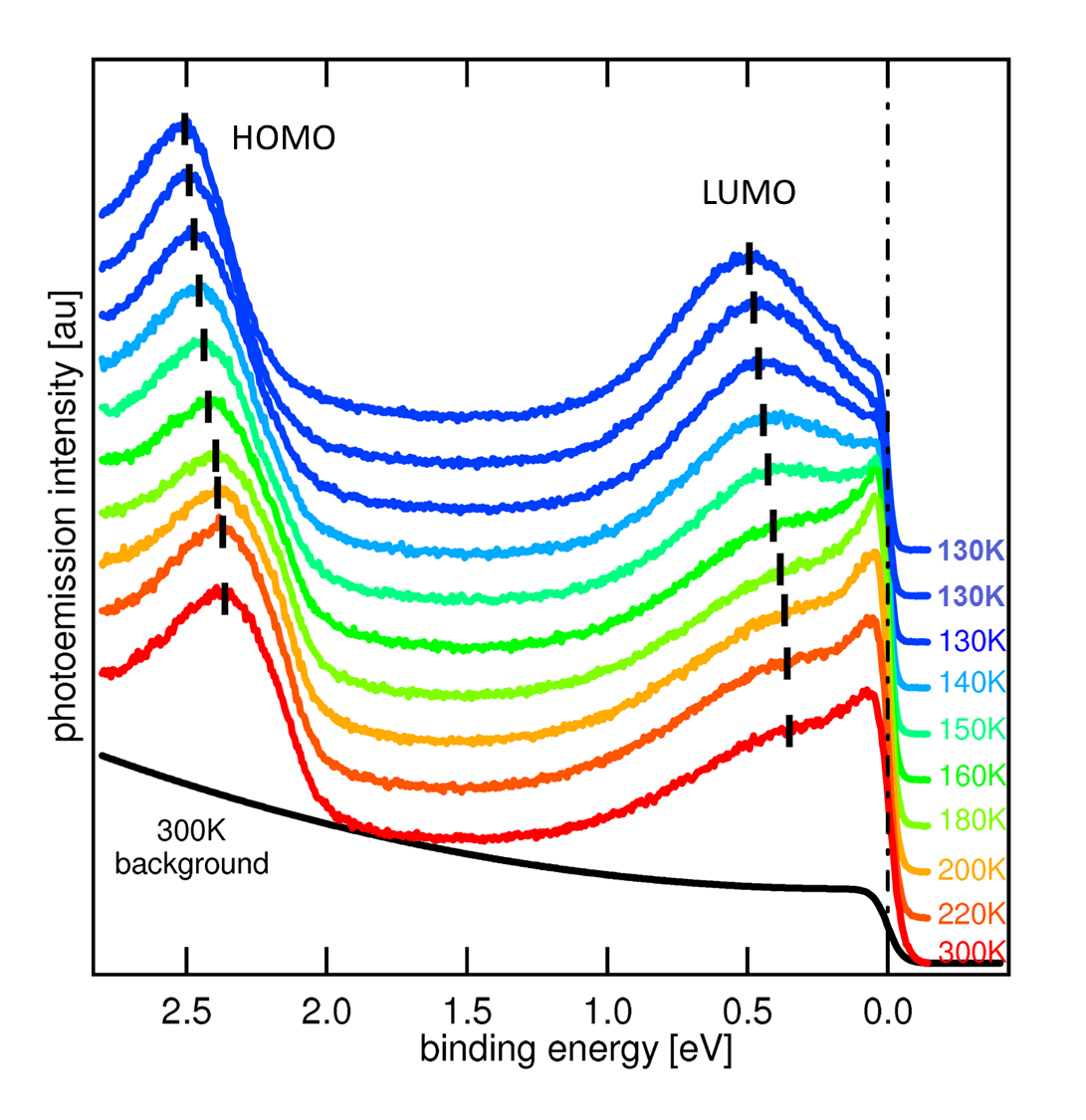
Fig. 1 shows a series of high-resolution, normal emission PES spectra of 1 ML NTCDA/Ag(111) for different temperatures. The intensity maxima of the two molecular features closest to the Fermi level, HOMO and LUMO, appear at approximately 2.4 eV and 0.4 eV, respectively. The typical line-width of these molecular states is of the order of 500 meV in monolayer systems. The background of the substrate is flat near the Fermi level and rises steeply at a binding energy of eV. Already in the room temperature (RT) spectrum, the most peculiar feature is an additional narrow peak right at the Fermi level, appearing on top of the comparatively broad LUMO. This new peak can be observed for all emission angles and shows the same angle dependence in its intensity as the LUMO (see supplement), which next to the energy shift described above Marks et al. (2011) is another clear evidence that it is not related to the Shockley-state appearing around the -points of the reconstructed surface Brillouin zones Forster et al. (2004) but a molecular feature. Furthermore, it can not be explained by the molecular states of the free NTCDA molecules alone. Therefore, it is reasonable to suppose that this state is a consequence of the close interplay between Bloch states of the substrate and molecular states at the interface.
Very important for the understanding of the new narrow feature is the analysis of its temperature dependence. Fig. 1 shows a series of normal emission spectra for temperatures between room temperature (bottom) and K (top three spectra). The sample was cooled slowly in this case, i.e. the time between first (RT) and last spectrum (130 K) amounts to 3 h. Note that the influence of contamination can be ruled out by core level PES Scholl et al. (2004) and X-ray absorption data Schöll et al. (2010). The spectra display several characteristic changes with decreasing temperature: 1.) HOMO and LUMO shift to higher binding energies by about 130 meV, 2.) the new peak at the Fermi level becomes narrower and 3.) looses rapidly in intensity at temperatures below 170 K until the peak has nearly completely vanished at 130 K. Keeping the temperature at constant 130 K, one can observe further changes with the same trend over several hours. Going back to RT, the initial spectrum will be restored again (not shown).
The described temperature dependence of the spectra is immediately related to an order-disorder transition Schöll et al. (2010). Below 180 K the long-range order within the NTCDA monolayer is distroyed by a thermally activated rearrangement of the molecules. The stable low-temperature phase is amorphous, which can be seen by the disappearance of both the LEED spots and the backfolding of the substrate bands in the photoemission data. However, since this order-disorder transition is connected to a thermally activated rearrangement of the molecules, the transition can be prevented if one cools down rapidly, leading to a “frozen” phase with the long-range ordered superstructure of the relaxed phase at RT. This frozen phase can be stabilized for several hours, which allows for a detailed study of the intrinsic line-width of the narrow peak in the ordered phase even down to low temperatures.
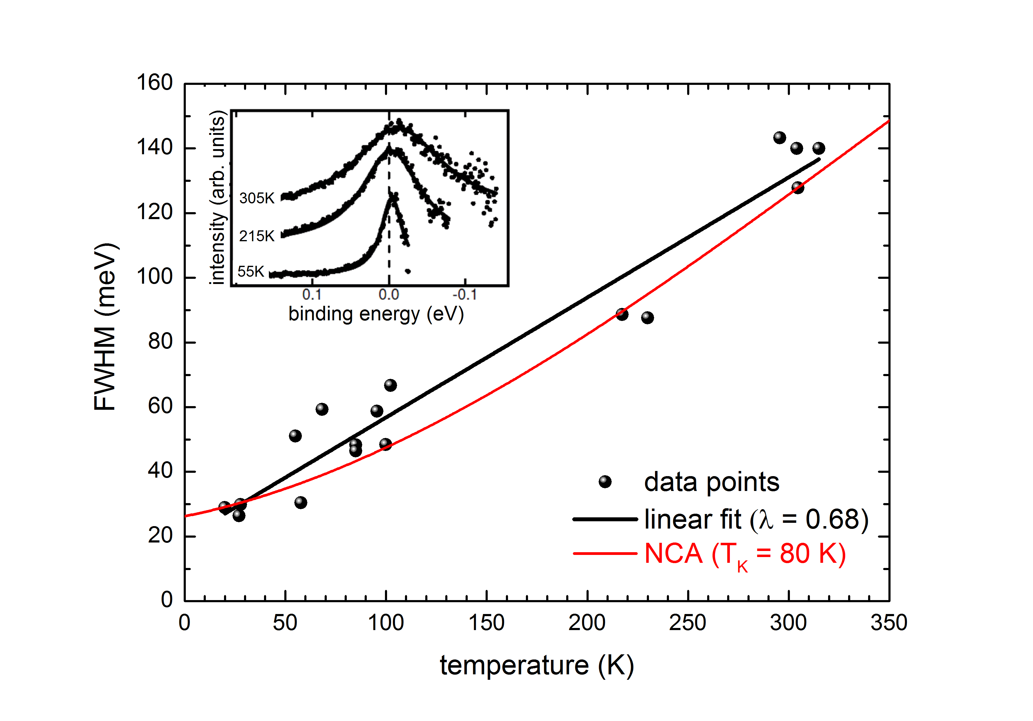
Fig. 2 shows the result of a quantitative line width analysis. The individual values for the full width at half maximum (FWHM) were obtained by a two step analysis: To restore the photoemission signal above , the spectra have been normalized to the Fermi-Dirac distribution (FDD) Klein et al. (2008); Greber et al. (1997); Reinert et al. (2001b); Reinert and Hüfner (2007); Ehm et al. (2007) first, and then fitted by a Lorentzian, which describes the line shape of the peak reasonably well (see inset of Fig. 2). Within the experimental errors, the narrow peaks appear exactly symmetrically to , independent from the temperature.
The temperature dependence of the line width follows roughly a linear behavior, down to minimum values of about 25 meV at the lowest temperatures accessible in our experiment. This value contains the energy resolution and contributions from extrinsic broadening effects as discussed in Reinert et al. (2001a). Such a linear temperature dependence is known from the high-temperature limit of the Debye model, where the slope is immediately related to the electron-phonon coupling parameter by Reinert and Hüfner (2007). From a linear least-squares fit of the temperature dependence we obtain here , which would indicate a very strong electron-phonon coupling. This is not far from characteristic values of typical superconducting metals () Reinert et al. (2003) or C60-systems Knupfer et al. (1993); GUNNARSSON et al. (1995); Yang et al. (2003) where also a characteristic satellite structure was observed. The coupling parameter within the metal substrate, on the other hand, is only of the order of Eiguren et al. (2002). Particularly for the similar PTCDA/Ag(111), electron energy loss spectroscopy Tautz et al. (2002) indicates substantially smaller coupling values than what one can derive from the slope in fig. 2. Obviously a strong electron-phonon coupling alone can not describe the observed peculiar spectral feature in our photoemission data. Note that the temperature dependence of the intensity would bear additional information but can not be evaluated in the present case since the peak is directly at and the normalisation to the FDD, which is crucial for the analysis of the line shape, can lead to artefacts in intensity Ehm et al. (2007).

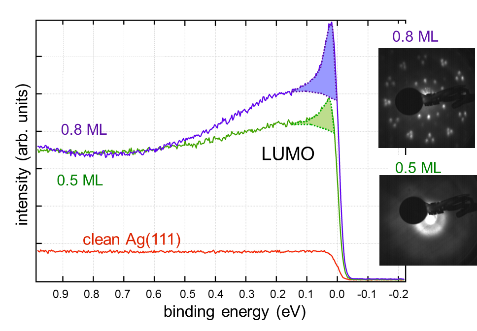
To clarify the importance of the long-range lateral order, we compare the results of NTCDA/Ag(111) to the data of CuPc submonolayers on Ag(111), which show many similarities. At constant temperature this system forms long-range ordered phases for high coverages, whereas the individual molecules remain separated and disordered for low coverages Kröger et al. (2010); Stadtmüller et al. (2011). The respective spectra at temperatures of K are shown in Fig. 3. For (green spectrum on bottom) the LEED-picture in the inset shows the disordered g-phase, while for (blue spectrum on top) the ordered c-phase is established. Note that the molecules do not form islands and the surface is thus completely covered in both cases. The overall photoemission intensity from the molecules is of course approximately a factor of two smaller than in the high coverage spectrum when normalized to the Ag background, simply because the number of molecules per area is smaller. Apart from that, the two spectra look identical: as in the case of NTCDA/Ag(111), the former LUMO is partly occupied and appears below the Fermi level ( eV). Most important, for both samples the narrow peak at the Fermi level is clearly observed. The width (FWHM) of this signal is FWHM meV and is the same for both coverages. Again, the line width is strongly temperature dependent and increases following a linear behavior with when interpreted as due to electron-phonon coupling.
Therefore it is evident, that long range lateral order of the adsorbate molecules is not necessary for the existence of the narrow peak. Instead, it is the position of the LUMO which is important for the development of this additional feature. In the case of NTCDA/Ag(111), there occurs a temperature and time dependent shift of the LUMO to higher binding energies, immediately connected to the disappearance of the narrow peak.
With other words, the exact position of the LUMO is determined by hybridisation between the Bloch states of the substrate and the localized molecular states of the individual molecule. Such a scenario is described by the very versatile single impurity Anderson model (SIAM), which is based on the interplay between a localized magnetic impurity, e.g. a transition metal or lanthanoid atom, with the conduction states of a metal Anderson (1961). The SIAM has been applied successfully for the explanation of the physical properties of many transition metal and rare-earth compounds Gschneidner, Jr. and Eyring (1982–2002), and even for the quantitative estimation of many-body effects in adsorbate systems (Newns-Anderson) Newns (1969). In particular, by use of numerical techniques Gunnarsson and Schönhammer (1983); Bickers et al. (1987) one can calculate the spectral function of such an impurity system, giving the so called Kondo resonance at the Fermi level. The maximum position and the width of the Kondo resonance reflect the small energy scale of the system, which determines all electronic and magnetic low-energy excitations in the system. Note that this model is based on a single impurity, coherence effects and the formation of an impurity lattice are not included and therefore not necessary for the appearance of the Kondo resonance. A typical temperature dependence for the SIAM line width derived from a non-crossing approximation (NCA) calculation Costi et al. (1996) is shown in fig. 2 (=80 K, off set by an additional broadening of 12 meV). A possible quantitative influence from coupling to phonons Mugarza et al. (2011); Fernández-Torrente et al. (2008) is not discussed here.
Indeed, the spectroscopic parallels between typical Kondo (actually Heavy Fermion) systems, as e.g. CeCu2Si2 Reinert et al. (2001b) or CeCu6 Klein et al. (2008) are striking: a narrow feature appears at the Fermi level with a strong temperature dependence in both line width and intensity.
A quantitative determination of the Kondo scale from the temperature dependence requires a more detailed knowledge of the model parameters Coulomb correlation energy , single particle energy and hybridization strength . However, a comparison with the spectra of inorganic systems allows a rough estimation of . For example, the Kondo temperature must be significantly larger than for CeCu6 ( K), where the Kondo resonance vanishes nearly completely in the raw data for temperatures above 100 K. From the comparison with other Ce compounds, particularly CeSi2 Ehm et al. (2007), we estimate to be on the scale of roughly 100 K. This is in accordance with the low-T limit of the experimental line width (25 meV), which represents an upper limit of , and with the NCA simulation in fig.2.
The interpretation in the frame of the SIAM has further important implications, namely the meaning of the position of the LUMO. As shown in Fig. 1, the narrow feature disappears, when the binding energy of the LUMO increases. Indeed, the investigation of other related organic adsorbate systems Bendounan et al. (2007); Kilian et al. (2008); Ziroff et al. (2009); Scholz et al. (2009); Stadtmüller et al. (2011) shows that only if the LUMO has its maximum below but close to the Fermi level — this might be seen as slightly above half filling — the narrow feature appears. This importance is known from the 2D Hubbard model Dagotto et al. (1992); Preuss et al. (1995) with a band filling in the range of 0.6–0.7.
Finally, for a “Kondo like” ground state, the correlation energy must be large in comparison to the other model parameters, in particular to the hybridization between conduction band and localized states and to the single particle binding energy , possibly given by the position of the LUMO maximum in this case, which is about 0.3 eV. An upper limit of 2 eV for U can be derived from the change in HOMO-LUMO separation from non-interacting molecules in multilayers to NTCDA at the interface. In addition, for the similar, yet larger, molecule PTCDA is estimated to about 400 meV if adsorbed on Ag(111) Greuling et al. (2011). Following simple size arguments this value can be regarded as a lower limit for of the smaller NTCDA. Moreover, a rough estimate of 0.2 eV can be derived for from the additional broadening that occurs for the LUMO in comparison to the HOMO (which shows weaker hybridisation Ziroff et al. (2010)).
In conclusion, we could demonstrate that photoemission excitation spectra of chemisorbed organic molecules show a narrow feature at the Fermi level with a strong characteristic temperature dependence, that can not be explained by a mere electron-phonon coupling process. The appearance of the feature is strongly dependent on the position of the LUMO, i.e. on the band filling of the system, but does not depend on the long-range order of the monolayer. Therefore, we conclude that the observed spectral resonance at the Fermi level is a result of the coupling between the individual molecule and the conduction electrons of the metallic substrate. For an interpretation within the SIAM, the model parameters can be estimated to reasonable values. However, further quantitative studies by numerical methods are required to understand the many-body properties of these molecular systems.
Acknowledgements.
We would like to thank Hans Kroha (University of Bonn) for helpful discussions. This work was supported by the Deutsche Forschungsgemeinschaft (FOR1162 and GRK 1221) and the Bundesministerium für Bildung und Forschung BMBF (grant no. 05K10WW2 and 3SF0356B).References
- TANG (1986) C. TANG, APPLIED PHYSICS LETTERS 48, 183 (1986).
- Schwoerer and Wolf (2006) M. Schwoerer and H. C. Wolf, Organic molecular solids (Wiley–VCH, 2006).
- Umbach and Fink (2002) E. Umbach and R. Fink, in Enrico Fermi CXLIX, edited by V. M. Agranovich and G. C. La Rocca, Societ’a Italiana di Physica (IOS Press, Amsterdam, 2002), pp. 233–259.
- Umbach et al. (1996) E. Umbach, M. Sokolowski, and R. Fink, APPLIED PHYSICS A-MATERIALS SCIENCE & PROCESSING 63, 565 (1996).
- Seki et al. (1997) K. Seki, E. Ito, and H. Ishii, SYNTHETIC METALS 91, 137 (1997).
- IG. Hill and Kahn (2000) J. S. IG. Hill, D. Milliron and A. Kahn, APPLIED SURFACE SCIENCE 166 (2000).
- Ueno and Kera (2008) N. Ueno and S. Kera, PROGRESS IN SURFACE SCIENCE 83 (2008).
- Fahlman et al. (2007) M. Fahlman, A. Crispin, X. Crispin, S. K. M. Henze, M. P. de Jong, W. Osikowicz, C. Tengstedt, and W. R. Salaneck, JOURNAL OF PHYSICS-CONDENSED MATTER 19 (2007).
- Kilian et al. (2008) L. Kilian, A. Hauschild, R. Temirov, S. Soubatch, A. Schöll, A. Bendounan, F. Reinert, T.-L. Lee, F. Tautz, M. Sokolowski, et al., Phys. Rev. Lett. 100, 136103 (2008).
- Temirov et al. (2008) R. Temirov, A. Lassise, F. B. Anders, and F. S. Tautz, Nanotechnology 19, 065401 (2008).
- Y. Wang and Hofer (2009) R. B. Y. Wang, J. Kroeger and W. Hofer, ANGEWANDTE CHEMIE-INTERNATIONAL EDITION 48, 1261 (2009).
- Tseng et al. (2010) T.-C. Tseng, C. Urban, Y. Wang, R. Otero, S. L. Tait, M. Alcami, D. Ecija, M. Trelka, J. Maria Gallego, N. Lin, et al., NATURE CHEMISTRY 2, 374 (2010).
- Krause et al. (2008) S. Krause, M. Casu, A. Schöl, and E. Umbach, New Journal of Physics 10, 085001 (2008).
- Scholz et al. (2009) M. Scholz, S. Krause, R. Schmidt, A. Schöll, F. Reinert, and F. Würthner, Apl. Phys. A 95, 285 (2009).
- Hüfner (2007) S. Hüfner, ed., Photoemission Spectroscopy with Very High Energy Resolution, vol. 715 of Lect. Notes Phys. (Springer-Verlag, Berlin–Heidelberg–New York, 2007).
- Fernández-Torrente et al. (2008) I. Fernández-Torrente, K. J. Franke, and J. I. Pascual, Phys. Rev. Lett. 101, 217203 (2008).
- Mugarza et al. (2011) A. Mugarza, C. Krull, R. Robles, S. Stepanow, G. Ceballos, and P. Gambardella, NATURE COMMUNICATIONS 2 (2011).
- Choi et al. (2010) T. Choi, S. Bedwani, A. Rochefort, C.-Y. Chen, A. J. Epstein, and J. A. Gupta, NANO LETTERS 10, 4175 (2010).
- Perera et al. (2010) U. G. E. Perera, H. J. Kulik, V. Iancu, L. G. G. V. D. da Silva, S. E. Ulloa, N. Marzari, and S. W. Hla, Phys. Rev. Lett. 105 (2010).
- Reinert and Hüfner (2005) F. Reinert and S. Hüfner, New J. Phys. 7, 97 (2005), focus Issue on ’Photoemission and Electronic Structure’.
- Klein et al. (2008) M. Klein, A. Nuber, F. Reinert, J. Kroha, O. Stockert, and H. v. Löhneysen, Phys. Rev. Lett. 101, 266404 (2008).
- Reinert et al. (2001a) F. Reinert, G. Nicolay, S. Schmidt, D. Ehm, and S. Hüfner, Phys. Rev. B 63, 115415 (2001a).
- Ziroff et al. (2009) J. Ziroff, P. Gold, A. Bendounan, F. Forster, and F. Reinert, Surf. Sci. 603, 354 (2009).
- Stahl et al. (1998) U. Stahl, D. Gador, A. Soukopp, R. Fink, and E. Umbach, Surf. Science 414, 423 (1998).
- Scholl et al. (2004) A. Scholl, Y. Zou, T. Schmidt, R. Fink, and E. Umbach, JOURNAL OF PHYSICAL CHEMISTRY B 108, 14741 (2004).
- Schöll et al. (2010) A. Schöll, L. Kilian, Y. Zou, J. Ziroff, S. Hame, F. Reinert, E. Umbach, and R. H. Fink, Science (2010), published.
- Hame (2007) S. Hame, Master’s thesis, Universität Würzburg, Würzburg (2007).
- Ziroff et al. (2010) J. Ziroff, F. Forster, A. Schöll, P. Puschnig, and F. Reinert, Phys. Rev. Lett. 104, 233004 (2010).
- Bendounan et al. (2007) A. Bendounan, F. Forster, A. Schöll, D. Batchelor, J. Ziroff, E. Umbach, and F. Reinert, Surf. Sci. 601, 4013 (2007).
- Zou et al. (2006) Y. Zou, L. Kilian, A. Scholl, T. Schmidt, R. Fink, and E. Umbach, SURFACE SCIENCE 600, 1240 (2006).
- Schwalb et al. (2008) C. H. Schwalb, S. Sachs, M. Marks, A. Schöll, F. Reinert, E. Umbach, and U. Höfer, Phys. Rev. Lett. 101, 146801 (2008), selected for Virtual Journal of Ultrafast Science, Nov. 2008 Vol. 7, Issue 11.
- Sachs et al. (2009) S. Sachs, C. H. Schwalb, M. Marks, A. Schöll, F. Reinert, E. Umbach, and U. Höfer, J. Chem. Phys. 131, 144701 (2009).
- Schwalb et al. (2010) C. H. Schwalb, M. Marks, S. Sachs, A. Schöll, F. Reinert, E. Umbach, and U. Höfer, European Physical Journal B 75, 23 (2010).
- Marks et al. (2011) M. Marks, N. L. Zaitsev, B. Schmidt, C. H. Schwalb, A. Schoell, I. A. Nechaev, P. M. Echenique, E. V. Chulkov, and U. Hoefer, Phys. Rev. B 84 (2011).
- Costi et al. (1996) T. Costi, J. Kroha, and P. Wolfle, Phys. Rev. B 53, 1850 (1996).
- Forster et al. (2004) F. Forster, S. Hüfner, and F. Reinert, J. Chem. Phys. B 108, 14692 (2004).
- Greber et al. (1997) T. Greber, T. J. Kreutz, and J. Osterwalder, Phys. Rev. Lett. 79, 4465 (1997).
- Reinert et al. (2001b) F. Reinert, D. Ehm, S. Schmidt, G. Nicolay, S. Hüfner, and J. Kroha, Phys. Rev. Lett. 87, 106401 (2001b).
- Reinert and Hüfner (2007) F. Reinert and S. Hüfner, in Photoemission Spectroscopy with Very High Energy Resolution, edited by S. Hüfner (Springer-Verlag, Berlin–Heidelberg–New York, 2007), vol. 715 of Lect. Notes Phys., chap. II, pp. 13–53.
- Ehm et al. (2007) D. Ehm, S. Hüfner, F. Reinert, J. Kroha, P. Wölfle, O. Stockert, C. Geibel, and H. von Löhneysen, Phys. Rev. B 76, 045117 (2007).
- Reinert et al. (2003) F. Reinert, B. Eltner, G. Nicolay, D. Ehm, S. Schmidt, and S. Hüfner, Phys. Rev. Lett. 91, 186406 (2003).
- Knupfer et al. (1993) M. Knupfer, M. Merkel, M. S. Golden, J. Fink, O. Gunnarsson, and V. P. Antropov, Phys. Rev. B 47, 13944 (1993).
- GUNNARSSON et al. (1995) O. GUNNARSSON, H. HANDSCHUH, P. BECHTHOLD, B. KESSLER, G. GANTEFOR, and W. EBERHARDT, Phys. Rev. Lett. 74, 1875 (1995).
- Yang et al. (2003) W. Yang, V. Brouet, X. Zhou, H. Choi, S. Louie, M. Cohen, S. Kellar, P. Bogdanov, A. Lanzara, A. Goldoni, et al., SCIENCE 300, 303 (2003).
- Eiguren et al. (2002) A. Eiguren, B. Hellsing, F. Reinert, G. Nicolay, E. V. Chulkov, V. M. Silkin, S. Hüfner, and P. M. Echenique, Phys. Rev. Lett. 88, 066805 (2002).
- Tautz et al. (2002) F. S. Tautz, M. Eremtchenko, J. A. Schaefer, M. Sokolowski, V. Shklover, and E. Umbach, Phys. Rev. B 65, 125405 (2002).
- Kröger et al. (2010) I. Kröger, B. Stadtmüller, C. Stadler, J. Ziroff, M. Kochler, A. Stahl, F. Pollinger, T.-L. Lee, J. Zegenhagen, F. Reinert, et al., New J. Phys. 12, 083038 (2010).
- Stadtmüller et al. (2011) B. Stadtmüller, I. Kröger, C. Kumpf, and F. Reinert, Phys. Rev. B 83, 085416 (2011).
- Anderson (1961) P. W. Anderson, Phys. Rev. 124, 41 (1961).
- Gschneidner, Jr. and Eyring (1982–2002) K. A. Gschneidner, Jr. and L. R. Eyring, eds., Handbook on the physics and chemistry of rare earths (North-Holland, Amsterdam–New York–Oxford, 1982–2002).
- Newns (1969) D. M. Newns, Phys. Rev. B 178, 1123 (1969).
- Gunnarsson and Schönhammer (1983) O. Gunnarsson and K. Schönhammer, Phys. Rev. Lett. 50, 604 (1983).
- Bickers et al. (1987) N. E. Bickers, D. L. Cox, and J. W. Wilkins, Phys. Rev. B 36, 2036 (1987).
- Dagotto et al. (1992) E. Dagotto, F. Ortolani, and D. Scalapino, Phys. Rev. B 46, 3183 (1992).
- Preuss et al. (1995) R. Preuss, W. Hanke, and W. von der Linden, Phys. Rev. Lett. 75, 1344 (1995).
- Greuling et al. (2011) A. Greuling, M. Rohlfing, R. Temirov, F. S. Tautz, and F. B. Anders, Phys. Rev. B 84 (2011).