Energy Levels of Few Electron Quantum Dots Imaged and Characterized By Atomic Force Microscopy
Abstract
Strong confinement of charges in few electron systems such as in atoms, molecules and quantum dots leads to a spectrum of discrete energy levels that are often shared by several degenerate quantum states. Since the electronic structure is key to understanding their chemical properties, methods that probe these energy levels in situ are important. We show how electrostatic force detection using atomic force microscopy reveals the electronic structure of individual and coupled self-assembled quantum dots. An electron addition spectrum in the Coulomb blockade regime, resulting from a change in cantilever resonance frequency and dissipation during tunneling events, shows one by one electron charging of a dot. The spectra show clear level degeneracies in isolated quantum dots, supported by the first observation of predicted temperature-dependent shifts of Coulomb blockade peaks. Further, by scanning the surface we observe that several quantum dots may reside on what topologically appears to be just one. These images of grouped weakly and strongly coupled dots allow us to estimate their relative coupling strengths.
The ability to confine single charges at discrete energy levels makes semiconductor quantum dots (QD) promising candidates as a platform for quantum computation Loss and DiVincenzo (1998); Tanamoto (2000) and single photon sources Dalacu et al. (2009). Tremendous progress has been made in not only understanding the properties of single electrons in QDs but also in controlling their quantum states which is an essential prerequisite for quantum computation Hanson et al. (2007). Single electron transport measurements have been the main experimental technique for investigating electron tunneling into quantum dots Kouwenhoven et al. (2001). Charge sensing techniques using built-in charge sensors, such as quantum point contacts Field et al. (1993), complement transport measurements as lower electron tunneling rates can be monitored with even real-time detection being possible Lu et al. (2003). It is instrumentally challenging to study self-assembled QDs via conventional transport and charge sensing methods due to the difficulty in attaching electrodes. Although progress is being made Ota et al. (2004); Jung et al. (2005); Igarashi et al. (2007); Vdovin et al. (2007); Amaha et al. (2008) these techniques have very small yield and therefore make it difficult to assess variation in QD electronic properties. Compared to typical QDs studied via transport measurements, in particular lithographically defined QDs, self-assembled QDs can be fabricated to have smaller sizes, stronger confinement potentials and a more scalable fabrication process, all of which make them attractive for practical applications.
In this paper, we focus on an alternative technique for studying QDs that is better suited for self-assembled QDs: charge sensing by atomic force microscopy (AFM). Charge sensing by AFM is a convenient method to study the electronic structure of QDs as nano-electrodes are not required and large numbers of QDs can be investigated in one experiment. Termed single-electron electrostatic force microscopy (-EFM), this technique relies on the high force-sensitivity of AFM to detect the electrostatic force resulting from single electrons tunneling into and out of the QD. It was first demonstrated on QDs formed in carbon nanotubes Woodside and McEuen (2002); Zhu et al. (2005), and later applied to self-assembled QDs Stomp et al. (2005); Dana and Yamamoto (2005) and also to gold nanoparticles Azuma et al. (2006); Zhu et al. (2008). We focus on epitaxially grown self-assembled InAs/InP QDs in the few-electron regime. Using a dissipation model, we find compelling evidence for the existence of electronic degeneracies (i.e. shell structure) by measuring an effective temperature-dependent level repulsion; although predicted for conductance measurements in 1991Beenakker (1991), we believe this to be the first observation of this effect. Further, we use the model to quantitatively extract various properties of both individual QDs, such as the tunneling rates and charging energy and coupled QDs, such as the strength of coupling.
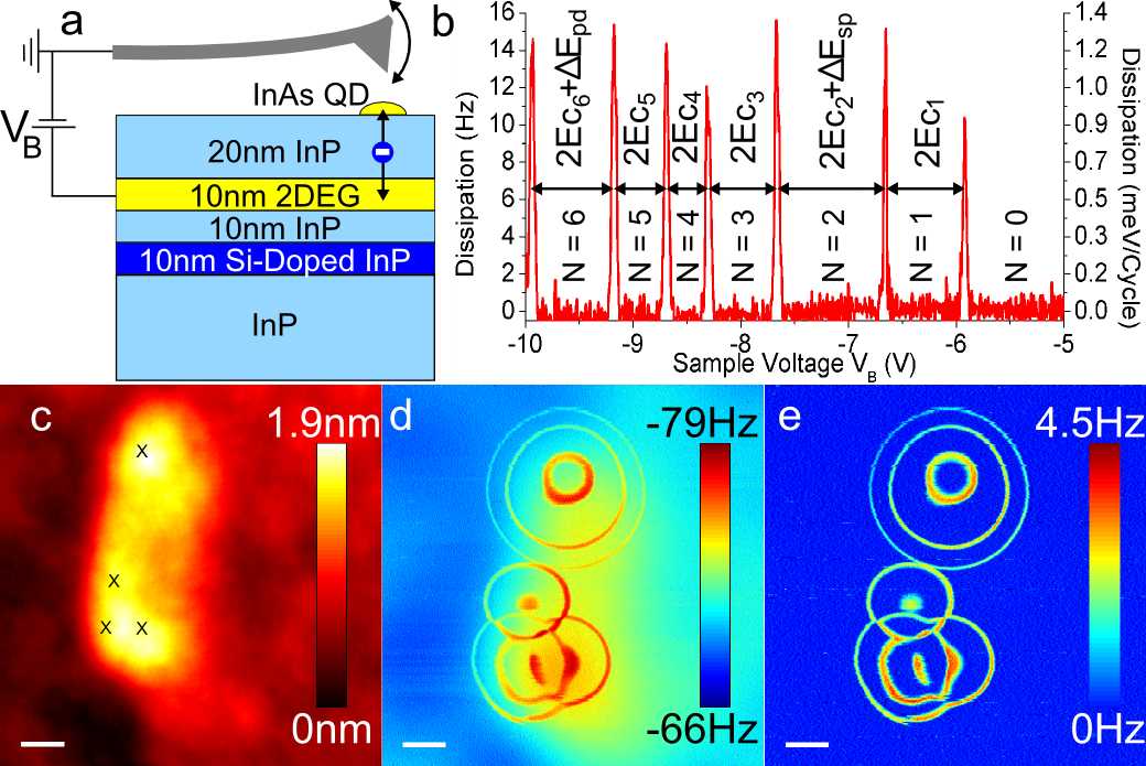
We study uncapped self-assembled InAs QDs grown on a 20 nm InP tunnel barrier below which a two-dimensional electron gas (2DEG) is formed in an In0.53Ga0.47As quantum well. A dc-bias voltage, , is applied to the 2DEG with respect to the grounded conductive AFM cantilever tip. Figure 1a shows the sample structure and experimental setup.
The AFM cantilever is driven at its mechanical resonance frequency, 166 kHz, with constant oscillation amplitude Albrecht et al. (1991). The voltage drop, , across the tunnel barrier between the QD and the 2DEG is only a fraction of , with being a function of the tip position. The tip-QD gap is tens of nanometers wide so that tip-QD tunneling is negligible. We thus have a single-electron box setup: the electrochemical potential of the 2DEG, , with respect to the QD, , is set by and a negative bias increases the number of electrons, , on the QD in integer steps whenever the electrochemical potentials are aligned (called a charge degeneracy point). Tunneling between 2DEG and QD is suppressed by the electrostatic energy cost, , of adding or removing an electron to the QD except near these charge degeneracy points (Coulomb blockade). The heart of the -EFM technique lies in the fact that oscillations of the AFM cantilever modulate , and hence are equivalent to an effective oscillating gate voltage applied to the QD. Thus, motion of the cantilever induces a modulation of which will be slightly out-of-phase with the cantilever’s motion (a result of the finite response time of electrons on the dot). The electrostatic coupling between QD and cantilever tip implies an electrostatic force proportional to acting on the tip, the net result being both a frequency shift, , and additional dissipation, , of the cantilever Hölscher et al. (2001). These effects are maximal at charge degeneracy points as here can easily change in response to the effective oscillating gate voltage.
Figure 1b is an example of at 4.5 K with the tip positioned over the upper QD imaged in Fig. 1d-e. The is equivalent to the energy addition spectra usually obtained from linear conductance or capacitance spectroscopy measurements Ashoori et al. (1992). Similar to those measurements, Coulomb blockade peaks in occur near charge degeneracy points of the QD. The peak positions are suggestive of the addition spectrum of a 2D circular QD with parabolic confinement potential; each peak is separated by twice the capacitive charging energy, , with a further splitting between peaks 2 and 3 and between peaks 6 and 7 corresponding to the energy difference between shells, . This type of shell structure has been previously observed in InAs QDs Drexler et al. (1994); Miller et al. (1997); Ota et al. (2004); Jung et al. (2005).
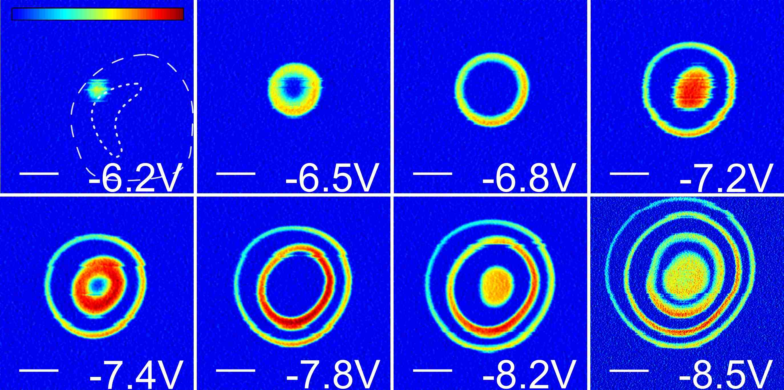
Figure 1c-e shows the topography, and images of an elongated InAs island. The peaks in the - spectra radially surround the QD center so that the ring furthest from the center corresponds to the first electron entering the QD; the rings themselves are contour lines of constant . Multiple sets of concentric rings appearing in the and images indicate multiple QDs in the island. Such observations would not be as easily identified via other experimental means Zhitenev et al. (1999). The tip-2DEG capacitive force adds a large background in the signal which locally varies due to topography Stomp et al. (2005); Cockins et al. (2007), making it advantageous to focus on the for image analysis.

Figure 2a shows and as a function of with the tip positioned over the center of the upper set of concentric rings in Fig. 1e at 30 K. While the energy addition spectra shown in Fig. 1b and 2a are consistent with the expected shell structure for a 2D circular QD with parabolic confinement potential, we obtain much stronger evidence of the shell structure from the temperature dependence of the peak positions. Theoretically, a temperature-dependent shift of Coulomb blockade peaks is expected whenever one has degenerate single particle levels, as predicted for the conductance peaks of a spin degenerate level Beenakker (1991). The result is an effective temperature-dependent energy level repulsion: the peaks in each shell move apart as temperature is increased. Furthermore, our theoretical analysis suggests that this effect should be enhanced in the tunneling-induced dissipation compared to the conductance due to an increased sensitivity to degeneracy. The same theory also leads to asymmetric lineshapes in the Coulomb blockade peaks, also enhanced compared to the conductance, but this is predicted to be small and could not be resolved in the present experiment. However, we were able to measure the temperature-dependent shifts of the peaks and find excellent agreement with theory. We are unaware of any experiments where these effects have been observed.
We model the dissipation on the cantilever using linear response and a master equation describing the charge state of the QD in the regime of weak coupling Beenakker (1991); Clerk and Bennett (2005). Details of the approach are provided in the Supplementary Information. Near a charge degeneracy point between two nondegenerate single particle levels, the dissipation is Zhu et al. (2008); Brink (2006)
| (1) |
where and are the intrinsic cantilever oscillation frequency and spring constant, is the measured resonance frequency due to forces on the cantilever, is the 2DEG-QD tunneling rate, and is the Fermi function evaluated at ( is the point of charge degeneracy). Lastly, is the sensitivity of the potential on the QD to the cantilever motion and is the tip-QD capacitance. We stress that equation (1) applies to each dissipation peak independently: and are obtained separately for each peak from the data with no assumption of constant .
Equation (1) only takes into account single nondegenerate levels. More generally, suppose we have a state with electrons, with in the valence shell. If this shell has a degeneracy , then near the charge degeneracy point between this state and the state with QD electrons, the dissipation is:
| (2) |
where
| (3) |
and the tunneling rate is assumed to be equal for each degenerate single particle level within a shell for a given peak Ste . Note that is the number of electrons occupying the given shell and not the total number of electrons on the dot, , and that because of the factor , is no longer symmetric about its maximum. The different coefficients of and in reflect the asymmetry between electron addition and removal processes caused by degeneracy, and corresponds to a nondgenerate level for which equation (2) reduces to equation (1). This asymmetry in equation (2) causes each peak in to be shifted in energy by an amount proportional to temperature. By fitting (e.g. Fig. 2a) to equation (2) we extract , allowing us to convert the axis into energy. This is done for all of the thermally limited peaks, yielding . Figure 2b shows at different temperatures together with the fitted curves.
The role of is further elucidated by the relation
| (4) |
where is the size of the frequency shift dip due to the single-electron tunneling. The ratio in equation (4) defines an inverse timescale set by the relative in-phase and out-of-phase parts of the electrostatic force; this is simply for a nondegenerate QD Zhu et al. (2008); Brink (2006), but modified by degeneracy through the factor . Using equations (2) and (4) and the measured values of and , we calculate the tunneling rates at the maxima of dissipation peaks 1-6, obtaining 70, 90, 160, 180, 230 and 330 kHz. As expected, increases with increasing as the height of the potential barrier between the 2DEG and the QD is reduced.
After extracting the tunneling rates, we fit each dissipation peak using equation (2) and measure the spacing between peaks as functions of temperature from 4.5-30 K and from 78-95 K. We focus on the relative shifts between peaks as these are less sensitive to slight offsets in peak positions due to small changes in the tip-QD distance. The size and direction of each peak shift is different (see Fig. 2a), in a manner that is completely captured by our model: the two peaks in the shell shift apart, as do the four peaks in the shell. The measured relative peak shifts of repelling pairs are shown in Fig. 2c-d and compared to the theoretical shift from equation (2) with a single fit parameter, . In addition, we expect that multiple shells, not just the valence shell, should play a role at high temperatures where is not satisfied. For the relative shift of peaks 1 and 2 (Fig. 2c) we plot a numerical calculation accounting for the possible occupation of the shell, showing that the high temperature correction agrees well with the data. Figure 2d shows that the relative shifts between peaks 3 and 6 and between 4 and 5 are well described by equation (2) up to 30 K. Finally we note that there is an overall shift of the and shells toward each other that we believe is a consequence of strong repulsion of the shell by the shell, predicted to be 6-fold degenerate.
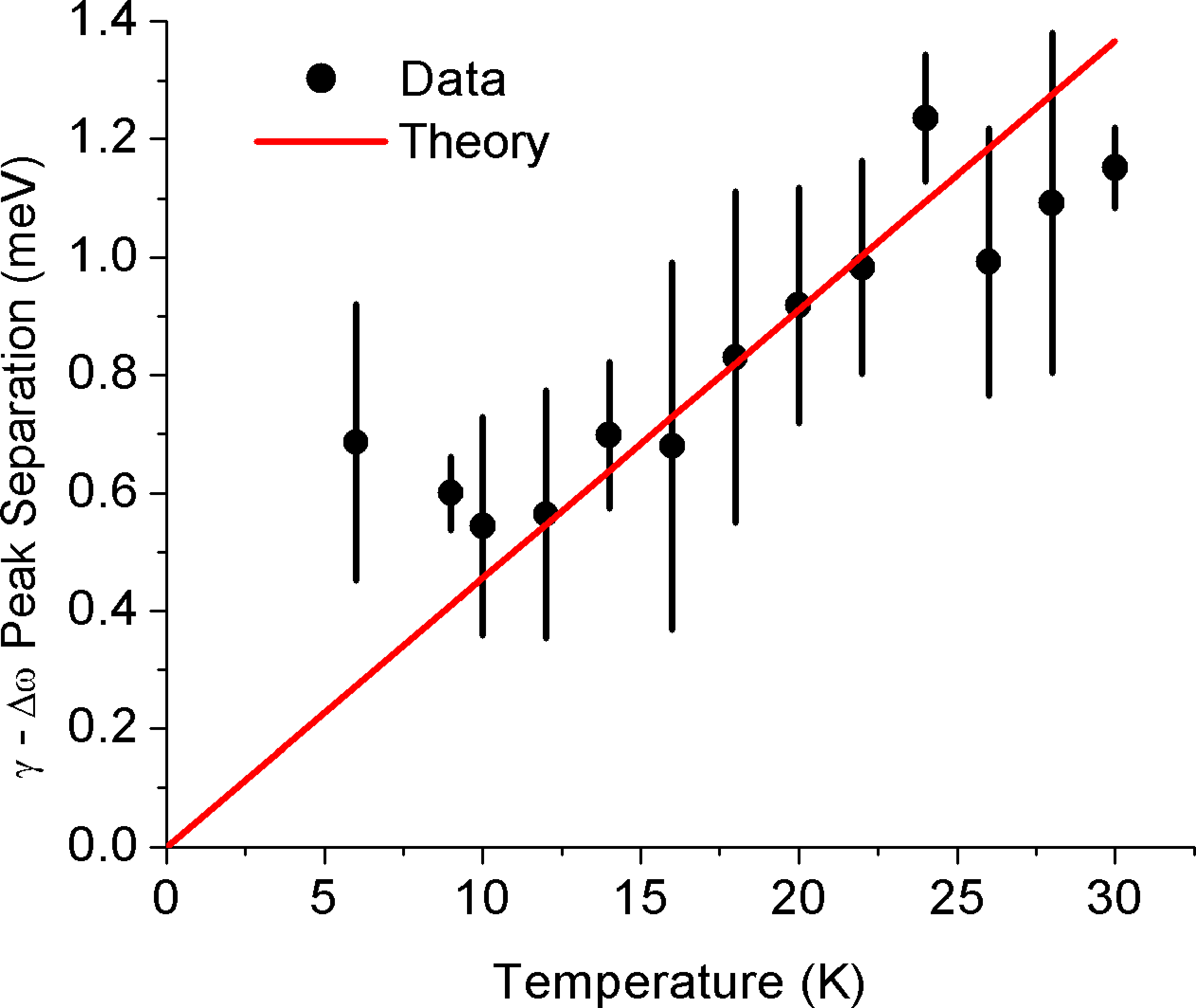
We performed several checks to support our conclusion that the peak shifts result from degeneracy. First, the predicted shifts are unchanged if the relevant electronic levels are not perfectly degenerate, but rather split by an amount smaller than ; thus, the effect only requires approximate shell degeneracy as is the reality in imperfect QDs. Second, if the level splitting is larger than we expect no temperature dependence of the peak shifts. Based on the observed peak shifts down to 4.5 K we thus conclude that the level splitting is smaller than this temperature, corresponding to roughly 0.4 meV. Lastly, the same degeneracy theory leads to small, but measurable, shifts between the dissipation peak and the frequency peak corresponding to the same charge degeneracy point. This is visible for the third peak (from the right) in Fig. 2a in which the and peaks do not exactly line up, with the measured shifts compared to theory in Fig. S2. These dissipation-frequency shifts strongly support our model and rule out the alternative of a temperature-dependent renormalization of or .
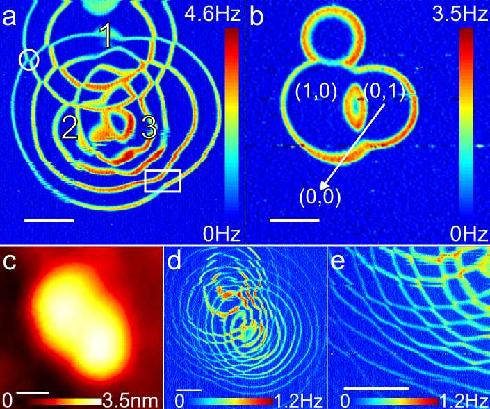
Significant efforts are ongoing towards understanding and controlling the properties of coupled QDs, in particular double QDs or “artificial molecules” van der Wiel et al. (2003). The images that we obtain for double QDs are equivalent to stability diagrams which depict the charge state of the double QD system. This is because of the position dependence of the lever arm for each QD which results in two electrochemical potentials, and . Scanning the AFM tip at constant height and corresponds to sweeping the - space through changing and even though only a single electrode (the tip) is being used.
In a conventional stability diagram, lines of constant electrochemical potential for each QD are plotted as a function of two gate voltages. When the two QDs are coupled, intersection points are split into two points (triple points), showing avoided-crossings van der Wiel et al. (2003). In the images the avoided-crossings are observed when the ring radii suddenly change at intersection points. Figure 3a,b show the same three QDs as in the lower part of Fig. 1e, now imaged at V and V, respectively. Such avoided-crossings are highlighted in the circle and box in Fig. 3a, representing an example of weak and strong coupling respectively.
We characterize the coupling strength by comparing the ratio of the change in ring radius to the separation between the first two rings () for QD2. This method is only valid when both rings are far enough from the QD center that the voltage drop between them is approximately linear. Following this procedure, the coupling of QD2 to QD1 (circle) and QD3 (box) can be compared. While the change in radius of QD2 is approximately 0.10 0.01 of due to QD1, it is 0.46 0.03 due to QD3, indicating a much stronger coupling between QD2 and QD3. We consider the former to be an example of weak coupling because the triple points are nearly joined. This is consistent with a small capacitive coupling between the two dots: the charging of one dot effectively gates the second dot, causing a sudden change in ring radius.
Conversely, the boxed region in Fig. 3a is an example of strong coupling since there is a large gap at intersections as in the triple points of a stability diagram. In Fig. 3b, the same 3 QDs as in Fig. 3a are imaged at smaller . This image allows for a more intuitive explanation of the coupling. Consider the diagonal line from the center of QD3 outwards; initially, the AFM tip is over QD3 in the =(0,1) state, but takes a path into QD2 in the (1,0) state. The ability to go continuously between these states without going through (0,0) or (1,1) necessarily indicates a large capacitive coupling between the dots. It also indicates evidence for an interesting charge transfer process as no dissipation is observed between circles. Lack of dissipation implies no change in the total dot charge; either there is a cotunneling process where two electrons simultaneously tunnel to and from the 2DEG, or there is coherent tunneling between the dots.
Figure 3c-e shows another example of coupled QDs at 4.5 K. The InAs structure (Fig. 3c) contains coupled QDs as shown in the image (Fig. 3d). Figure 3e zooms up on the region in Fig. 3d showing many avoided crossings. Within this distance range from the QD centers, each is approximately linearly dependent on the tip position so that scanning the tip more closely resembles sweeping two gate voltages, resulting in the image resembling a conventional stability diagram. Figure 3 also highlights how advantageous it is to have images in addition to the - spectra as the spectra alone will contain the peaks from nearby QDs which can be identified using the images.
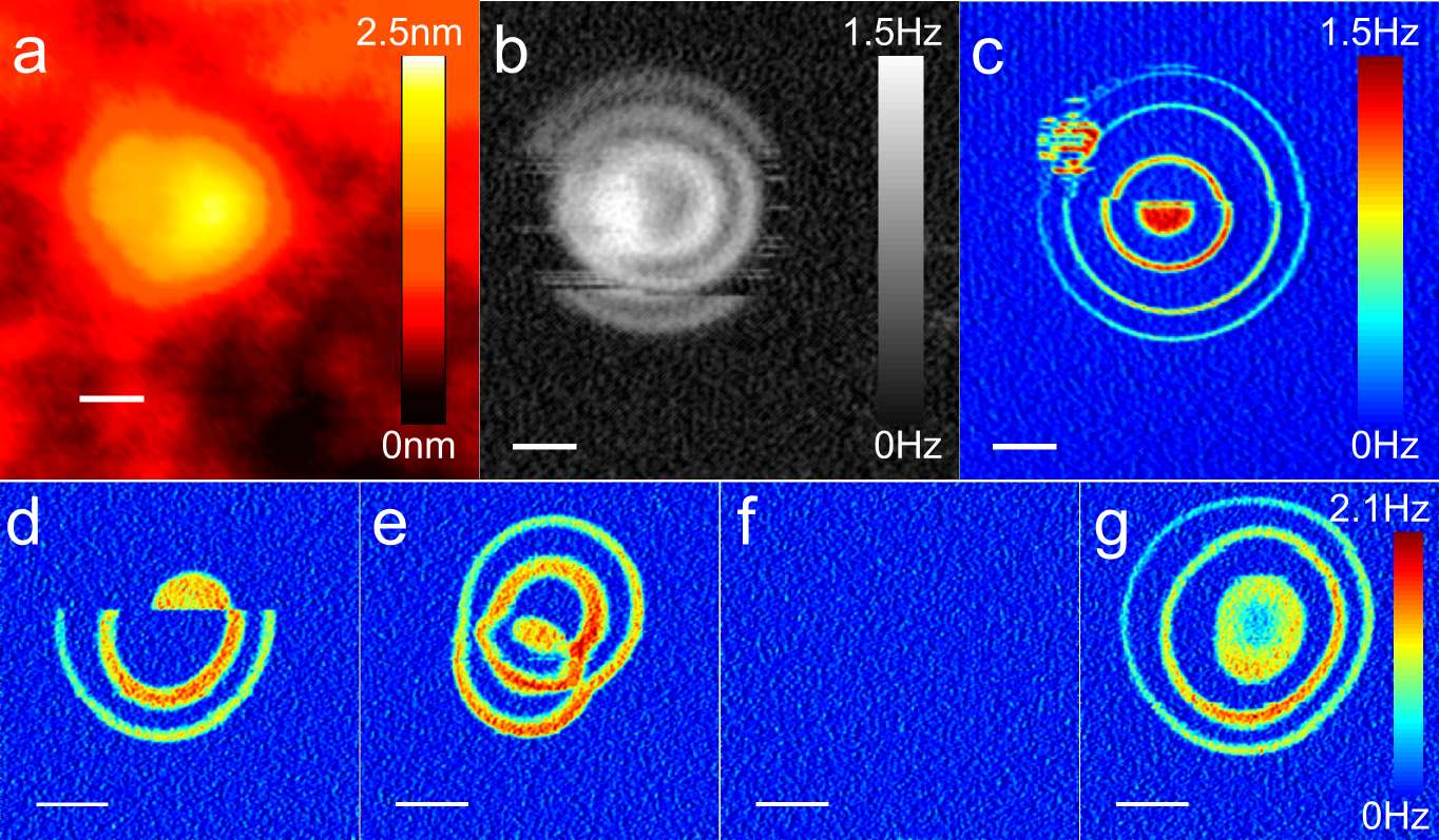
Yet another advantage of our technique is the ability to spatially resolve the effects of background charge fluctuations; for self-assembled QDs, no other technique is capable of doing this. The AFM images show how the QD confinement potential is being influenced by charge reconfiguration. Figure 4 shows two such cases. The structure in Fig. 4a shows a fluctuation in electron population due to nearby fluctuations in the electrostatic background at both 78 K (Fig. 4b) and 4.5 K (Fig. 4c). The missing sections of the first ring in Fig. 4b indicate that the number of electrons in the QD is fluctuating by one in this region. Interestingly, depending on the scan direction (left to right or right to left) over the QD, the missing section may appear. Similar reconfiguration was observed in the - spectra. While all the peaks appear in the reverse sweep (positive to negative), the first peak disappears in the forward sweep. Figure 4d-g shows a more dramatic change. During the scan, a sudden switch in the confinement potential occurs, leading to the transition from the single QD (Fig. 4d) to a coupled double QD (Fig. 4e). This double QD state could be switched back to the single state (Fig. 4g) by scanning over the same area with a positive (Fig. 4f). Although both of these changes are readily identified in the images, having a spectrum alone may cause confusion as is the case in conventional transport measurements. These observations indicate that the QD confinement potentials are very sensitive to the electrostatic background and can be modified, or switched, controllably.
Charge sensing with AFM can be used to investigate the electronic structure of single and coupled self-assembled QDs. The technique enables the quantitative extraction of the tunneling rate, charging energy, and the QD interaction energies; further, we have used it to perform the first measurement of temperature-dependent Coulomb blockade peak shifts confirming the shell degeneracy of the QD. The dissipation images proved especially useful in analyzing multiple QDs and changes in QD confinement potential resulting from nearby charge fluctuations. The images also revealed that what looked like a single QD structure topographically can actually contain multiple QDs. Additionally, the imaging capability of AFM provides insight into the link between QD electronic structure and topography which is of great importance in developing electronic devices based on QDs.
Methods
The sample, grown by chemical beam epitaxy Poole et al. (2001), consists of the following layers: 460 nm undoped InP grown on top of an insulating InP substrate, followed by a 10 nm Si-doped InP layer, 10 nm undoped layer, 10 nm In0.53Ga0.47As layer, 20 nm undoped layer and a 1.82 ML InAs layer that results in the formation of InAs QDs by Stranski-Krastanow growth. The QDs cover the surface with at a density of 2.5 QDs per having diameters in the range of 30-95 nm and heights of 0.5-6 nm. The 2DEG layer formed in the InGaAs well serves as a back electrode and an Ohmic contact to the 2DEG is made by indium diffusion.
Our home-built cryogenic AFM Roseman and Grutter (2000) includes an RF- modulated fiber optic interferometer Rugar et al. (1989) with 1550 nm wavelength for cantilever position detection. We coat Si AFM cantilevers (Nanosensors PPP-NCLR) with 10 nm titanium (adhesion layer) and 20 nm platinum. The cantilevers typically have a 160 kHz resonance frequency and a quality factor between 100,000 and 200,000 at 4.5 K.
All of the images were taken in frequency modulation mode Albrecht et al. (1991). In this mode, the cantilever is self-oscillated at its resonance frequency with a constant amplitude. The frequency shift and dissipation were measured with a commercially available phase-locked loop frequency detector (Nanosurf, easyPLL plus). The topography images were taken in constant frequency shift mode where a constant frequency shift is maintained by regulating the cantilever tip-sample distance using a feedback controller. The frequency shift and dissipation images were taken in constant-height mode with a typical tip height of 20 nm. Dissipation images are shown in Fig. S1 as a function of . More negative results in adding more electrons to the quantum dot. Areas of increased dissipation mark 2DEG-QD tunneling events. Each time a ring is crossed when traveling towards the quantum dot center marks the addition of an electron to the dot. More details of the AFM images are listed in Supplementary Table 1.
The amplitude of the cantilever excitation signal, , is provided as the dissipation signal from the Nanosurf oscillator controller. It is converted to units of 1/s via: . is the excitation amplitude independent of the tunneling process, in other words the background dissipation. This conversion is independent of cantilever oscillation amplitude. Similarly, the signal is converted to units of eV/cycle by multiplying by the factor where is the cantilever oscillation amplitude Hug and Baratoff (2002).
The spectra shown in Fig. 2a was originally superposed onto a large parabolic background arising from the capacitive force between the 2DEG and cantilever tip. Over several volts, at typical cantilever-sample gaps of 20 nm, the curve can be fit with a single parabola. In Fig. 2a this parabola was subtracted from the frequency shift data.
The exact positions of the peaks (dips) in the dissipation (frequency shift) are sensitive to the distance between cantilever tip and quantum dot. In particular, slight changes in cantilever tip lateral position with respect to the quantum dot center can lead to slight shifts in the peaks as can be deduced from the images where the rings can have different spacing depending on location. The shift in peaks as a function of height, however, is linearly dependent over the distances used in this experiment (12-22 nm). We took the differences in peak positions in the data displayed in Fig. 2b to be caused by small height differences (sub 1 nm) and so the voltage axis was rescaled to align the peaks with the data but the peak heights were not rescaled. The mean factor involved in the voltage rescaling is 1.011 with the most extreme factor being 1.088. The temperature data above 22 K had thermally limited peaks for a cantilever oscillation amplitude of 0.4 nm, but needed to be reduced to 0.2 nm at 4.5 K. The errorbars in Fig. 2c and d represent how well the measurement over a single location was reproduced.
References
References
- Loss and DiVincenzo (1998) D. Loss and D. P. DiVincenzo, Phys. Rev. A 57, 120 (1998).
- Tanamoto (2000) T. Tanamoto, Phys. Rev. A 61, 022305 (2000).
- Dalacu et al. (2009) D. Dalacu, M. Reimer, S. Frederick, D. Kim, J. Lapointe, P. J. Poole, G. Aers, R. L. Williams, W. R. McKinnon, M. Korkusinski, et al., Laser & Photon. Rev. (2009), dOI:10.1002/lpor.200810077.
- Hanson et al. (2007) R. Hanson, L. P. Kouwenhoven, J. R. Petta, S. Tarucha, and L. M. K. Vandersypen, Rev. Mod. Phys. 79, 1217 (pages 49) (2007).
- Kouwenhoven et al. (2001) L. P. Kouwenhoven, D. G. Austing, and S. Tarucha, Rep. Prog. Phys. 64, 701 (2001).
- Field et al. (1993) M. Field, C. G. Smith, M. Pepper, D. A. Ritchie, J. E. Frost, G. A. Jones, and D. G. Hasko, Phys. Rev. Lett. 70, 1311 (1993).
- Lu et al. (2003) W. Lu, Z. Ji, L. Pfeiffer, K. W. West, and A. J. Rimberg, Nature 423, 422 (2003), ISSN 0028-0836.
- Ota et al. (2004) T. Ota, K. Ono, M. Stopa, T. Hatano, S. Tarucha, H. Z. Song, Y. Nakata, T. Miyazawa, T. Ohshima, and N. Yokoyama, Phys. Rev. Lett. 93, 066801 (2004).
- Jung et al. (2005) M. Jung, T. Machida, K. Hirakawa, S. Komiyama, T. Nakaoka, S. Ishida, and Y. Arakawa, Appl. Phys. Lett. 87, 203109 (pages 3) (2005).
- Igarashi et al. (2007) Y. Igarashi, M. Jung, M. Yamamoto, A. Oiwa, T. Machida, K. Hirakawa, and S. Tarucha, Phys. Rev. B 76, 081303 (pages 4) (2007).
- Vdovin et al. (2007) E. E. Vdovin, Y. N. Khanin, P. L. Shabel’nikova, A. Levin, L. Eaves, S. V. Dubonos, and M. Henini, J. Exp. Theor. Phys. 105, 145 (2007).
- Amaha et al. (2008) S. Amaha, T. Hatano, S. Teraoka, A. Shibatomi, S. Tarucha, Y. Nakata, T. Miyazawa, T. Oshima, T. Usuki, and N. Yokoyama, Appl. Phys. Lett. 92, 202109 (pages 3) (2008).
- Woodside and McEuen (2002) M. T. Woodside and P. L. McEuen, Science 296, 1098 (2002).
- Zhu et al. (2005) J. Zhu, M. Brink, and P. L. McEuen, Appl. Phys. Lett. 87, 242102 (2005).
- Stomp et al. (2005) R. Stomp, Y. Miyahara, S. Schaer, Q. Sun, H. Guo, P. Grutter, S. Studenikin, P. Poole, and A. Sachrajda, Phys. Rev. Lett. 94, 056802 (pages 4) (2005).
- Dana and Yamamoto (2005) A. Dana and Y. Yamamoto, Nanotechnology 16, S125 (2005).
- Azuma et al. (2006) Y. Azuma, M. Kanehara, T. Teranishi, and Y. Majima, Phys. Rev. Lett. 96, 016108 (2006).
- Zhu et al. (2008) J. Zhu, M. Brink, and P. L. McEuen, Nano Lett. 8, 2399 (2008).
- Beenakker (1991) C. W. J. Beenakker, Phys. Rev. B 44, 1646 (1991).
- Albrecht et al. (1991) T. R. Albrecht, P. Grütter, D. Horne, and D. Rugar, J. Appl. Phys. 69, 668 (1991).
- Hölscher et al. (2001) H. Hölscher, B. Gotsmann, W. Allers, U. D. Schwarz, H. Fuchs, and R. Wiesendanger, Phys. Rev. B 64, 075402 (2001).
- Ashoori et al. (1992) R. C. Ashoori, H. L. Stormer, J. S. Weiner, L. N. Pfeiffer, S. J. Pearton, K. W. Baldwin, and K. W. West, Phys. Rev. Lett. 68, 3088 (1992).
- Drexler et al. (1994) H. Drexler, D. Leonard, W. Hansen, J. P. Kotthaus, and P. M. Petroff, Phys. Rev. Lett. 73, 2252 (1994).
- Miller et al. (1997) B. T. Miller, W. Hansen, S. Manus, R. J. Luyken, A. Lorke, J. P. Kotthaus, S. Huant, G. Medeiros-Ribeiro, and P. M. Petroff, Phys. Rev. B 56, 6764 (1997).
- Zhitenev et al. (1999) N. B. Zhitenev, M. Brodsky, R. C. Ashoori, L. N. Pfeiffer, and K. W. West, Science 285, 715 (1999).
- Cockins et al. (2007) L. Cockins, Y. Miyahara, R. Stomp, and P. Grutter, Rev. Sci. Instrum. 78, 113706 (2007).
- Clerk and Bennett (2005) A. A. Clerk and S. Bennett, New J. Phys. 7, 238 (2005).
- Brink (2006) M. Brink, Ph.D. thesis, Cornell University (2006).
- (29) We checked that taking distinct rates for each degenerate state leads to no qualitative change in the results.
- van der Wiel et al. (2003) W. G. van der Wiel, S. De Franceschi, J. M. Elzerman, T. Fujisawa, S. Tarucha, and L. P. Kouwenhoven, Rev. Mod. Phys. 75, 1 (2003).
- Poole et al. (2001) P. J. Poole, J. McCaffrey, R. L. Williams, J. Lefebvre, and D. Chitrani, J. Vac. Sci. Technol. B19, 1467 (2001).
- Roseman and Grutter (2000) M. Roseman and P. Grutter, Rev. Sci. Instrum. 71, 3782 (2000).
- Rugar et al. (1989) D. Rugar, H. J. Mamin, and P. Guethner, Appl. Phys. Lett. 55, 2588 (1989).
- Hug and Baratoff (2002) H. J. Hug and A. Baratoff, in Noncontact Atomic Force Microscopy, edited by S. Morita, R. Wiesendanger, and E. Meyer (Springer, 2002), chap. 20, pp. 395–432.
Acknowledgement
Funding for this research was provided by the Natural Sciences and Engineering Research Council of Canada, le Fonds Québécois de le Recherche sur la Nature et les Technologies, the Carl Reinhardt Fellowship, and the Canadian Institute for Advanced Research.
SUPPLEMENTARY INFORMATION
I Figure Details
The tip height for the voltage spectra and all of the constant height images was 19 nm 1 nm, with the exception of Fig. 4d-g and Fig. S1 where the height was nm. Additional image details are listed in Supplementary Table 1. The acquisition time of the majority of spectra was 15 seconds.
| Fig. | T (K) | (Hz) | Oscillation Amplitude (nm) | (V) | Acquisition Time (min.) |
|---|---|---|---|---|---|
| 1c | 78 | -9.4 | 1.6 | -0.35 | 6 |
| 1d | 4.5 | – | 0.4 | -8.0 | 119 |
| 1e | 4.5 | – | 0.4 | -8.0 | 119 |
| 3a | 4.5 | – | 0.4 | -9.0 | 51 |
| 3b | 4.5 | – | 0.4 | -7.6 | 17 |
| 3c | 78 | -9.4 | 1.6 | -0.35 | 14 |
| 3d-e | 4.5 | – | 0.4 | -8.0 | 51 |
| 4a | 78 | -9.4 | 1.6 | -0.35 | 9 |
| 4b | 78 | – | 1.6 | -8.0 | 68 |
| 4c | 4.5 | – | 0.4 | -8.0 | 51 |
| 4d | 4.5 | – | 0.8 | -8.0 | 9 |
| 4e | 4.5 | – | 0.8 | -9.0 | 9 |
| 4f | 4.5 | – | 0.8 | +6.8 | 9 |
| 4g | 4.5 | – | 0.8 | -8.0 | 9 |
| 5 | 4.5 | – | 0.8 | – | 17 (last image 9) |
II Details of dissipation with degenerate shells
Here we outline the approach used to derive the general expression for the dissipation in Eq. (2). The charging Hamiltonian for small cantilever tip motion may be written
| (S1) | ||||
where is a state with electrons on the QD, is the dimensionless gate voltage, is the QD-tip capacitance and is the QD-2DEG capacitance. In the second line, is the cantilever-independent part of the charging Hamiltonian, describes an electrostatic modification of the cantilever potential, and the QD-cantilever coupling strength for given is . We emphasize that and may be different for each electron added, as indicated by the index . From the second equality in equation (S1), plays the role of a force on the cantilever. As a result, the dissipation and frequency shift may be found from the linear response coefficient describing the response of to changes in Clerk and Bennett (2005).
Consider the charge degeneracy point between and electrons on the QD. This may always be viewed as or electrons occupying a shell of degeneracy (even for a nondegenerate single level, for which and ). Neglecting interactions, the charge state with () electrons in the shell is -fold (-fold) degenerate, with
| (S2) |
where denotes a binomial coefficient. These arise simply from the different ways to put or electrons into single particle states. Let be the probability to find electrons occupying the shell in configuration , and be the probability to find electrons occupying the shell in configuration . In general, these probabilities will satisfy the master equations Beenakker (1991)
| (S3) | ||||
| (S4) |
where is the rate to add an electron to configuration producing configuration , and vice versa for (note that these rates are nonzero only for configurations and that differ by the addition or removal of one electron). We calculate the rates using Fermi’s golden rule.
The master equations ( S3) and ( S4) may be solved for given values of and , but the solutions are cumbersome for highly degenerate shells. To simplify the equations we assume that for a given charge degeneracy point (i.e. a single dissipation peak), the tunneling matrix elements from Fermi’s golden rule are equal for all single particle states within the relevant shell. This is an approximation, since degenerate states may indeed have different wavefunctions leading to different tunneling rates. However, we expect the rates to be similar since the tunnel barrier between the QD and the 2DEG extends over the entire QD area, minimizing the effects of the spatial variations of different wavefunctions. Moreover, we checked that significantly unequal rates lead only to very small corrections in the peak shifts. For example, taking distinct rates for the two degenerate orbital states in the shell, we find that rates differing by a factor of 2 lead to a correction of 1.5% for the shift of the 3rd dissipation peak (i.e. the 1st peak in the shell). We thus neglect these possible differences here. Taking the rates to be equal we arrive at the simplified master equation for the total probability to find electrons in the shell,
| (S5) |
where
| (S6) |
are the rates to add (+) or remove () an electron to or from a single particle state, and is the Fermi function. Note that the master equation for is not independent in our approximation of equal rates; this is a result of . The stationary solution of equation (S5) is
| (S7) | ||||
| (S8) |
where is defined in equation (3).
The quantity we need is the linear response coefficient . To find this, we assume that the cantilever is oscillating at frequency . This causes the chemical potential difference between the QD and the 2DEG to oscillate,
| (S9) |
and this leads to a change in the probabilities,
| (S10) | ||||
| (S11) |
Inserting equations (S7)–(S11) into equation (S5) and linearizing in , we solve for . Its real and imaginary part yield the dissipative and conservative parts of the electrostatic force from and . The dissipation for arbitrary degeneracy is given in equation (2). and for the frequency shift we obtain
| (S12) |
Note that we recover the single level result [i.e. equation (1) for the dissipation] by taking and as expected. Finally, we point out that the temperature-dependent level repulsion discussed in the paper is contained in a symmetry of equations (2) and (S12), from which we find that taking is equivalent to .
The peak shifts of and are proportional to temperature and we can solve for the coefficients analytically. However, in general the coefficients are complicated and unenlightening. To show how the peak shifts depend on degeneracy, we provide the coefficients in the low and high frequency limits where they are greatly simplified. Note that our experiment is in the intermediate regime , so the peak shifts measured and calculated in the main text lie between these two limits. For , the peak shifts in the low and high frequency limits are
| (S13) |
where
| (S14) |
For a nondegenerate level, and there is no peak shift at any frequency. For the peak shifts in the same two limits are
| (S15) |
Comparing these limits, we see that the shell degeneracy results in a greater peak shift in than in . This is a direct consequence of equation (4), from which we see that, aside from an energy-independent prefactor, differs from by a factor of .
We measured the separation between the peak in and the peak in for each charge degeneracy point. This is shown in Fig. S2 for the third peak in Fig. 2a as a function of temperature and compared to theory with no fit parameters. As argued in the main text, this provides strong evidence that the observed peak shifts are indeed a result of shell degeneracy.
