Manipulation of Colloids by Nonequilibrium Depletion Force in Temperature Gradient
Abstract
The non-equilibrium distribution of colloids in a polymer solution under a temperature gradient is studied experimentally. A slight increase of local temperature by a focused laser drives the colloids towards the hot region, resulting in the trapping of the colloids irrespective of their own thermophoretic properties. An amplification of the trapped colloid density with the polymer concentration is measured, and is quantitatively explained by hydrodynamic theory. The origin of the attraction is a migration of colloids driven by a non-uniform polymer distribution sustained by the polymer’s thermophoresis. These results show how to control thermophoretic properties of colloids.
pacs:
82.70.Dd, 66.10.Cb, 47.57.J-Introduction.— Gradients of thermodynamic variables such as temperature, chemical potential, and osmotic pressure cause migration of molecules and small particles in fluidsde Groot and Mazur (1984). For example in biological cell, coupling of two gradients is often used to promote molecular transport against one of the gradients as in chemiosmosisMitchell (1961). In physics and chemistry, novel methods to utilize phoretic properties (electro-, thermo-, and diffusiophoresis) are proposed for transporting and screening particles in lab-on-chipbazant:2004 ; Janča et al. (2003) or designing active materialsHowse:2007 . In thermophoresis, the speed and direction of migration along a temperature gradient are characterized by Soret coefficient, which is generally material-dependentGiglio and Vendramini (1977); de Gans:2003 ; Ning et al. (2006); Duhr and Braun (2006); braibanti:2008 . Diffusiophoresis is a similar phenomenon where colloids move along a gradient of solute moleculesEbel et al. (1988); Anderson (1989); Nardi (1999). The benefits of them have been used for efficient and amplified transport in microfuidicsBraun and Libchaber (2002); Staffeld and Quinn (1989); Abécassis et al. (2008). However, strong material dependence of Soret coefficients, and difficulty in keeping a stationary solute gradient of diffusiophoresis prevent further development of application of nonequilibrium force by the phoretic effects. Understanding physical mechanism of thermophoresis has made marked progress recentlydeGennes (1981); Piazza and Parola (2008); Würger (2007), however, it is still challenging to control magnitude of the force to overcome these problems.
In this paper, we demonstrate that a suitable coupling of thermophoresis for polymer solute molecules and diffusiophoresis for a target particle (colloid) largely resolve the problems. More specifically, we report experimental and theoretical studies on a phoretic motion of colloidal particles in a polymer solution under a temperature gradient. We find that a Soret coefficient of a colloid is sensitive to the polymer (PEG) concentration; as increasing the amount of polymer, the Soret coefficient reverses its sign and its magnitude outweighs by far its intrinsic value at the highest polymer concentration studied. The dependence of the Soret coefficient on the polymer is experimentally determined and is corrobolated by our hydrodynamic calculations. This novel effect allows us to transport and trap colloids at any desired position by suitably controlling a temperature distribution and the polymer concentration.

Experiment.— In a thin chamber containing a solution, a steep temperature gradient up to 1 [K/m] was created while keeping the local temperature rise quite small (K) by focusing an infrared laser (Nd:YAG, 1064 nm, power 4mW before an objective lens) on a light absorbing metal coated surface of the thin glass chamber (Setup I) as shown in Fig.1a Jiang and Sano (2007). Using this setup, we measured the distributions of polystyrene beads. Figure 1b shows fluorescence intensity of 100 nm diameter beads (F8803; Invitrogen) in a water solution. The fluorescence intensity became lower at the hot region around laser focus, indicating polystyrene beads escaped from the hot region due to thermophoresis. The effects of laser trapping and convection are negligible compared with thermophoresis of the beads in this setup. Polystyrene beads and typical biomolecules such as DNA migrate to colder regions at room temperatureDuhr and Braun (2006). However, when we added a small amount of neutral polymer, polyethylene glycol (PEG6000, MW7500, 3.5wt%) in 10mM Tris buffer, beads and DNA moleculesjia migrated and were trapped at the hot region regardless of the sign of thermophoresis (Fig.1c). The range of attraction reached 5 to 10m, corresponding to the size of the temperature distribution (see Fig.2a), exceeding by far about 1m in optical tweezers. As we moved the laser or chamber at several micrometers per second, the trapped polystyrene particles moved with the hot region (see the supporting information movie ). This indicated that the sign and the magnitude of thermophoresis were modified in the presence of polymer under a temperature gradient. This effect was also observed in a bulk heating configuration as shown in Fig.1d (Setup II). Direct absorption of laser light with longer wavelength(1480nm, 25mW, Furukawa) in the water produced a local temperature gradient, which creates a 3D colloidal aggregation within the hot region (Fig. 2e)


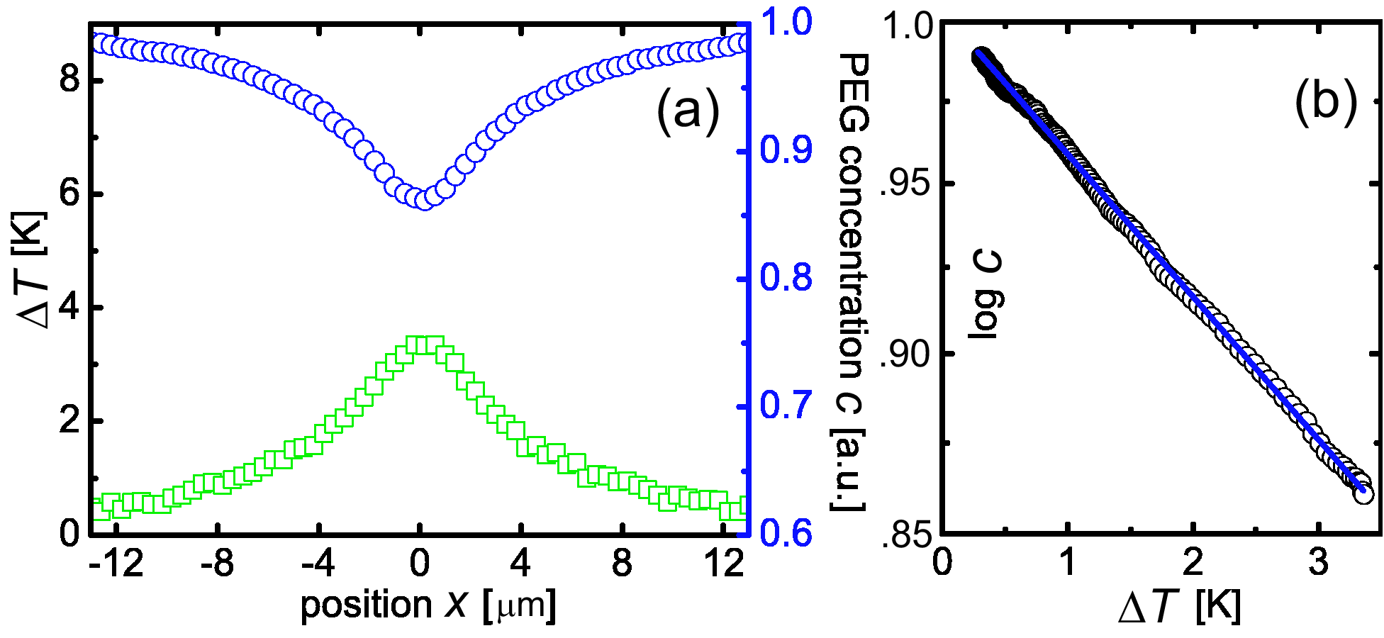
Polymer Distribution.— To elucidate the mechanism of the attraction, we measured a profile of temperature and density distribution of PEG near the laser focus in the Setup I. The temperature profile and the density of PEG molecules in chamber were shown in Fig.2amet . In this setup, temperature profile and density profile were axisymmetric. Thus the data shown below are averaged along azimuthal direction. The temperature in the laser heating spot increased approximately 1 K with a 1 mW increase in the laser power. We found that the PEG molecules were depleted by about 15% due to PEG thermophoresis when the temperature increment was about 3K in the center (Fig. 2a,b). The depletion of PEG can be described as a steady state distribution by the balance between thermodiffusion and diffusion as follows. The flux of PEG molecules obeys the relation,
| (1) |
where are the polymer density, flux, diffusion constant, and thermodiffusion constant, respectively. In the steady state (), the density satisfies
| (2) |
where the Soret coefficient defined by is measured by . We obtain [K-1] for PEG5000 from the slope in Fig.2(b), in agreement with ref.Chan et al. (2004).
Trapping Ability.— Next we examined how the accumulation of beads is dependent on the concentration of polymer and on the local temperature increase by measuring the density distribution of beads. The fluorescence images of beads were recorded for different PEG6000 concentrations. In Fig. 3, spatial distributions of fluorescent polystyrene beads (100 nm, 0.05%) were displayed. Each image was integrated for 300 frames in 20 seconds and was linearly scaled to 256 grey levels. In 0% solution, the density decreased from the outer region to the center (Fig.3a). This depletion of beads is explained quantitatively by thermophoresis, as described below. In 1% solution (Fig. 3b), there was still no trapping, but the degree of depletion is diminished compared to the 0% solution. In 2% solution, the trapping phenomenon began to appear and the fluorescence intensity roughly doubled in the hot region (Fig.3c). In the 3.5% and 5% solutions, the density increased approximately 10- and 100-fold at the center compared with the surrounding concentrations, respectively (Fig.3d,e).
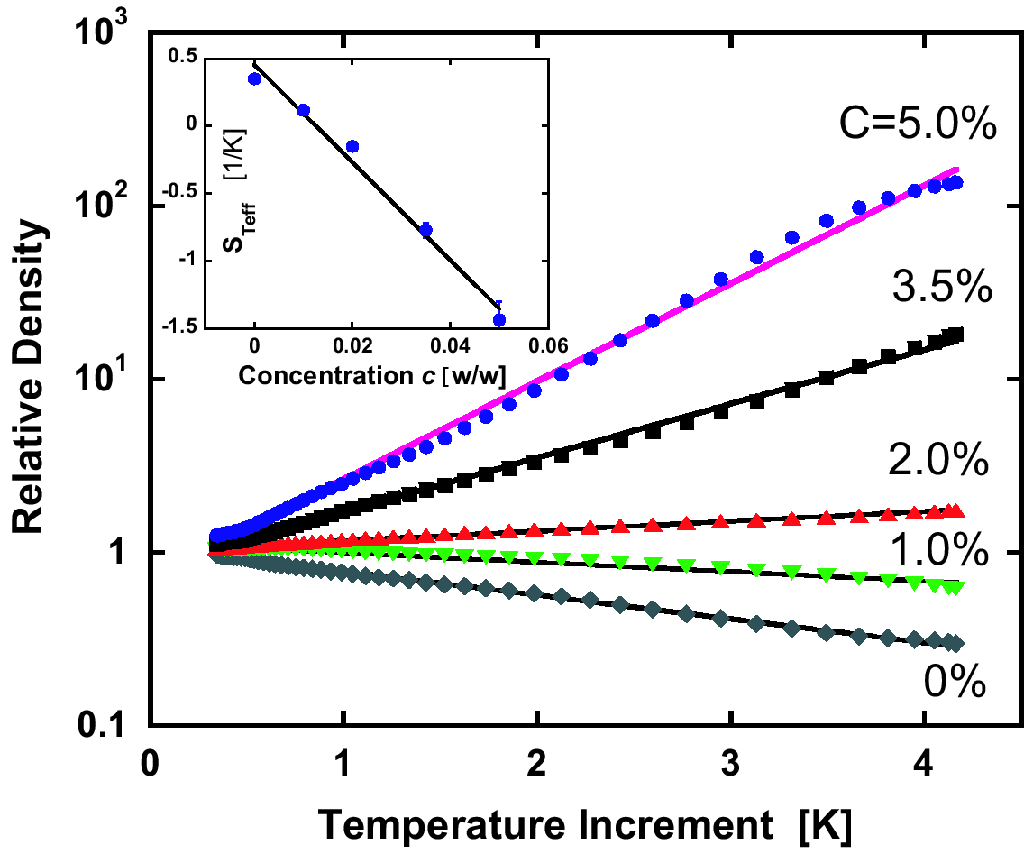
In Fig.4, the particle density at each distance from the center is plotted against temperature at the same radius (Fig.4). The result for 0% solution can be described as a steady state distribution due to thermophoresis. The density of beads, , obeys Eq. (2) with replacement of by and by , where is the Soret coefficient of the beads. Neglecting temperature dependence of , an exponential distribution is obtainedBraun and Libchaber (2002)
| (3) |
The slope of the curve in Fig.4 gives the Soret coefficient as [K-1] in agreement with previous resultsDuhr and Braun (2006). Importantly, even in the presence of polymer, the particle density varied exponentially as a function of the temperature increment (see Fig.4). Therefore a slope of each curves gives an effective Soret coefficient, . We plot as a function of PEG concentration in the inset of Fig.4. As a first-order approximation, decreased linearly with the increase of polymer concentration according to the relation
| (4) |
with [K-1] as the best fit, and where is the concentration of PEG in weight fraction, or [nm3K-1] for in volume density. Hence, the sign and the magnitude of the Soret coefficient was controlled by varying the concentration of polymer.
Theory.— The main results obtained in our experiment are quantitatively explained by the hydrodynamic theoriesAnderson (1989); Piazza and Parola (2008); Würger (2007). We consider a spherical particle of radius in a polymer solution, see the inset of Fig. 5. In a dilute regime valid to our experiments, PEG distribution around the particle obeys Boltzmann distribution, , where is the concentration at infinity, is the Boltzmann constant, and is a short range potential for PEG molecules. As a PEG is non-ionic and inactive, represents an entropic repulsion from the colloid surface with its interaction distance . Note that also defines a length scale of the depletion interaction between colloids at equilibriumAsakura and Oosawa (1958); Crocker et al. (1999). In a temperature gradient applied along direction, a PEG gradient is developed according to Eq. (3). As changes only gradually at a scale of , the steady state distribution is given by . Neglecting interactions between polymers, an osmotic force density is given by , which is written in, , where , and was used. The velocity field around the colloidal sphere, , is obtained by solving the Stokes equation, , with the incompressibility condition, , where is the viscosity of the solution and is the hydrostatic pressure. The Stokes equation is rewritten in the following transparent form,
| (5) |
where and . In Eq. (5), the osmotic pressure, , is compensated by a hydrostatic pressure . (This ensures the absence of osmotic force on the particle proportional to its volume, .) The nonequilibrium force, in Eq. (5), on the other hand, is balanced with the shear viscous force, , leading to a fluid flow relative to the colloid surface and a phoretic motion of the colloid at a velocity relative to the fluid at infinity. Solving Eq. (5) in the colloid-reference frame with the no-slip boundary condition, and summing up the resulting fluid stress over the colloidal surface, we obtain the total force acting on the colloid as Piazza and Parola (2008). This must be zero because no external force is acting on the colloid. Choosing a specific potential of hard-core type (i.e., for and zero otherwise), we finally arrive at
| (6) |
Plugging then Eq. (6) into an expression of the density current of colloids, Würger (2007), and rewriting it in a form , we see that an effective Soret coefficient is given by
| (7) |
which confirms the linear dependence on in Eq. (4), where .

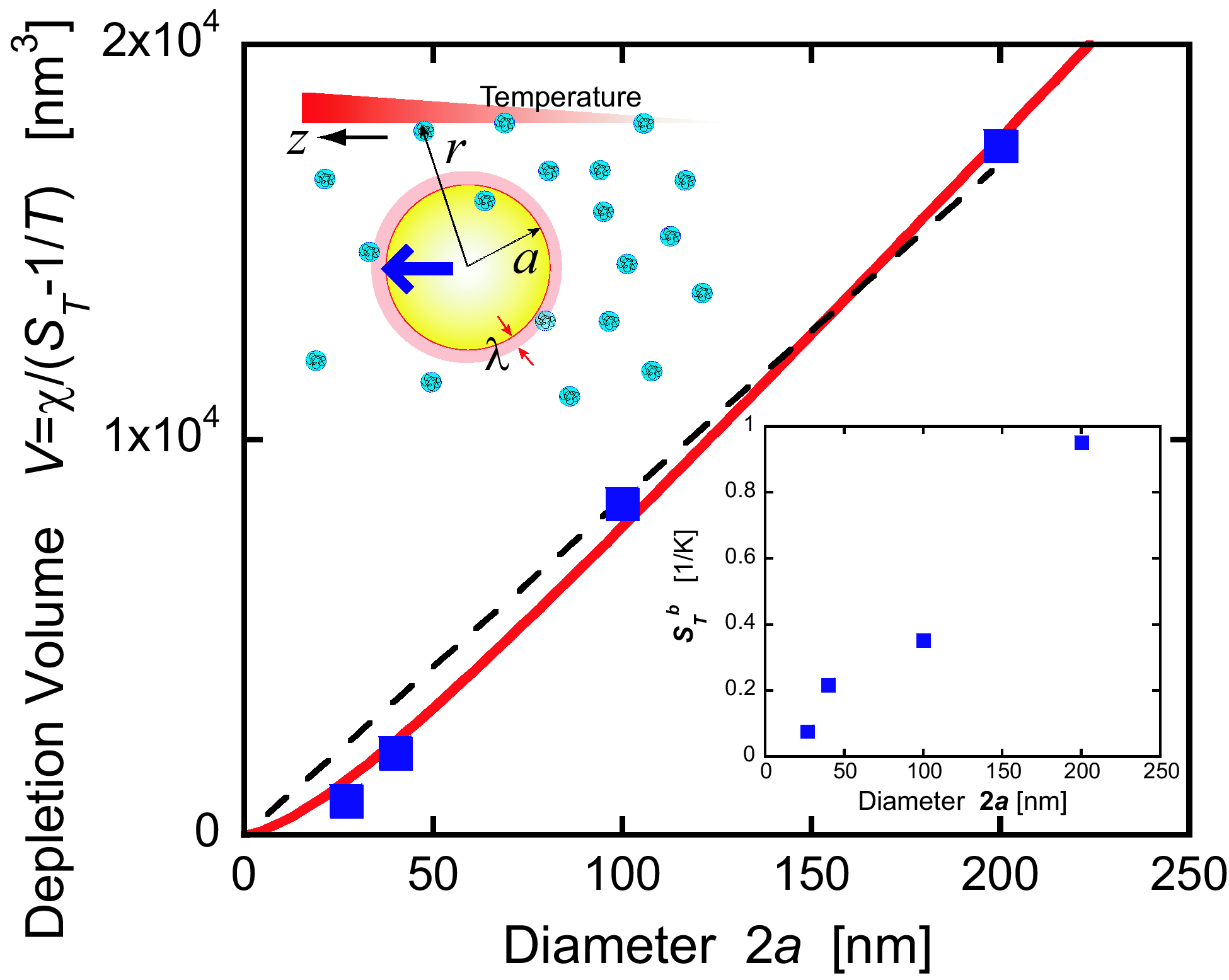
Equation (7) is compared to our experimental data in Fig. 4 inset by employing for PEG with MW=7500 estimeted from Chan et al. (2004). The best fit gives nm, which is close to the PEG gyration radius nm Kawaguchi et al. (1997). We consider the agreement satisfactory, as should be of the order of a PEG size. A further consistency check was made by looking at the dependence of on the particle radius . To focus only on this dependence of , we define an effective depletion volume at nonequilibrium . First, we performed the series of experiments for four different colloid sizes, and extracted from the data on , which are plotted in Fig. 4 as a function of the diameter . The data shows an overall linear dependence, and the agreement with the theoretical prediction for no-slip condition, , obtained from Eq. (7) is good. Note that we used nm obtained before, and thus no adjustable parameter is assumed. Nevertheless, it would be important to examine effects of slip of the polymer solution upon the enhancement of the Soret effect, since the perfect slip condition predicts a different scaling, Würger (2007). A boundary condition for a general slip case is a so-called Navier condition Happel and Brenner (1965), given by at , where and are the tangential components of the fluid stress and velocity. The parameter defines a slip length on the surface (the no-slip or perfect slip limit is attained for or )Ajdari and Bocquet (2006). In this Navier case we obtain . This formula, with nm and nm as the best fit, actually improves the agreement with the data for nm, where the deviation from the linear dependence is observable (see Fig. 5). In our system, therefore, a fluid slip might be relevant for nm (with a relatively large slip lengh nm).
Summary.— We have developed a micro-manupulation technique allowing us to invert and amplify the movement of colloid particles due to thermophoresis of polymers induced by laser focusing. The polymer concentration gradient created by thermophoresis causes migration of a particle with a speed determined by the balance between the driving force and the viscous force. This new method and its application to trapping of molecules based on nonequilibrium effects does not rely on a specific character of particles and polymers, and thus provides further applications for manipulating a diverse range of colloidal particles as well as biological cells and DNA moleculesjia ; ichikawa:2005 . Our technique is also applicable with other water-soluble molecules111We confirmed that polyvinylpyrrolidone (PVP) and sodium polystyrene sulfonate (NaPSS) also work, yet the effect is strongest for PEG.. We also note that the effect is stronger for PEG with a larger molecular weight at the same monomer concentration as far as it is lower than the overlap concentration.
This works is supported by Grant-in-Aid for Scientific Research from MEXT Japan.
References
- (1)
- de Groot and Mazur (1984) S. de Groot and P. Mazur, Non-Equilibrium Thermodynamics (Dover Publications, 1984).
- Mitchell (1961) P. Mitchell, Nature 191, 144 (1961), see also P. Nelson et al., Biological Physics: Energy, Information, Life, (W H Freeman, 2007).
- (4) M.Z. Bazant and T.M. Squires, Phys. Rev. Lett 92, 066101 (2004).
- Janča et al. (2003) J. Janča, J.-F. Berneron, and R. Boutin, J. Coll. Int. Sci. 260, 317 (2003).
- (6) H.R. Howse et al., Phys. Rev. Lett. 99, 048102 (2007).
- Giglio and Vendramini (1977) M. Giglio and A. Vendramini, Phys. Rev. Lett. 38, 26 (1977).
- (8) B-J. de Ganset al., Phys. Rev. Lett. 91, 245501 (2003).
- Ning et al. (2006) H. Ning, et al., J. Chem. Phys. 125, 204911 (2006).
- Duhr and Braun (2006) S. Duhr and D. Braun, Proc. Nat. Acad. Sci. USA 103, 19678 (2006).
- (11) M. Braibanti, D. Vigolo, and R. Piazza, Phys. Rev. Lett. 100, 108303 (2008).
- Ebel et al. (1988) J. P. Ebel, J. L. Anderson, and D. C. Prieve, Langmuir 4, 396 (1988).
- Anderson (1989) J. Anderson, Ann. Rev. Fluid Mech. 21, 61 (1989).
- Nardi (1999) J. Nardi, R. Bruinsma, and E. Sackmann, Phys. Rev. Lett. 82, 5168 (1999).
- Braun and Libchaber (2002) D. Braun and A. Libchaber, Phys. Rev. Lett. 89, 188103 (2002).
- Staffeld and Quinn (1989) P. O. Staffeld and J. A. Quinn, J. Coll. Int. Sci. 130, 69 (1989).
- Abécassis et al. (2008) B. Abécassiset al., Nature Materials 7, 785 (2008).
- deGennes (1981) F. Brochard and P.G. de Gennes, CR. Acad. Sc. Paris 293, 1025 (1981).
- Piazza and Parola (2008) R. Piazza and A. Parola, J. Phys. Cond. Mat. 20, 153102 (2008), id. Eur. Phys. J. E 15, 255 (2004).
- Würger (2007) A. Würger, Phys. Rev. Lett. 98, 138301 (2007).
- Jiang and Sano (2007) H.-R. Jiang and M. Sano, Appl. Phys. Lett. 91, 154104 (2007).
- (22) H.-R. Jiang et al. (to be published).
- (23) http://daisy.phys.s.u-tokyo.ac.jp/articles/JiangSp.mpg for the supplemental movie.
- (24) The temperature profile in the solution was measured from the intensity profiles of a temperature-sensitive fluorescent dye, 2’,7’ Bis(2-carboxyethyl)-5(6) carboxyfluorescein (BCECF)Braun and Libchaber (2002); Duhr and Braun (2006). The density of PEG molecules was measured by using fluorescently labeled PEG molecules (rhodamin-PEG, MW5000, Nanos).
- Chan et al. (2004) J. Chan et al., J. Sol. Chem. 32, 197 (2004).
- Asakura and Oosawa (1958) S. Asakura and F. Oosawa, J. Poly. Sci. 33, 183 (1958).
- Crocker et al. (1999) J. C. Crocker et al., Phys. Rev. Lett. 82, 4352 (1999).
- Kawaguchi et al. (1997) S. Kawaguchiet al., Polymer 38, 2885 (1997).
- Happel and Brenner (1965) J. Happel and H. Brenner, Low Reynolds Number Hydrodynamics (Noordhoff International Pub., Leyden, 1965).
- Ajdari and Bocquet (2006) A. Ajdari and L. Bocquet, Phys. Rev. Lett. 96, 186102 (2006).
- (31) M. Ichikawa, Y. Matsuzawa, K. Yoshikawa, J. Phys. Soc. Jpn. 74, 1958 (2006).