Uncovering the triplet ground state of triangular graphene nanoflakes
engineered with atomic precision on a metal surface
Abstract
Graphene can develop large magnetic moments in custom crafted open-shell nanostructures such as triangulene, a triangular piece of graphene with zigzag edges. Current methods of engineering graphene nano-systems on surfaces succeeded in producing atomically precise open-shell structures, but demonstration of their net spin remains elusive to date. Here, we fabricate triangulene-like graphene systems and demonstrate that they possess a spin ground state. Scanning tunnelling spectroscopy identifies the fingerprint of an underscreened Kondo state on these flakes at low temperatures, signaling the dominant ferromagnetic interactions between two spins. Combined with simulations based on the meanfield Hubbard model, we show that this -paramagnetism is robust, and can be manipulated to a state by adding additional H-atoms to the radical sites. Our results demonstrate that -paramagnetism of high-spin graphene flakes can survive on surfaces, opening the door to study the quantum behaviour of interacting -spins in graphene systems.
In spite of their apparent simplicity, custom-crafted graphene nanoflakes (GNF) are predicted to exhibit complex magnetic phenomenology Yazyev (2010) with promising possibilities as active components of a new generation of nanoscale devices Wang et al. (2009); Han et al. (2014); Bullard et al. (2015). As predicted by Lieb’s theorem for bipartite lattices Lieb (1989), certain shapes of graphene structures may accommodate a spin imbalance in the electron cloud, resulting in GNFs with a net magnetic moment. Graphene -paramagnetism is more delocalized, mobile, and isotropic than conventional magnetism from or states Trauzettel et al. (2007), and can be electrically addressable Li et al. (2019); Mishra et al. (2020a). Furthermore, the magnetic moments and their correlations in GNFs can be precisely engineered through their sizes, edge topology, or chemical doping Fernández-Rossier and Palacios (2007); Wang et al. (2008); Kan et al. (2012); Sandoval-Salinas et al. (2019).
The fabrication of such GNFs has been hindered due to their high reactivity Clar and Stewart (1953). As open-shell structures, the presence of unpaired electrons (radicals) makes the synthesis difficult. First unsubstituted triangulene was synthesized by dehydrogenating precursor molecules with the tip of a scanning tunneling microscope (STM) Pavliček et al. (2017). Very recently, triangular GNFs with larger sizes have been synthesized through an on-surface synthetic (OSS) approach Mishra et al. (2019); Su et al. (2019); Mishra et al. (2020b), an strategy for fabricating atomically precise graphene flakes on a metallic surface Cai et al. (2010); Treier et al. (2011); Ruffieux et al. (2016); Clair and de Oteyza (2019). Despite such progresses in fabrication, their magnetic properties on a surface have not been demonstrated experimentally.
Here we report the OSS of triangulene-like GNFs and demonstrate that they have a triplet ground state. The synthesized GNFs have reduced symmetry compared to triangulene, which increases the localization of the magnetic moments. High-resolution STM images and spectroscopy allow us to identify the two spin centers on the GNFs and map their localization. Their ferromagnetic correlations are characterized by the spin-1 Kondo effect Sasaki et al. (2000); Roch et al. (2009); Parks et al. (2010), which happens due to the incomplete screening of two coupled spins by conducting electrons of the metal substrate. Our results also show that the strength of correlations between spins depends on the distance between them.
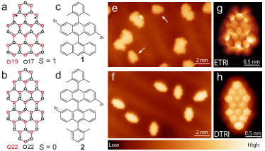
Figure 1a shows the chemical structures of the GNF characterized in the experiments, named here extended-triangulene (ETRI). This GNF has 19 carbon atoms in one sub-lattice (highlighted by red circles) and 17 carbon atoms in the other (black circles), which results in a total spin according to Lieb’s theorem Lieb (1989). For comparison, we also produced the GNF in Fig. 1b (named as double-triangulene (DTRI)), which has 22 carbon atoms in both sub-lattices and, thus, a spin ground state. These GNFs we synthesized by depositing the respective molecular precursors, shown in Fig. 1c,d (details in supplementary material (SM) SI ), on a Au(111) surface at 330 ∘C. The hot surface activates a simultaneous debromination and cyclodehydogenation step that planarizes them into their final structures.
Low-temperature STM overview images of the Au (111) surface after deposition of precursors 1 and 2, are shown in Figs. 1e and 1f, respectively. While the reacted precursor 1, ETRI, appear on the surface mostly as monomers and dimers in a ratio of approximately 1:7, nearly all deposited precursors 2, DTRI, remain as monomer species. In every case, the monomers adopt a planar configuration, as expected for the the structures in Fig. 1a,b. Furthermore, high-resolution STM current images using a CO-terminated tip Gross et al. (2009); Kichin et al. (2011) (Fig. 1g,h) reproduce the chemical bond structures of Fig. 1a,b, indicating the successful synthesis of ETRI and DTRI. It is worth noting that the current image of DTRI shows merely the backbone structure, as also observed in similar symmetric systems Beyer et al. (2019), while the current image of ETRI shows additional bright features at the edges. Considering that these images were recorded at 2 mV, the bright features surrounding the backbone structure indicate an enhancement of the local density of states (LDOS) close to Fermi level for the ETRI molecule.
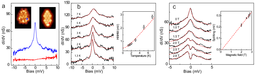
The different shape in the images is better manifested in differential conductance () spectra measured on both types of GNFs (Fig. 2a). The spectrum on ETRI shows a pronounced and narrow (FWHM 1 meV) peak centered at zero bias. The zero-bias peak broadens anomalously fast with temperature (as described in Fig. 2b) and splits with magnetic fields (Fig. 2c), demonstrating that it is a manifestation of the Kondo effect Ternes et al. (2009). A (zero-bias) Kondo-derived resonance reflects the screening of a local spin by conduction electrons Ternes (2015) and, hence, is a direct proof of the presence of localized magnetic moments on ETRI even when it lays on a metal surface. On the contrary, the spectra taken on DTRI is featureless. Furthermore, both wide bias range spectra and maps reveal their closed-shell ground state (see SM).
The temperature and magnetic field dependence of the Kondo resonances provides further insight on the nature of the spin state of ETRI. Although the Kondo effect is frequently described on systems, it also occurs for higher spin configurations Roch et al. (2009); Parks et al. (2010). For , a zero bias resonance reflects a partly screened spin, i.e. with only one interaction channel with the substrate Pustilnik and Glazman (2001). Similar to the spin-1/2 case, the resonance broadens with temperature (with a Kondo temperature K, after Fig. 2b, see SM), but shows a larger sensitivity to the magnetic field Roch et al. (2009); Parks et al. (2010). While the Kondo resonance of a spin-1/2 system in the strong coupling regime () splits linearly with magnetic fields only above a critical field Costi (2000); Li et al. (2019), an underscreened conserves some magnetic moment, and its zero bias state splits already with Parks et al. (2010); Roch et al. (2009). In tunneling spectra, such peak split should become visible as soon as the Zeeman energy is greater than the thermal broadening (), this is above one Tesla at the 1.2 K of our experimental setup. If the Kondo resonance in Fig. 2b were caused by a state, it should appear split in the spectra only for magnetic fields above 3.5 T. However, the peak appears split already at T, proving that the ETRI molecule has a spin in an underscreened Kondo state on the gold substrate SI .
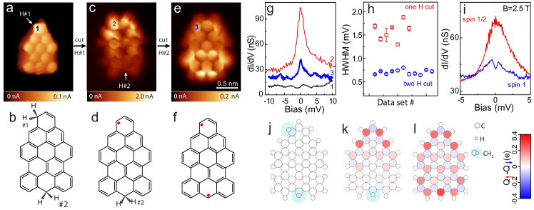
The spin-1 configuration of the triangular GNFs was further supported by tip-induced manipulation experiments. Due to their bi-radical character, the zigzag sites show some reactivity, and are frequently found passivated by hydrogen atoms produced during the OSS reactions Talirz et al. (2013); Li et al. (2019). The passivated carbon sites can be identified in high-resolution images by their larger bond length Gross et al. (2012) due to the change from to hybridization. The STM image in Fig. 3a shows the bare backbone structure of an ETRI with two H-atoms passivating the zigzag sites indicated with arrows (as in Fig. 3b). The corresponding spectrum is featureless around zero bias (black curve in Fig. 3g), explaining the absence of bright features in the STM image.
We first cut off one of the passivated H atoms by placing the STM tip on top of site #1 in Fig. 3a and ramping up the sample bias above 1.5 V Talirz et al. (2013); Li et al. (2019). The H removal was monitored by a sudden step in the tunnelling current Li et al. (2019). The STM image afterwards appeared with an enhanced LDOS signal around the #1 site (Fig. 3c) and the spectrum on site #1 presented a pronounced zero-energy peak (red Fig. 3g). We identify this to a spin-1/2 Kondo resonance Li et al. (2019), resulting from the single radical state recovered by the removal of the extra H #1 atom (Fig. 3d). Following a similar process to cleave the second H-CH bond at site #2, recovered bright current features all around the ETRI backbone, similar to the reference bi-radical structure of Fig. 1g. The spectrum measured again over site #1 shows now the Kondo resonance with smaller amplitude and line-width, similar to the one in Fig. 2a, agreeing with its underscreened S=1 state. As quantified in Fig. 3h, the Kondo resonance of the doubly dehydrogenated GNFs appeared repetitively with line-width about FWHM 0.7 meV, significantly smaller than in the singly-passivated species (Fig. 3g,h), hinting to their different Kondo states. The spin assignment of each species was further corroborated by comparing their response to a magnetic field of 2.5 T. While the intermediate specie did not present any detectable split of the Kondo resonance, the bi-radical one exhibited a clear split, as in Fig. 2c, demonstrating the larger spin polarization of its underscreened ground state.
The emergence of magnetism in ETRI is reproduced by both mean field Hubbard (MFH) and density functional theory (DFT) simulations (see SM). Similar to the case of triangulene Yazyev (2010); Ortiz et al. (2019), this modified GNF present two (singly-occupied) zero-energy modes, with a triplet ground state clearly preferred over a singlet one by more than 60 meV (see Figs. S9 and S16). The spin polarization (Fig. 3l) shows two spin centers localized at opposite sides of the triangular GNF, with a distribution that resembles the experimental current maps at zero bias (Fig. 1g and Fig. 3e). We note that their ferromagnetic exchange interaction is much larger than the Kondo energy scale ( meV), thus explaining the underscreened Kondo ground state found in the experiments. MFH simulations also shows that hydrogen passivation of the radical states quenches sequentially the spins, turning ETRI into a doublet (Fig. 3k) or completely non-magnetic (Fig. 3j), as demonstrated by the electron-induced removal of extra H-atoms in the experiments.
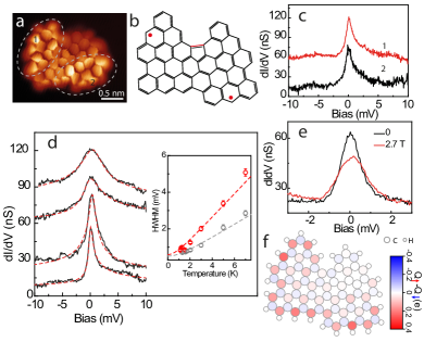
A paramagnetic ground state was also found on molecular dimers formed during the OSS process (as shown in Fig. 1e), but their larger size crucially affects their magnetic properties. Fig. 4a shows a high resolution image of a dimer. Two ETRI moieties are covalently linked together (Fig. 4b) following the Ullmann-like C-C coupling of their halogenated sites De Oteyza et al. (2016). During the cyclodehydrogenation step, an extra pentagonal ring is created between them, as highlighted by a red bond in Fig. 4b, reducing the number of radicals of the dimer to only two. The bi-radical state is experimentally demonstrated in the supplementary material SI .
The STM image of Fig. 4a also reproduces bright features around the dimer backbone (indicated with dashed ellipses) corresponding to the localization of the Kondo effect: spectra measured on either of the two show pronounced peaks centered at zero bias (Fig. 4c), which broaden with temperature (Fig. 4d) and magnetic field (Fig. 4e). However, these Kondo resonances broaden faster with than for the case of ETRI (both compared in the inset of Fig. 4d), and show no split at T, signaling a different magnetic ground state.
Our MFH simulations of ETRI dimers like in Fig. 4b confirm their bi-radical state, but triplet and singlet solutions lay now closer in energy (see SM). On a surface, magnetic ground state probably behaves as two non-interacting spins. This explains the lack of -induced split and the faster broadening with , expected for s=1/2 Kondo systems Parks et al. (2010). The spin polarization maps obtained from MFH simulations reproduce well the bright current regions on the dimers (Fig. 4f), further corroborating that this signal can be associated to the spin distribution. The origin of such small magnetic exchange between the two radical states is related to the presence of a pentagon between them, and to the larger separation between the two spin centers. In fact, in the absence of this extra C-C bond MFH simulations find a robust ground state. Thus, pentagonal rings embedded in certain sites of an open-shell GNF can affect critically its magnetic state by quenching a radical state and modifying the total spin (here by S=1) Mishra et al. (2020), just as extra hydrogen atoms do Li et al. (2019), but their placement on a specific site can designed precisely during the OSS process.
In summary, we have demonstrated the magnetic ground state of graphene flakes fabricated deterministically with a triangular-like shape. The survival of the state on the metal surface is identified first through the net spin polarization of their Kondo state, which reacts to magnetic fields as an underscreened spin triplet. The state was further confirmed through removal of hydrogen atoms by tip manipulation experiments, which revealed the stepwise emergence of two spins localized at different sides of the flakes. We note that our findings here contrast with the absence of magnetic signals in previous studies of larger triangulene-flakes Mishra et al. (2019); Su et al. (2019). It is therefore an interesting subject for future work to unveil the precise interplay between size and symmetry of GNFs for their magnetic state over a metal surface. Nevertheless, the existence of GNFs with a net spin on a surface opens the door to new investigations of their spin dynamics and coherence over an inorganic supports, which are crucial aspects for utilizing graphene nanosystems Lombardi et al. (2019) in quantum spintronics applications.
Acknowledgements.
We are indebted to Carmen Rubio for fruitful discussions. We acknowledge financial support from Spanish AEI (MAT2016-78293-C6, FIS2017-83780-P, and the Maria de Maeztu Units of Excellence Programme MDM-2016-0618), the European Union’s Horizon 2020 (FET-Open project SPRING Grant. no. 863098), the Basque Departamento de Educación through the PhD fellowship No. PRE201920218 (S.S.), the Xunta de Galicia (Centro singular de investigación de Galicia accreditation 2016-2019, ED431G/09), and the European Regional Development Fund (ERDF).References
- Yazyev (2010) O. V. Yazyev, Rep. Prog. Phys. 73, 56501 (2010).
- Wang et al. (2009) W. L. Wang, O. V. Yazyev, S. Meng, and E. Kaxiras, Phys. Rev. Lett. 102, 157201 (2009).
- Han et al. (2014) W. Han, R. K. Kawakami, M. Gmitra, and J. Fabian, Nat. Nano. 9, 794 (2014).
- Bullard et al. (2015) Z. Bullard, E. C. Girão, J. R. Owens, W. A. Shelton, and V. Meunier, Sci. Rep. 5, 7634 (2015).
- Lieb (1989) E. H. Lieb, Phys. Rev. Lett. 62, 1201 (1989).
- Trauzettel et al. (2007) B. Trauzettel, D. V. Bulaev, D. Loss, and G. Burkard, Nat. Phys. 3, 192 (2007).
- Li et al. (2019) J. Li, S. Sanz, M. Corso, D. J. Choi, D. Peña, T. Frederiksen, and J. I. Pascual, Nat. Commun. 10, 200 (2019).
- Mishra et al. (2020a) S. Mishra, D. Beyer, K. Eimre, S. Kezilebieke, R. Berger, O. Gröning, C. A. Pignedoli, K. Müllen, P. Liljeroth, P. Ruffieux, X. Feng, and R. Fasel, Nat. Nanotechnol. 15, 22 (2020a).
- Fernández-Rossier and Palacios (2007) J. Fernández-Rossier and J. J. Palacios, Phys. Rev. Lett. 99, 177204 (2007).
- Wang et al. (2008) W. L. Wang, S. Meng, and E. Kaxiras, Nano Letters 8, 241 (2008).
- Kan et al. (2012) E. Kan, W. Hu, C. Xiao, R. Lu, K. Deng, J. Yang, and H. Su, J. Am. Chem. Soc. 134, 5718 (2012).
- Sandoval-Salinas et al. (2019) M. E. Sandoval-Salinas, A. Carreras, and D. Casanova, Phys. Chem. Chem. Phys. 21, 9069 (2019).
- Clar and Stewart (1953) E. Clar and D. G. Stewart, J. Am. Chem. Soc. 75, 2667 (1953).
- Pavliček et al. (2017) N. Pavliček, A. Mistry, Z. Majzik, N. Moll, G. Meyer, D. J. Fox, and L. Gross, Nat. Nanotechnol. 12, 308 (2017).
- Mishra et al. (2019) S. Mishra, D. Beyer, K. Eimre, J. Liu, R. Berger, O. Gröning, C. A. Pignedoli, K. Müllen, R. Fasel, X. Feng, and P. Ruffieux, J. Am. Chem. Soc. 141, 10621 (2019).
- Su et al. (2019) J. Su, M. Telychko, P. Hu, G. Macam, P. Mutombo, H. Zhang, Y. Bao, F. Cheng, Z.-Q. Huang, Z. Qiu, S. J. R. Tan, H. Lin, P. Jelínek, F.-C. Chuang, J. Wu, and J. Lu, Science Advances 5 (2019).
- Mishra et al. (2020b) S. Mishra, D. Beyer, K. Eimre, R. Ortiz, J. Fernandez-Rossier, R. Berger, O. Groening, C. A. Pignedoli, R. Fasel, X. Feng, and P. Ruffieux, “Collective all-carbon magnetism in triangulene dimers,” (2020b), arXiv:2003.00753 [cond-mat.mes-hall] .
- Cai et al. (2010) J. Cai, P. Ruffieux, R. Jaafar, M. Bieri, T. Braun, S. Blankenburg, M. Muoth, A. P. Seitsonen, M. Saleh, X. Feng, K. Mullen, and R. Fasel, Nature 466, 470 (2010).
- Treier et al. (2011) M. Treier, C. A. Pignedoli, T. Laino, R. Rieger, K. Müllen, D. Passerone, and R. Fasel, Nature Chem. 3, 61 (2011).
- Ruffieux et al. (2016) P. Ruffieux, S. Wang, B. Yang, C. Sánchez-Sánchez, J. Liu, T. Dienel, L. Talirz, P. Shinde, C. A. Pignedoli, D. Passerone, T. Dumslaff, X. Feng, K. Müllen, and R. Fasel, Nature 531, 489 (2016).
- Clair and de Oteyza (2019) S. Clair and D. G. de Oteyza, Chem. Rev. 119, 4717 (2019).
- Frota (1992) H. O. Frota, Phys. Rev. B 45, 1096 (1992).
- Zhang et al. (2013) Y.-H. Zhang, S. Kahle, T. Herden, C. Stroh, M. Mayor, U. Schlickum, M. Ternes, P. Wahl, and K. Kern, Nat. Commun. 4, 2110 (2013).
- Nagaoka et al. (2002) K. Nagaoka, T. Jamneala, M. Grobis, and M. F. Crommie, Phys. Rev. Lett. 88, 77205 (2002).
- (25) Online supplementary material, which includes Refs. [40-47]..
- Ternes (2015) M. Ternes, New J. Phys. 17, 63016 (2015).
- Sasaki et al. (2000) S. Sasaki, S. De Franceschi, J. M. Elzerman, W. G. van der Wiel, M. Eto, S. Tarucha, and L. P. Kouwenhoven, Nature 405, 764 (2000).
- Roch et al. (2009) N. Roch, S. Florens, T. A. Costi, W. Wernsdorfer, and F. Balestro, Phys. Rev. Lett. 103, 197202 (2009).
- Parks et al. (2010) J. J. Parks, A. R. Champagne, T. A. Costi, W. W. Shum, A. N. Pasupathy, E. Neuscamman, S. Flores-Torres, P. S. Cornaglia, A. A. Aligia, C. A. Balseiro, G. K. L. Chan, H. D. Abruña, and D. C. Ralph, Science 328, 1370 (2010).
- Gross et al. (2009) L. Gross, F. Mohn, N. Moll, P. Liljeroth, and G. Meyer, Science 325, 1110 (2009).
- Kichin et al. (2011) G. Kichin, C. Weiss, C. Wagner, F. S. Tautz, and R. Temirov, Journal of the American Chemical Society 133, 16847 (2011).
- Beyer et al. (2019) D. Beyer, S. Wang, C. A. Pignedoli, J. Melidonie, B. Yuan, C. Li, J. Wilhelm, P. Ruffieux, R. Berger, K. Müllen, R. Fasel, and X. Feng, J. Am. Chem. Soc. , 8 (2019).
- Ternes et al. (2009) M. Ternes, A. J. Heinrich, and W.-D. Schneider, J. Phys.: Condens. Matter 21, 053001 (2009).
- Pustilnik and Glazman (2001) M. Pustilnik and L. I. Glazman, Phys. Rev. Lett. 87, 216601 (2001).
- Costi (2000) T. A. Costi, Phys. Rev. Lett. 85, 1504 (2000).
- Talirz et al. (2013) L. Talirz, H. Söde, J. Cai, P. Ruffieux, S. Blankenburg, R. Jafaar, R. Berger, X. Feng, K. Müllen, D. Passerone, R. Fasel, and C. A. Pignedoli, J. Am. Chem. Soc. 135, 2060 (2013).
- Gross et al. (2012) L. Gross, F. Mohn, N. Moll, B. Schuler, A. Criado, E. Guitián, D. Peña, A. Gourdon, and G. Meyer, Science 337, 1326 (2012).
- Ortiz et al. (2019) R. Ortiz, R. Á. Boto, N. García-Martínez, J. C. Sancho-García, M. Melle-Franco, and J. Fernández-Rossier, ArXiv , 1906.08544 (2019).
- De Oteyza et al. (2016) D. G. De Oteyza, A. García-Lekue, M. Vilas-Varela, N. Merino-Díez, E. Carbonell-Sanromà, M. Corso, G. Vasseur, C. Rogero, E. Guitián, J. I. Pascual, J. E. Ortega, Y. Wakayama, and D. Peña, ACS Nano 10, 9000 (2016).
- Mishra et al. (2020) S. Mishra, D. Beyer, R. Berger, J. Liu, O. Gröning, J. I. Urgel, K. Müllen, P. Ruffieux, X. Feng, and R. Fasel, J. Am. Chem. Soc. (2020).
- Lombardi et al. (2019) F. Lombardi, A. Lodi, J. Ma, J. Liu, M. Slota, A. Narita, W. K. Myers, K. Müllen, X. Feng, and L. Bogani, Science 366, 1107 (2019).
- Hancock et al. (2010) Y. Hancock, A. Uppstu, K. Saloriutta, A. Harju, and M. J. Puska, Phys. Rev. B 81, 245402 (2010)..
- Papior (2019) N. Papior, sisl: v0.9.6 (2019)..
- Hirshberg and Fischer (1953) Y. Hirshberg and E. Fischer, J. Chem. Soc. pp. 629–636 (1953)..
- Goldhaber-Gordon et al. (1998) D. Goldhaber-Gordon, J. Göres, M. A. Kastner, H. Shtrikman, D. Mahalu, and U. Meirav, Phys. Rev. Lett. 81 (1998)..
- Daroca et al. (2018) D. P. Daroca, P. Roura-Bas, and A. A. Aligia, Phys. Rev. B 98, 1 (2018)..
- Ortiz et al. (2018) R. Ortiz, N. A. García-Martínez, J. L. Lado, and J. Fernández-Rossier, Phys. Rev. B 97, 195425 (2018)..
- Soler et al. (2002) J. M. Soler, E. Artacho, J. D. Gale, A. García, J. Junquera, P. Ordejón, and D. Sánchez-Portal, J. Phys. Cond. Matter 14, 2745 (2002)..
- Perdew et al. (1996) J. P. Perdew, K. Burke, and M. Ernzerhof, Phys. Rev. Lett. 77, 3865 (1996)..